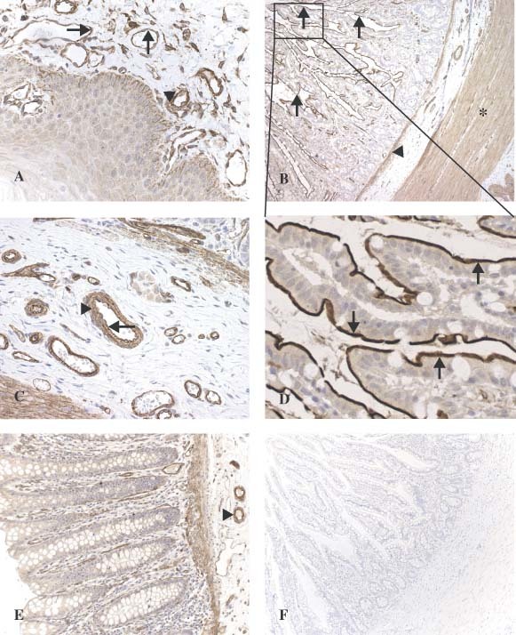
Figure 2.

Overview of oral mucosa (A). Strong staining is observed in vascular endothelium (arrow) and vascular smooth muscle cells (arrow‐head). Granular ACE2 staining is present in the basal layer of the epithelium. In the small intestine (ileum) (B), strong staining can be seen in the villous brush border (arrow), the muscularis mucosae (arrow‐head), and the muscularis propria (star). In a larger magnification of the submucosa (C), strong staining is present in vascular endothelium (arrow) and vascular smooth muscle cells (arrow‐head). In a larger magnification of the villi (D), abundant staining is seen on the brush border of the enterocytes (arrow). In the colon (E), ACE2 staining is present in endothelium and vascular smooth muscle cells from the blood vessels (arrow‐head) and in the muscular layers. Control section stained with anti‐ACE2 in the presence of the synthetic ACE2 peptide shows no staining in the small intestine (ileum) (F)
