Abstract
The axial resolving power of a light-sheet microscope is determined by the thickness of the illumination beam and the numerical aperture of its detection optics. Bessel-beam based optical lattices have generated significant interest owing to their reportedly narrow beam waist and propagation-invariant characteristics. Yet, despite their significant use in lattice light-sheet microscopy and recent incorporation into commercialized systems, there are very few quantitative reports on their physical properties and how they compare to standard Gaussian illumination beams. Here, we measure the beam properties in the transmission of dithered square lattices, which is the most commonly used variant of lattice light-sheet microscopy, and Gaussian-based light-sheets. After a systematic analysis, we find that square lattices are very similar to Gaussian-based light-sheets in terms of thickness, confocal parameter, propagation length and overall imaging performance.
1. Introduction
Light-sheet fluorescence microscopy (LSFM) is a rapidly growing volumetric imaging technique that offers life scientists a unique combination of speed, sensitivity, optical sectioning, and low phototoxicity and photobleaching [1]. In LSFM, a biological specimen is illuminated with a focused sheet of light, and fluorescence originating from this sheet of light is captured in a widefield format using modern scientific cameras. Thus, unlike traditional epi-fluorescence imaging methods that nonproductively illuminate regions above and below the detection objective depth-of-focus, a LSFM restricts the potentially damaging illumination to only the in-focus region of the specimen. This not only decreases phototoxicity and photobleaching, but delivers higher contrast imaging owing to its inherent optical sectioning capability [2].
Several factors critically influence the performance of a LSFM [3]. Like conventional epifluorescence widefield microscopes, the lateral resolution of a LSFM scales linearly with the fluorescence emission wavelength () and inversely with the numerical aperture () of the detection objective, . However, the axial resolution of a LSFM depends upon the wavelengths of the illumination sheet () and fluorescence emission (), and the numerical aperture of the excitation light-sheet () and detection objective (). In the Fourier domain, the resolution can be estimated from the highest axial frequencies that contribute to image formation, where is the solid angle over which the detection objective collects light and n is the refractive index of the medium. As a result, the axial resolution can be described as: [3]. Of note, this is an optimistic estimate that assumes no attenuation of higher axial frequencies (e.g., optical transfer function roll-off), but it nevertheless serves as an upper bound for the axial resolution of a LSFM. Consequently, for thick light-sheets, the numerical aperture of the detection objective largely dictates the axial resolution of the imaging system. In contrast, if the light-sheet can be made thinner than the detection objective depth of focus, the axial resolution approaches that of the illumination beam waist.
Critically, for Gaussian optics, the field of view of a LSFM is only as large as the region over which the illumination beam waist is narrow () and thus approximates a sheet of light. Commonly, this is estimated from the confocal parameter, which is 2 times the Rayleigh length (ZR) of the beam, where . However, because the Rayleigh length decreases nonlinearly as the beam waist gets smaller, one must make a critical tradeoff between the field of view and the thickness of the illumination beam. Importantly, for very narrow beam waists (e.g., those approaching the wavelength of light), diffraction renders the Gaussian approximation inaccurate. An alternative description of light-sheet thickness and propagation length can be obtained by using the scalar diffraction theory for a laser focus. Here, the thickness and propagation length of an illumination beam can be estimated as and , respectively, where is the half opening angle over which the illumination objective delivers light (more detail provided in Appendix A, derived in [4]). Nonetheless, these too are subject to the same tradeoff between propagation length and beam waist thickness.
In an effort to overcome the tradeoff between the size of the field of view and light-sheet thickness, several labs adopted propagation-invariant illumination beams (e.g., Bessel and Airy beams), which in theory, maintain a tight 2D (e.g., circularly symmetric) focus throughout an infinite propagation length [5–8]. In practice, such beams are finite in length and accompanied by sidelobe structures that grow in magnitude with the propagation length of the beam [9]. For many cases, these sidelobe structures contain more excitation energy than the tightly focused central lobe, which results in heightened image blur and diminished optical sectioning [9,10]. As such, in an effort to partially reduce blur and restore image contrast, iterative deconvolution routines become increasingly necessary. Thus, while propagation-invariant beams overcome the divergence of Gaussian optics, they do so by compromising optical sectioning (which is a key feature of LSFMs), potentially increasing photobleaching and introducing significant computational overhead.
More recently, carefully crafted coherent superpositions of Bessel beams have been reported to provide a compromise between light-sheet thickness and sidelobe energy density [11]. Two major variants of the technique, which is referred to as lattice light-sheet microscopy (LLSM), exist. First, the hexagonal lattice, which has a narrow propagation-invariant beam waist surrounded by a series of sidelobes that grow in strength with propagation length, and secondly the square lattice, which consists of a thicker central beam lobe that grows in size with propagation length but has small sidelobes. Although the former provides superior axial resolution, the latter is the most widely adopted, as it achieves a more favorable balance between optical sectioning and axial resolution [11,12]. Indeed, the square lattice has served as the bedrock for a number of stunning publications that reveal the beauty and complexity of biological systems [13–16].
While LLSM has received significant interest and has been hailed for its unique combination of gentle illumination, high-resolution, and high-speed imaging, many key optical parameters have not been independently investigated, including light-sheet thickness and propagation length. Indeed, a recent study based on numerical simulations challenged the notion that the optical lattices adopted by LLSM, in particular the square lattice, are superior to Gaussian light-sheets [17]. Nonetheless, owing to the complexities in modeling high NA optical lattices, we saw a strong need to experimentally verify these predictions.
In this paper, we set out to compare the optical characteristics of Gaussian beams and dithered square lattices, which are the most commonly used light-sheet in LLSM (see supporting tabular data S1 in [12]). Importantly, we find that Gaussian beams and square optical lattices share surprisingly similar optical properties, including beam waist thickness, confocal parameter, point spread function (PSF), and optical transfer function (OTF). Thus, we stipulate that LSFMs equipped with Gaussian beams will perform indistinguishably to a LLSM operating with a square lattice. Furthermore, we find that at the numerical apertures used here, which are needed for high axial resolution, the usual approximations for Gaussian light-sheets are inaccurate. And lastly, we explore different LLSM tuning parameters that have not been discussed in detail in the literature and document how they affect light-sheet performance.
2. Materials and methods
2.1. Experimental setup for light-sheet generation
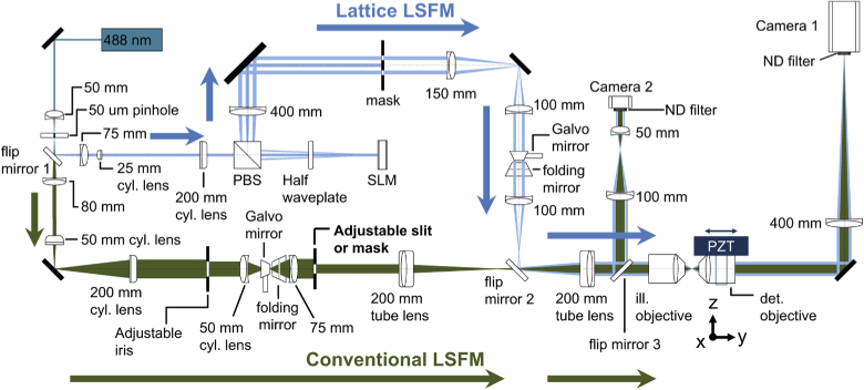
A detailed schematic of the experimental setup is shown in Appendix B. The system allows the generation of optical lattices and Gaussian light-sheets and possesses a detection path with which these light-sheets can be imaged in transmission. In general, all lenses were placed in a 4f arrangement, which was confirmed with a shear plate interferometer, and the lateral and rotational alignment of each element was confirmed by measuring residual back-reflections from each optical surface. For all experiments described here, a 488-nm continuous wave laser (Sapphire 488-300 LP, Coherent) was used as the light source, which was spatially filtered by focusing it through a 50-µm pinhole (P50D, Thorlabs) with a 50-mm focal length achromatic lens (AC254-050-A, Thorlabs) prior to being diverted down one of two optical paths with a flip mirror.
The first path is a modified version of a LLSM [11], which has been described in detail elsewhere [12]. Here, the laser is recollimated with a 75-mm achromatic doublet (AC254-075-A, Thorlabs) prior to asymmetric expansion into a line profile with a pair of achromatic cylindrical lenses (68-160, Edmund Optics, and ACY254-200-A, Thorlabs). This line-shaped beam uniformly illuminates a narrow strip on a spatial light modulator (SLM, SXGA-3DM, Forth Dimension Displays) where binary phase patterns are displayed to generate the desired optical lattice. A polarized beam splitter (10FC16PB.3, Newport) and a half-wave plate (AHWP10M-600, Thorlabs) are placed in front of the SLM, and together with the SLM, form a reconfigurable lattice light-sheet generator. The polarization of the input laser light was adjusted to maximize the light-throughput through this unit. The diffracted light from the SLM is focused by an achromatic lens (AC508-400-A, Thorlabs) onto a custom-designed binary transmission mask (Photo Sciences, Inc.), which consists of multiple annuli of various sizes that serve to block the unwanted zeroth and higher-order (e.g., N > 1) diffracted beams, and to specify the inner and outer numerical apertures of the lattice light-sheet. After passing through the mask, the desired diffraction orders are de-magnified through two achromatic lenses (AC254-150-A and AC254-100-A, Thorlabs) and projected onto a mirror galvanometer (6215H, Cambridge Technology) for rapid lateral dithering of the illumination light-sheet. Thereafter, the light reflects off of a folding mirror, and is relayed to the back pupil of the 40X NA 0.8 illumination objective (CFI Apo 40XW NIR, Nikon Instruments) with an achromatic lens (AC-254-100-A, Thorlabs), a second flip mirror, and a tube lens (ITL-200, Thorlabs).
The second optical path is recollimated with an 80-mm achromatic doublet (AC254-080-A, Thorlabs), and is used to create conventional Gaussian-based illumination light-sheets. Here, the laser light is asymmetrically magnified into a line profile by a pair of achromatic cylindrical lenses (ACY254-050-A and ACY254-200-A, Thorlabs), and focused with a cylindrical lens (ACY254-050-A, Thorlabs) onto a mirror galvanometer (6215H, Cambridge Technology) that is conjugate to the focal plane of the illumination objective for rapid light-sheet pivoting [18]. Thereafter, the light is reflected with a folding mirror, and relayed to the back focal plane of the illumination objective with an achromatic lens (AC254-075-A, Thorlabs) and two tube lenses (ITL-200, Thorlabs). An adjustable slit, which is placed conjugate to the back focal plane of the illumination objective, is used to control the numerical aperture of the Gaussian illumination light-sheet. Besides the slit, we also used annular masks and placed the line beam at specific positions to generate Gaussian light-sheets (see Appendix C). We used these datasets to supplement our light-sheet measurements with a variable slit. We note that in either way (slit or annular mask), only the central portion of the Gaussian input beam is used (i.e., the intensity profile in the back focal plane is relatively uniform, instead of possessing a Gaussian profile). The non-Gaussian input profile, and the aforementioned effects of diffraction at the employed , cause the light-sheet to deviate from a true Gaussian beam. Nonetheless, given that they share many key characteristics with Gaussian beams (e.g., the tradeoff between beam waist and confocal parameter), we refer to them as Gaussian light-sheets here, as is common in the LSFM literature.
Both LLSM and conventional Gaussian-based light-sheets were evaluated with a camera conjugate to the back pupil of the illumination objective. Here, a third flip mirror directs the laser light towards the camera (DCC1545M, Thorlabs), and two achromatic doublets (AC254-100-A and AC254-050-1, Thorlabs), and a neutral density filter (ND10A, or ND20A, Thorlabs), served to relay and attenuate the laser power, respectively. For LLSM, the correct placement of the diffraction orders in relation to the annular mask were monitored and the inner and outer numerical apertures of the illumination beam were measured. For conventional Gaussian-based LSFM, the width of the line profile in the back focal plane was measured to determine the numerical aperture of the illumination.
2.2. Light-sheet measurements
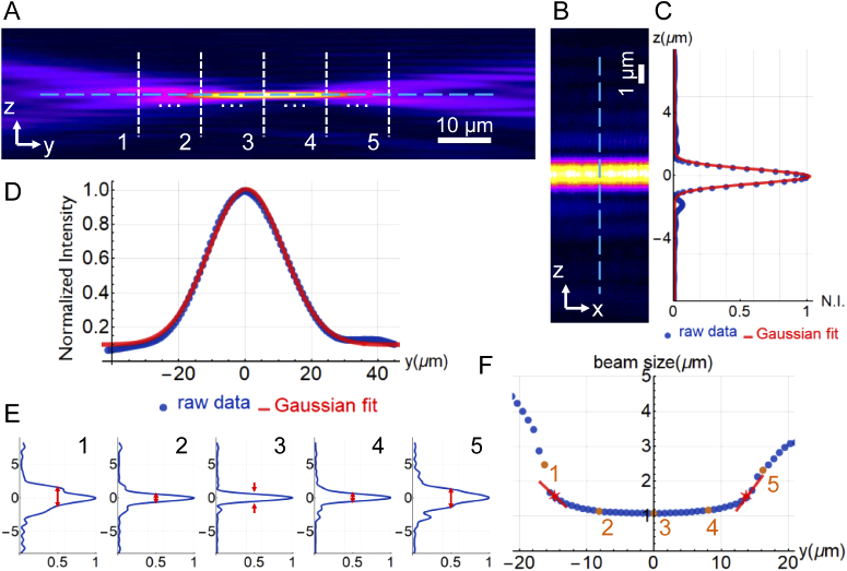
To measure the characteristics of the light-sheets in the front focal plane of the illumination objective, the light-sheets were imaged in a direct transmission mode using another 40X NA 0.8 objective (CFI Apo 40XW NIR, Nikon Instruments). Here, a 400-mm focal length achromatic lens (AC508-400-A) served as the tube lens, and the laser light was detected with a scientific CMOS camera (Orca Flash 4.0 V2, Hamamatsu Corp.) after attenuation with a neutral density filter (ND40A, Thorlabs). The combination of a 400-mm tube lens and the 40X detection objective offers a final magnification of 80X, which results in a pixel size of 81.25 nm. To acquire three-dimensional data sets of the light-sheets, the detection objective was scanned along the direction of laser propagation with a piezoelectric stage (P621.1CD, Physik Instrumente). The data acquired encompassed the main in-focus region of the light-sheet, as well as the more diffuse out-of-focus regions before and after it (See Fig. 1).
Fig. 1.
Details of the light-sheet quantification. (A) A YZ image of the light-sheet averaged along the X-axis. (B) XZ image at the waist of the light sheet. (C) The average intensity profile along the Z direction in (B), and a Gaussian fit. The FWHM of the fitted curve is used to represent the thickness of the light-sheet. (D) The intensity profile of the blue dashed line in (A) and the Gaussian fit. The FWHM of the fitted curve is used to represent the propagation length of the light-sheet. (E) Intensity profiles of the five dashed white lines in (A). (F) The beam size along the propagation direction (Y) of the light-sheet. The red lines go through the points that are close to beam waist, and the star points show exactly where the beam size increased to beam waist by interpolation from the red lines. The distance between the star points represents the confocal parameter. The data set shown here is a Gaussian light-sheet generated with an NA of approximately 0.2. Slices from (A) and (E) are represented by the yellow numbers in (F).
Importantly, it should be noted that all optical systems generate a slightly blurred image of the real object (here, the light-sheets). However, because the light-sheets that we have measured have a lower numerical aperture (0.19–0.64) than our detection objective (0.80) and hence were significantly thicker than the diffraction limit, we expect this effect to be negligible. Furthermore, both the Gaussian and LLSM light-sheets were measured identically, and thus subject to the same amount of optical blurring.
2.3. Light-sheet analysis
All light-sheet measurements were performed in a fully automatic fashion using publicly available software written in MATLAB (https://github.com/AdvancedImagingUTSW/Systematic-Comparison-of-Lattice-and-Gaussian-Light-Sheets). Since the light-sheet stays constant over hundreds of µm in the transverse direction (e.g., the X-direction in this manuscript), we subdivided the acquired data into 6 sub-volumes along the X-direction and analyzed each block individually. This allowed us to confirm that the light-sheet was uniform in the X-direction and allowed for averaging of the measured parameters.
For each light-sheet, three parameters are measured. 1) The full-width half-maximum (FWHM) of the light-sheet thickness in the Z-direction at the waist of the beam. 2) The FWHM of the light-sheet propagation length in the Y-direction. 3) The confocal parameter, for which we measure the distance between the two points along the Y-direction where the light-sheet thickness increases by a factor of from its smallest value.
To measure the light-sheet thickness, we first evaluated each XZ plane along the Y-axis to find the focus of the light-sheet, i.e. the location of the highest beam intensity (Fig. 1(A)). Next, the image dataset was rotated (usually less than 1°) such that the light-sheet was perfectly aligned perpendicular to the Z-axis (Fig. 1(B)). Figure 1(C) shows a line profile of the intensity along the Z-axis at the center of the light-sheet, for which we measured its thickness from the standard deviation (σ) of a Gaussian function fit (). Importantly, a Gaussian function might be a poor approximation for light-sheets with strong sidelobes (e.g., Bessel and hexagonal lattices) [17]. However, we found that at the beam waist, the profile was well approximated by a Gaussian function for both the Gaussian and square lattice light-sheets (Fig. 1(C)).
To measure the propagation length, we first average the plane-by-plane image (YZ slice) along the X-axis for each block, and then we rotate the image slightly (usually also less than 1°) to make sure the propagation of the light-sheet is parallel to the Y-axis. Figure 1(D) shows a line profile of the intensity along the Y-axis at the center of the light-sheet. We measured the beam length from the standard deviation from the fit of a Gaussian function in the same way as we do for the light-sheet thickness.
To measure the confocal parameter of the light-sheet, we measured the width of the light sheet in the Z-direction at multiple positions along the Y-axis, i.e. the intensity profiles of the white dashed lines in Fig. 1(A). Although a Gaussian curve fit provided satisfactory results near the beam waist, it failed in regions away from the light-sheet focus as the intensity distribution becomes increasingly non-Gaussian. Thus, we numerically estimated the FWHM of the light-sheet thickness by interpolating where the intensity is 50% of its peak value (Fig. 1(E)). As can be seen in Fig. 1(F), the light-sheet thickness remained relatively constant over a length ∼20 µm but grows rapidly outside of this range. From this curve, we then numerically identified the confocal parameter, which is defined as two times the distance over which it takes for the light-sheet thickness to increase by a factor of (red stars in Fig. 1(F)). We want to emphasize that for a purely Gaussian beam, the confocal parameter would be twice the Rayleigh length, which could be computed analytically from the beam waist thickness. However, we observed that our light-sheets do not behave exactly like Gaussian light-sheets (i.e., their beam waist stays constant in the main lobe of the light-sheet, and then increases rather rapidly outside), and thus yielded poor results if fitted with a quadratic function. As such, we decided to report the confocal parameter as defined by the thickness increase.
2.4. Point spread and optical transfer function measurements
To measure PSFs and the corresponding OTFs of Gaussian and lattice light-sheets, we earranged the detection path into a LSFM format. A 40X NA0.8 objective (CFI Apo 40XW NIR, Nikon Instruments) was used for detection, oriented at 90 degrees to the optical axis of the illumination objective, followed by a 500 mm focal length tube lens (AC508-500-A, Thorlabs), a fluorescence filter (ET525/50m, Chroma) and a scientific CMOS camera (Orca Flash 4.0 V2, Hamamatsu Corp.). 100-nm fluorescent beads (Polysciences, PA) were either embedded in 2% agarose, or sparsely coated on a 5 mm coverslip. Acquisition of z-stacks was performed by moving the sample along the detection axis with a piezoelectric actuator (P621.1CD, Physik Instrumente). Care was taken to achieve high signal-to-noise ratios, and the image volume spanned ∼33 × 33 × 20 µm3. To evaluate the OTF, we cropped the raw data into a smaller volume with an isolated bead at the center, performed rotational averaging around the detection axis, background corrected the data, and then computed the 3D Fourier Transform of the PSF using MATLAB.
2.5. Light-sheet simulations
For numerical simulations of Gaussian and square lattice light-sheets, we used the publicly available code from Remacha et al. (see https://github.com/remachae/beamsimulator) [17]. For all simulations, a refractive index of and an excitation wavelength of was assumed. We decided to adopt their definitions for beam length and main-lobe thickness (), which is described as the beam size when the intensity drops by a factor of . Thus, the factor is applied to the simulation results. As previously noted, the square lattice contains small sidelobes. However, because these sidelobes remain below the 1/e threshold, regardless of the beam lengths evaluated here, accurately describes the beam thickness. We also include the theoretical values for Gaussian light-sheets using an analytical formula for Gaussian optics (see section 8.5 in [19]). The beam waist, , used in Gaussian optics is the radius of the beam when the intensity drops to , and relates to the FWHM in the following way: . By applying these relationships, the thickness (given as FWHM) and the confocal parameter () are obtained.
3. Results
3.1. Systematic evaluation of light-sheet properties
To evaluate the optical properties of the square lattice light-sheet in LLSM, we systematically varied the inner and outer numerical apertures of the illumination annulus and measured the thickness and propagation length of the light-sheet in transmission. In each case, a matching hologram was applied to the SLM, and the correct diffraction pattern was confirmed with the camera that is conjugate to the back focal plane of the illumination objective. We further evaluated parameters linked to the hologram generation, which among other things influenced the relative positioning of the diffraction orders relative to the annular mask. Lattice light-sheets were rapidly dithered in the X-direction with a mirror galvanometer to average the lateral interference patterns. To facilitate a side-by-side comparison, we report measurements on nearly all of the square lattice light-sheets from the original lattice light sheet microscopy manuscript [11]. We then acquired an exhaustive series of Gaussian light-sheets where the excitation NA was varied in small steps. From this data set, Gaussian light-sheets were selected that closely matched their lattice light-sheet equivalents. A list of the NAs for both the Gaussian and square lattice light-sheets is provided in Appendix D.
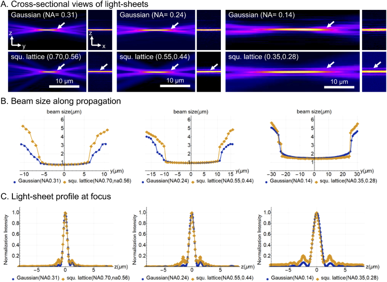
Qualitative differences are readily visualized in the cross-sectional views for the dithered square lattice and Gaussian light-sheets (Fig. 2(A)). For example, the dithered square lattice, while having a similar shaped central lobe compared to the Gaussian sheet, exhibits some structural differences in the transition zones before and after the light-sheet focus (white arrows). Further, the dithered square lattice features two distinct sidelobes (white arrows in XZ-view), whereas the Gaussian sheet is almost free of sidelobes. Further, in Fig. 2(A), one can see two faint beams coming from much steeper angles in the cross-sectional view for the square lattice, which are not present in the Gaussian sheet. These beams originate from the central diffraction order of the hologram used for lattice generation. Interestingly, we experimentally blocked these orders in Appendix E and found that they negligibly influence light-sheet properties.
Fig. 2.
PSFs and OTFs of dithered square lattice and Gaussian light-sheet microscopes. (A) Beam profiles for Gaussian light-sheets of NA = 0.31, 0.24, and 0.14, and square light-sheets of NA 0.70 and na 0.56, NA 0.55 and na 0.44, and NA 0.35, na 0.28. YZ and XZ cross-sectional views are shown. The XZ view is located at the center (thinnest waist) of each light-sheet. Arrows point to differences at the edges of the useful light-sheet and at sidelobes in the lattice light-sheet. NA: outer numerical aperture of annulus used for squared lattice; na: inner numerical aperture for annulus used for the squared lattice. (B) Measurements of the beam size along propagation direction. (C) Measurements of the light-sheet thickness.
In an effort to facilitate a more detailed assessment of light-sheet properties, Figs. 2(B) and 2(C) provide detailed profiles for three square lattices and three Gaussian light-sheets with matching confocal parameters. Strikingly, Fig. 2(B) shows that both light sheets maintain a nearly constant thickness throughout their optical focus (e.g., within the range dictated by their confocal parameters). This is a property that was expected for the lattice light-sheets, as they derive from Bessel beams that theoretically maintain a constant intensity profile throughout their propagation length. It is however a surprise for the Gaussian light-sheets, which were expected to show a quadratic increase in beam waist thickness over their length. Instead, owing to their sufficiently large NA, these Gaussian light-sheets displayed a point spread function-like intensity distribution that advantageously maintains a narrow beam waist throughout its propagation length.
The cross-sectional profiles (Fig. 2(B)) show a close correspondence in their main lobe thickness, which is one of the primary determinants for the axial resolution of a LSFM. However, as the confocal parameter increases, the sidelobe structures in square lattice light-sheets grow more prominently than those seen in the Gaussian light-sheets. Importantly, because these structures exist below the FWHM threshold used here to characterize light-sheet thicknesses, their existence is not reflected in our measurements. Nonetheless, these structures do illicit out-of-focus excitation. Thus, for a given light-sheet confocal parameter, Gaussian beams appear to provide similar axial resolution, albeit with reduced image blur, and greater optical sectioning.
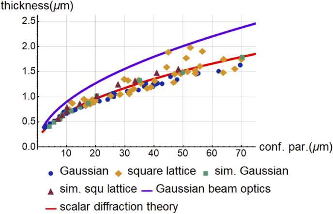
A more quantitative analysis of the relationship between light-sheet thickness and propagation length is summarized in Fig. 3. Here, each point represents the average of multiple measurements obtained independently from different image sub-volumes of the light-sheet image data, and detailed values of the average and standard deviation are provided in Appendix D. As can be seen, and in agreement with previous numerical simulations [17], the tradeoff between light-sheet propagation length and thickness is similar for both square lattices and Gaussian light-sheets. Of note, the data points for the square lattice show greater variability than those for the Gaussian light-sheets. This arises owing to the larger number of degrees of freedom for generating square lattices. Furthermore, the SLM hologram has parameters that can be tuned, most notably the cropping factor and the spacing, and varying these factors does slightly alter light-sheet properties (Appendix F). Nonetheless, in our hands, square lattice light-sheets (i.e. possessing a minimal thickness for a given length) did not outperform the Gaussian light-sheets but instead followed an almost identical trend for thickness increase with increasing confocal parameter. Importantly, these data also suggest that the inherent complexity of LLSM likely results in greater performance fluctuations than a LSFM using comparatively simple Gaussian light-sheets.
Fig. 3.
(A) Light-sheet thickness versus light-sheet propagation length. Thickness is measured as described in Fig. 1(C) and propagation length is measured as described in Fig. 1(D). (B) Light-sheet thickness versus confocal parameter. The confocal parameter is measured as described in Figs. 1(E) and 1(F).
3.2. Comparison of simulated and experimentally measured light-sheets
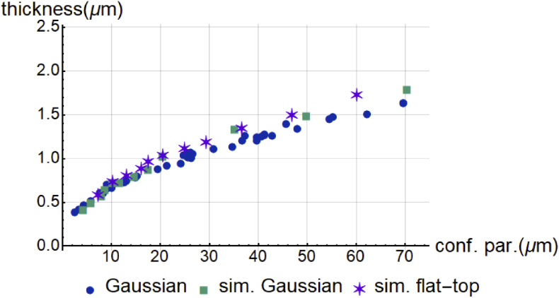
Recently, Remacha et al. numerically evaluated the optical characteristics for a diverse array of light-sheets (including Gaussian, Bessel, dithered lattice, airy, and more) and concluded that Gaussian beams provided superior image contrast [17]. However, these data were not experimentally confirmed, which is of particular concern for lattice light-sheets since the simulations assumed that each diffraction order in the back focal plane of the illumination objective had constant phase and electric field strength. As such, it has remained an open question if such simulations correctly predicted the properties of experimental lattices. Here we address these concerns and compare our experimental data to theoretical Gaussian beam optics, scalar diffraction theory, and simulated lattice and Gaussian light-sheets. In an effort to maintain consistency, the simulations were performed as described previously [17]. As can be seen in Fig. 4, our experimental Gaussian light-sheets are thinner than the theoretical values from Gaussian beam optics but match closely with the scalar diffraction theory for a laser focus. As mentioned earlier, the intensity distribution in the back focal plane of our illumination objective is more uniform than a traditional Gaussian beam and thus resembles a “flat-top” beam, which could potentially contribute to this discrepancy. Nevertheless, previous simulations by Remacha et al. demonstrate that Gaussian and “flat-top” beams behave similarly [17], which we have performed as well; Our experimental Gaussian light-sheets are in close correspondence with both the simulated Gaussian and “flat-top” beams, and overall, the difference appears small (Appendix G). Furthermore, both the simulated and experimentally measured square lattice light-sheets are in near-perfect agreement, suggesting that the numerical simulations performed by Remacha et al. were accurate. And lastly, our Gaussian beams are indeed largely indistinguishable from the lattice light-sheets in terms of light-sheet thickness. Importantly, this analysis only takes into account the thickness of the main lobe, while ignoring the sidelobes that are present in lattice light-sheets.
Fig. 4.
Comparison of experimentally measured Gaussian and square lattice light-sheets with numerically simulated Gaussian and square lattice light-sheets. The solid lines depict theoretical Gaussian light-sheets derived from an analytical formula (magenta line), and the scalar diffraction theory for a laser focus (red line), respectively.
3.3. Experimental measurements of PSFs and OTFs
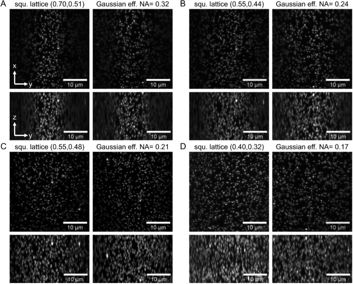
To evaluate the functional resolution of a microscope equipped with square lattice and Gaussian light-sheets, we experimentally measured PSFs in a LSFM format (Appendix H). Measurements were carried out on 100 nm fluorescent beads immobilized in agarose, for light-sheets with four different propagation lengths (12, 19, 32 and 40 µm). As can be seen in Table 1, for a given propagation length, Gaussian and square lattice light-sheets have statistically indistinguishable axial resolutions for propagation lengths of 12, 19, and 32 µm, as measured with a two-sample T-test. However, when the propagation length increases to 40 µm, the Gaussian light-sheet provides a statistically significant improved axial resolution.
Table 1. Lateral and axial resolutions of square and Gaussian light-sheets as measured from 100 nm fluorescent nanospheres. Statistical significance was evaluated with a two-sample T-test.
| Light-Sheet (NA) | Prop. Length (µm) | Lateral Resolution (nm) | Axial Resolution (nm) | P-Value | n |
|---|---|---|---|---|---|
| Square (0.7, 0.51) | 12 | 363 ± 13.1 | 649 ± 24.4 | 0.43 | 15 |
| Gaussian (0.32) | 12 | 370 ± 12.9 | 641 ± 26.5 | 15 | |
| Square (0.55, 0.44) | 19 | 365 ± 18.0 | 800 ± 25.8 | 0.79 | 15 |
| Gaussian (0.24) | 19 | 369 ± 14.5 | 798 ± 19.0 | 15 | |
| Square (0.55, 0.48) | 32 | 360 ± 20.0 | 942 ± 65.0 | 0.26 | 10 |
| Gaussian (0.21) | 32 | 365 ± 12.5 | 927 ± 33.3 | 15 | |
| Square (0.40, 0.32) | 40 | 364 ± 16.2 | 1152 ± 86.6 | 1.9 × 10⁻⁴ | 15 |
| Gaussian (0.17) | 40 | 370 ± 15.8 | 1051 ± 27.9 | 15 |
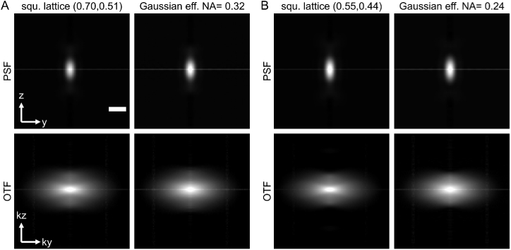
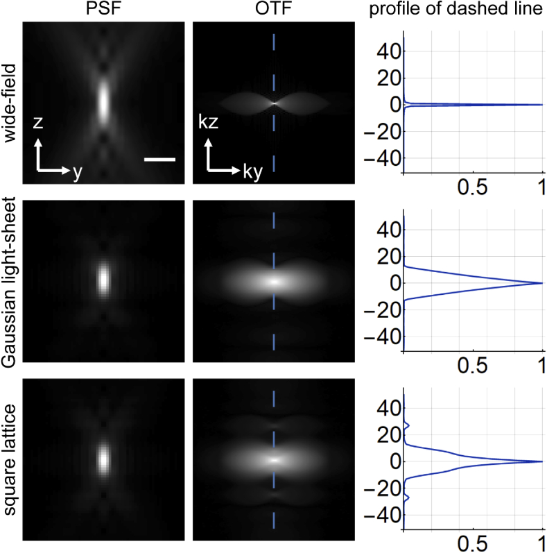
Lastly, we sought to evaluate the frequency support of the OTF for square lattice and Gaussian light-sheets. Here, isolated and coverslip immobilized 100 nm fluorescent beads were imaged for light-sheet propagation lengths of 12 and 19 µm. As can be seen in Figs. 5(A) and 5(B), both the PSFs and OTFs for square lattices and Gaussian light-sheets are nearly identical. Thus, not only do they possess similar lateral and axial resolution (Table 1), but also provide indistinguishable optical sectioning capacity. Nevertheless, to exclude the possibility that insufficient SNR could obscure some high-frequency OTF support, we also performed numerical convolution of the experimental light-sheets with a simulated detection PSF. This allowed us to generate OTFs with higher SNR. As can be seen in Appendix I, even these semi-synthetic OTFs have similar frequency support. Thus we conclude that square lattice and Gaussian light-sheets provide indistinguishable levels of imaging performance.
Fig. 5.
PSFs and OTFs of dithered square lattice and Gaussian light-sheet microscopes. A Gamma correction of 0.8 was applied to all PSF and OTF images to increase the contrast of weak features. The Gaussian light-sheets were chosen to have similar confocal parameters to the lattice light-sheets. Scale bar in the PSF is 1 µm.
4. Summary
LLSM has generated significant interest in the biological imaging community, with several high-profile publications referring to its illumination optics as “ultrathin” [11,13,14,20] and its resolution as “unparalleled” [21]. Yet, as we have shown previously [12], the time-averaged equivalent of these illumination beams can be generated incoherently, and therefore their performance is not a unique consequence of the coherent superposition of multiple Bessel beams. Furthermore, as we show here, the most popular lattice light-sheet, the dithered square lattice, does not appear to provide a significant performance improvement over Gaussian light-sheets, as measured from their confocal parameter and their thickness. In the spirit of thoroughness, we also investigated the OTFs and PSFs for both Gaussian and square lattice light-sheets. Given that PSFs or OTFs are sufficient to fully characterize a shift-invariant imaging system, these measurements should reveal any performance differences between these illumination light-sheets. Nonetheless, with the exception of one Gaussian light-sheet that actually provided better axial resolution, the differences between these PSFs were statistically insignificant. And likewise, their OTFs shared similar support in the frequency domain. Thus, for all practical matters, square lattice and Gaussian light-sheets are indistinguishable.
According to both numerical simulations [9,10,17] and the experimental measurements, the square lattice does not behave like a propagation-invariant beam, but rather a divergent beam. Indeed, its tendency to increase in thickness as the confocal parameter grows is in close agreement to a simple Gaussian light-sheet. Likewise, in contrast to the behavior anticipated for canonical Bessel beams, we found a square lattice light-sheet generated from a low NA and thick annulus that is similar in terms of thickness and propagation length to one generated from a high NA and thin annulus (Appendix J). These observations are indeed a surprise, as square optical lattices are produced by a coherent superposition of Bessel beams, which are de facto propagation invariant. Therefore, we conclude that for certain periodicities of the lattice (i.e. corresponding to the square lattice), the propagation-invariant nature of the parent beam can be lost, and a clear explanation for this phenomenon is still under investigation.
Our results also stipulate that at the numerical apertures used within this manuscript, Gaussian light-sheets are better described as a diffraction limited laser focus (Fig. 4). Importantly, these light-sheets have a more favorable aspect ratio (propagation length to beam waist thickness) than the Gaussian approximation suggests. This advantage seems not to depend too much on the input profile of the laser (i.e. flat-top or Gaussian profile), but more on diffraction effects that cannot be ignored at these low to moderate numerical apertures. This in itself is a finding that seems to have been underappreciated in LSFM.
For higher resolution imaging, the coherent modulation present in static (e.g., non-dithered) lattice light-sheets can be used for structured illumination microscopy (SIM) [11]. However, owing to the LSFM geometry, this mode improves the resolution only in the axial dimension and one of the two lateral dimensions (e.g., the X-direction). As such, we believe that the main benefit of optical lattices for SIM is that they provide greater frequency support in the axial dimension which cannot otherwise be achieved with one dimensional beam engineering. Yet, the use of the SIM mode of LLSM has been rare so far, owing to the need to acquire 5 images for one SIM reconstruction, and the fact that the lateral resolution gain is modest. Alternatively, hexagonal lattices provide a thinner main lobe than square lattices at the cost of much stronger sidelobes that grow drastically with the light-sheet propagation length. Indeed, sidelobes that approach 50% strength of the main lobe are notoriously hard to remove computationally [22,23], which is likely the main reason why hexagonal lattice light-sheets have seen little use. And lastly, it appears to us that for both the square and hexagonal lattice light-sheets, the reported axial resolution of ∼350 nm could have only been achieved following aggressive and non-linear deconvolution of their data. Indeed, this hypothesis is supported by the data presented here, as well as one LLSM manuscript that reports axial resolutions that vary from 649 - 947 nm [20].
In conclusion, our results stipulate that the vast majority of LLSM experiments could have been performed with conventional Gaussian light-sheets. This would be a welcome simplification as the assembly, alignment, and operation of a LLSM requires significant optical expertise. Furthermore, by dispensing with the SLM, much less input laser power is needed, and simultaneous multicolor excitation is easily achieved. Simply put, we interpret these results as a poignant reminder that there is no free lunch in linear optics; as the confocal parameter increases, one must accept a thicker light-sheet, stronger sidelobes, or both. LLSM was heralded as a breakthrough that overcomes this tradeoff but nonetheless remains subject to the same physical laws as Gaussian beams.
Acknowledgments
The authors would like to thank Dr. Florian Fahrbach for help with the simulation code and critical reading of the manuscript. We would also like to thank Dr. James Manton for discussions about diffraction theory. Furthermore, we are grateful to Dr. Kim Reed for her continued support, as well as all the employees at the University of Texas Southwestern Medical Center who make our research possible.
Appendix A. Thickness and propagation length of a light-sheet
For a light-sheet centered on the back pupil of the illumination objective (Fig. 6(A)), we can estimate the thickness and propagation length of the light-sheet using the well-known Abbe formulations for a diffraction limited laser focus. Here, the lateral and axial resolution are given by:
| (1) |
| (2) |
with tlightsheet being the thickness and dlightsheet the depth of focus of the light-sheet, respectively [4].
Fig. 6.
(A) Schematic representation of a light-sheet centered on the pupil. (B) Schematic illustration of the pupil plane and position of the lattice bands (blue bars). The two circles depict the inner (na) and outer (NA) edges of the annulus. (C) Schematic representation of the Ewald sphere corresponding to the excitation objective, upon which the pupil plane is mapped. The axial spread along the ky axis for the central band (1/dax-center) corresponds to the highest and lowest points on the Ewald-sphere in the ky direction. (D) Construction of the axial spread of the sideband (1/dax-side) along the ky axis.
But what happens if the illumination is not centered on the back pupil, as is the case for square lattice light-sheets? Figure 6(B) shows schematically the pupil for generating a square lattice light-sheet, which consists of two side bands and two smaller central bands (termed “side” and “center”, respectively). These bands each form a light-sheet that interferes with the other light-sheets to generate the final square lattice pattern. Below, we derive a theoretical derivation based on scalar diffraction theory of their depth of focus, which allows us to estimate their propagation length. For a detailed explanation of the method, please see [4]. In short, the pupil plane is mapped onto an Ewald’s sphere whose radius equals to the wavenumber of the laser light. The full Ewald’s sphere cannot be accessed with an objective; instead only subset of the wavevectors on a spherical cap are utilized (a cross-section of this cap is shown in Figs. 6(C) and 6(D)). The depth of focus is the reciprocal value of the spread of the “illumination wavevectors” in the axial ky direction [24]. As one can see in Figs. 6(C) and 6(D), the central and side bands have similar axial spreads, but slight differences remain.
For the central band, as shown in Fig. 6(C), we can find the following spread of the wavevectors on the ky axis:
| (3) |
Here, λ0 is the vacuum wavelength of light, n is the refractive index of the immersion media, and the angle is defined by the numerical aperture . By taking the reciprocal value of Eq. (3), we can describe the depth of focus for the central band as follows, where NA and na are the outer and the inner numerical apertures of the annulus, respectively:
| (4) |
For the sideband, placed in the annulus as shown in Fig. 6(D) and using a similar derivation as above, the depth of focus can be calculated as follows:
| (5) |
One can see analytically from the two formulas that the depth of focus of the side and central bands are not identical. This difference becomes smaller if one moves the sidebands inwards (and disappears if the sidebands touch the inner edge of the annulus) and gets larger if one moves the sidebands outwards. We present experimental results for different placement of the sidebands in Appendix F and Fig. 10.
The thickness of the light-sheet stemming from the sidebands is governed by the spread in the kz direction in the pupil, and can be found using the effective NA ( and the Abbe diffraction limit for lateral resolution:
| (6) |
| (7) |
has a close correspondence to NAEX of a centered light-sheet, and we suggest that it can be used as a key number to characterize square lattice light-sheets. Both numbers describe the thickness in the same way (Eqs. (1) and (6)). If is used in Eq. (2), the depth of focus is slightly overestimated compared to the proper computation using Eq. (5). As an example, using an annulus with NA=0.55 and na=0.44 gives an of 0.24. These values result in a depth of focus of 22.5 µm using in Eq. (2) and 20.8 µm when correctly using NA and na of the annulus in Eq. (5). Thus, serves as an approximation of the characteristics of a square lattice light-sheet and as such can serve as a single key number for comparisons.
Appendix B. Experimental system
The experimental setup consists of two paths, one for lattice light-sheet generation, and one for generating conventional Gaussian light-sheets. A schematic representation of the setup is shown in Fig. 7.
Fig. 7.
Schematic representation of the experimental setup, which consists of two illumination paths, one to produce optical lattices (blue arrow “Lattice LSFM”) and one to produce conventional Gaussian light-sheets (green arrow “Conventional LSFM”). Both light-sheets are ultimately injected into the illumination objective (ill. objective). A secondary objective (det. objective) observes the image of the light-sheets in transmission and projects them onto a scientific CMOS camera (Camera 1). Flip mirror 1 and 2 are used to select between the lattice LSFM or the conventional LSFM light path. An image of the back focal plane of the illumination objective can be observed on Camera 2 by directing the laser light with flip mirror 3. SLM: Spatial light modulator; ND: neutral density filter; cyl. lens: cylindrical lens.
Appendix C. Effective NA of Gaussian light-sheets generated with an annulus
Some Gaussian light-sheets were created using an annulus, for which we computed the effective numerical aperture as shown in Fig. 8.
Fig. 8.
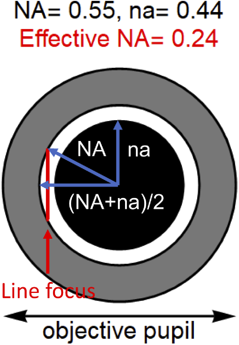
While we primarily used an adjustable slit to create the different Gaussian light-sheets, some datasets were acquired using an annular mask, and placing the line focus at the periphery of the annulus. This generated a Gaussian light-sheet in the same fashion as a single side band for a square lattice light-sheet. The effective NA is calculated as Eq. (7) in Appendix A, which results in = 0.24 using an annular mask with NA = 0.55 and na = 0.44 as shown here.
Appendix D. Comparison of Gaussian and square lattice light-sheets
This appendix summarizes all measurements carried out here. In total 39 and 52 experiments were performed for Gaussian and square lattice light-sheets, respectively (see also Table 2). These data cover a wide range to illustrate the characteristics of both Gaussian and square lattice light-sheets. In the square lattice, NA and na represent the outer and inner numerical apertures of the annulus, respectively. Bold indicates that the light-sheet was, or approximates, a light-sheet in the original LLSM manuscript [17]. For most Gaussian beams, the effective NA used to generate the light-sheet was controlled with a micrometer slit conjugate to the back-pupil of the illumination objective. In some cases, we replaced the micrometer slit with an annulus that served as the slit (as described in Appendix C).
Table 2. Summary of all experimental data shown in Figs. 3 and 4. The table is sorted with thickness and then the confocal parameter. Effective NA() corresponds to the NA of the slit or the theoretical NA of the annulus used to create the Gaussian beam. In the square lattice, sp and cp represents spacing and cropping factor, respectively. n represents the number of measurements. For Gaussian measurements, n equals to 24 for all measurements. In the square lattice measurements, n equals 6 unless it is specified otherwise.
| Gaussian | Square lattice | ||||||
|---|---|---|---|---|---|---|---|
| Thickness | Prop. Length | Confocal Parameter | eff. NA | Thickness | Prop. Length | Confocal Parameter | NA, na (sp., cp., (n)) |
| 0.4 ± 0.01 | 3.3 ± 0.09 | 2.6 ± 0.04 | 0.61 | 0.8 ± 0.02 | 10.3 ± 0.16 | 11.2 ± 0.20 | 0.70,0.56 |
| 0.4 ± 0.00 | 4.0 ± 0.15 | 3.5 ± 0.08 | 0.54 | 0.8 ± 0.01 | 14.3 ± 0.11 | 16.7 ± 0.24 | 0.70,0.60 |
| 0.5 ± 0.01 | 4.9 ± 0.27 | 4.5 ± 0.14 | 0.49 | 0.9 ± 0.04 | 10.9 ± 0.30 | 11.9 ± 0.46 | 0.70,0.51 |
| 0.5 ± 0.00 | 6.3 ± 0.26 | 5.9 ± 0.28 | 0.42 | 0.9 ± 0.05 | 11.8 ± 0.44 | 12.1 ± 0.68 | 0.70,0.56 |
| 0.6 ± 0.01 | 7.7 ± 0.24 | 7.8 ± 0.31 | 0.35 | 0.9 ± 0.02 | 17.7 ± 0.52 | 17.5 ± 1.41 | 0.55,0.44 |
| 0.6 ± 0.01 | 8.5 ± 0.12 | 8.4 ± 0.25 | 0.36 | 0.9 ± 0.00 | 16.5 ± 0.15 | 17.9 ± 0.44 | 0.55,0.44 |
| 0.7 ± 0.02 | 9.0 ± 0.38 | 9.2 ± 0.10 | 0.32 | 0.9 ± 0.01 | 18.2 ± 0.17 | 17.9 ± 0.35 | 0.55,0.44 |
| 0.7 ± 0.02 | 9.9 ± 0.40 | 9.2 ± 0.88 | 0.35 | 0.9 ± 0.02 | 17.7 ± 0.64 | 18.2 ± 1.07 | 0.55,0.44 |
| 0.7 ± 0.02 | 9.5 ± 0.34 | 10.2 ± 0.69 | 0.32 | 0.9 ± 0.01 | 18.2 ± 0.15 | 18.3 ± 0.32 | 0.55,0.44 |
| 0.7 ± 0.01 | 11.4 ± 0.45 | 12.3 ± 0.27 | 0.28 | 0.9 ± 0.01 | 10.4 ± 0.10 | 18.5 ± 0.34 | 0.55,0.44 |
| 0.7 ± 0.01 | 12.4 ± 0.08 | 12.7 ± 0.23 | 0.31 | 0.9 ± 0.01 | 16.9 ± 0.18 | 18.6 ± 0.26 | 0.55,0.40 |
| 0.7 ± 0.04 | 12.0 ± 0.78 | 13.2 ± 0.98 | 0.28 | 0.9 ± 0.01 | 17.9 ± 0.28 | 18.6 ± 0.33 | 0.55,0.44 |
| 0.8 ± 0.02 | 14.1 ± 0.61 | 15.0 ± 0.77 | 0.26 | 0.9 ± 0.02 | 18.1 ± 0.65 | 18.6 ± 0.81 | 0.55,0.52 |
| 0.8 ± 0.02 | 14.4 ± 0.77 | 15.3 ± 1.21 | 0.26 | 0.9 ± 0.01 | 17.5 ± 0.19 | 18.7 ± 0.38 | 0.55,0.44 |
| 0.9 ± 0.02 | 18.0 ± 0.93 | 19.5 ± 0.73 | 0.22 | 0.9 ± 0.01 | 17.3 ± 0.24 | 19.0 ± 0.68 | 0.55,0.44 |
| 0.9 ± 0.02 | 19.8 ± 0.27 | 21.4 ± 0.24 | 0.24 | 0.9 ± 0.01 | 19.0 ± 0.46 | 19.2 ± 0.91 | 0.55,0.44 |
| 0.9 ± 0.03 | 22.0 ± 1.46 | 24.2 ± 1.59 | 0.22 | 0.9 ± 0.02 | 17.9 ± 0.23 | 19.8 ± 0.67 | 0.55,0.44 |
| 1.0 ± 0.03 | 23.9 ± 1.20 | 24.9 ± 1.01 | 0.20 | 0.9 ± 0.01 | 18.0 ± 0.12 | 19.8 ± 0.05 | 0.55,0.44 |
| 1.0 ± 0.03 | 24.3 ± 1.90 | 25.7 ± 1.87 | 0.20 | 0.9 ± 0.01 | 18.3 ± 0.26 | 19.8 ± 0.65 | 0.55,0.44 |
| 1.0 ± 0.03 | 24.4 ± 2.00 | 26.4 ± 2.41 | 0.20 | 0.9 ± 0.02 | 18.9 ± 0.23 | 19.8 ± 0.45 | 0.55,0.44 |
| 1.1 ± 0.02 | 23.6 ± 1.17 | 25.9 ± 1.11 | 0.20 | 1.0 ± 0.04 | 13.6 ± 0.21 | 16.1 ± 0.26 | 0.70,0.60 |
| 1.1 ± 0.02 | 24.7 ± 1.61 | 26.3 ± 2.27 | 0.20 | 1.0 ± 0.01 | 17.0 ± 0.24 | 19.0 ± 0.32 | 0.55,0.40 |
| 1.1 ± 0.04 | 25.2 ± 1.96 | 26.7 ± 1.46 | 0.20 | 1.0 ± 0.01 | 25.3 ± 0.61 | 25.3 ± 0.82 | 0.51,0.43 (n=18) |
| 1.1 ± 0.03 | 30.5 ± 1.76 | 31.0 ± 0.74 | 0.18 | 1.0 ± 0.01 | 25.7 ± 0.41 | 28.5 ± 0.61 | 0.51,0.43 |
| 1.1 ± 0.05 | 32.0 ± 1.73 | 34.8 ± 2.06 | 0.18 | 1.1 ± 0.02 | 26.5 ± 0.23 | 28.5 ± 0.77 | 0.51,0.43 |
| 1.2 ± 0.02 | 34.7 ± 0.83 | 36.9 ± 2.41 | 0.16 | 1.1 ± 0.01 | 29.3 ± 0.30 | 31.7 ± 0.57 | 0.55,0.48 (n=12) |
| 1.2 ± 0.02 | 37.0 ± 0.4 | 39.8 ± 0.92 | 0.18 | 1.1 ± 0.01 | 27.5 ± 0.34 | 31.9 ± 0.60 | 0.55,0.48 |
| 1.3 ± 0.09 | 35.4 ± 2.79 | 37.5 ± 1.17 | 0.16 | 1.2 ± 0.01 | 30.9 ± 1.36 | 33.2 ± 0.22 | 0.55,0.48 |
| 1.3 ± 0.04 | 36.8 ± 1.89 | 39.8 ± 2.16 | 0.16 | 1.2 ± 0.02 | 35.3 ± 0.42 | 37.8 ± 1.27 | 0.40,0.32 |
| 1.3 ± 0.05 | 38.8 ± 2.36 | 40.8 ± 3.35 | 0.16 | 1.2 ± 0.01 | 36.1 ± 0.85 | 39.1 ± 1.01 | 0.40,0.32 (n=18) |
| 1.3 ± 0.02 | 38.2 ± 0.86 | 41.4 ± 2.36 | 0.16 | 1.3 ± 0.02 | 28.4 ± 0.35 | 31.2 ± 0.52 | 0.70,0.67 |
| 1.3 ± 0.02 | 40.4 ± 0.75 | 41.4 ± 2.43 | 0.16 | 1.3 ± 0.01 | 29.4 ± 0.44 | 34.9 ± 0.93 | 0.70,0.67 |
| 1.3 ± 0.03 | 40.8 ± 1.64 | 42.9 ± 2.43 | 0.16 | 1.3 ± 0.02 | 36.9 ± 1.05 | 35.6 ± 1.35 | 0.55,0.52 |
| 1.3 ± 0.06 | 44.2 ± 3.36 | 48.0 ± 0.81 | 0.14 | 1.3 ± 0.02 | 40.2 ± 0.86 | 40.4 ± 0.99 | 0.55,0.52 (sp0.93, cp0.05) |
| 1.4 ± 0.09 | 42.3 ± 4.55 | 45.8 ± 1.56 | 0.16 | 1.3 ± 0.01 | 42.6 ± 0.48 | 47.0 ± 0.84 | 0.35,0.28 |
| 1.5 ± 0.08 | 48.2 ± 2.55 | 54.7 ± 3.34 | 0.14 | 1.4 ± 0.02 | 28.0 ± 0.79 | 30.0 ± 1.19 | 0.70,0.67 |
| 1.5 ± 0.03 | 50.6 ± 1.66 | 55.4 ± 1.30 | 0.14 | 1.5 ± 0.02 | 44.3 ± 0.85 | 47.2 ± 1.67 | 0.55,0.52 (sp0.93, cp0.10) |
| 1.5 ± 0.04 | 53.4 ± 1.53 | 62.4 ± 4.05 | 0.14 | 1.5 ± 0.01 | 44.5 ± 2.46 | 49.4 ± 1.99 | 0.55,0.52 |
| 1.6 ± 0.03 | 54.2 ± 2.67 | 69.8 ± 3.80 | 0.13 | 1.5 ± 0.02 | 44.2 ± 1.16 | 49.9 ± 0.44 | 0.55,0.52 |
| - | - | - | - | 1.5 ± 0.03 | 46.0 ± 0.71 | 51.6 ± 0.73 | 0.55,0.52 |
| - | - | - | - | 1.5 ± 0.02 | 46.7 ± 0.63 | 54.4 ± 2.19 | 0.55,0.52 (sp0.93, cp0.15) |
| - | - | - | - | 1.5 ± 0.08 | 47.5 ± 2.43 | 54.4 ± 3.10 | 0.55,0.52 |
| - | - | - | - | 1.5 ± 0.02 | 48.0 ± 2.38 | 63.8 ± 2.20 | 0.55,0.52 (sp0.92, cp0.05) |
| - | - | - | - | 1.6 ± 0.02 | 44.7 ± 1.17 | 44.5 ± 1.25 | 0.55,0.52 (sp0.93, cp0.25) |
| - | - | - | - | 1.6 ± 0.16 | 45.5 ± 2.09 | 56.0 ± 11.75 | 0.55,0.52 (sp0.93, cp0.15) |
| - | - | - | - | 1.6 ± 0.11 | 52.1 ± 2.35 | 67.8 ± 8.24 | 0.55,0.52 |
| - | - | - | - | 1.8 ± 0.13 | 48.3 ± 1.59 | 51.2 ± 7.98 | 0.55,0.52 (sp0.92, cp0.15) |
| - | - | - | - | 1.8 ± 0.02 | 53.5 ± 1.22 | 64.1 ± 2.33 | 0.55,0.52 (sp0.92, cp0.15) |
| - | - | - | - | 1.8 ± 0.14 | 57.5 ± 1.74 | 70.2 ± 7.37 | 0.55,0.52 (sp0.91, cp0.15) |
| - | - | - | - | 1.9 ± 0.16 | 44.1 ± 4.31 | 45.1 ± 6.44 | 0.55,0.52 (sp0.92, cp0.25) |
| - | - | - | - | 1.9 ± 0.16 | 50.0 ± 3.20 | 56.8 ± 8.31 | 0.55,0.52 (sp0.91, cp0.25) |
| - | - | - | - | 2.0 ± 0.22 | 47.6 ± 1.97 | 52.6 ± 6.36 | 0.55,0.52 (sp0.93, cp0.25) |
Appendix E. Contribution of central band to square lattice properties
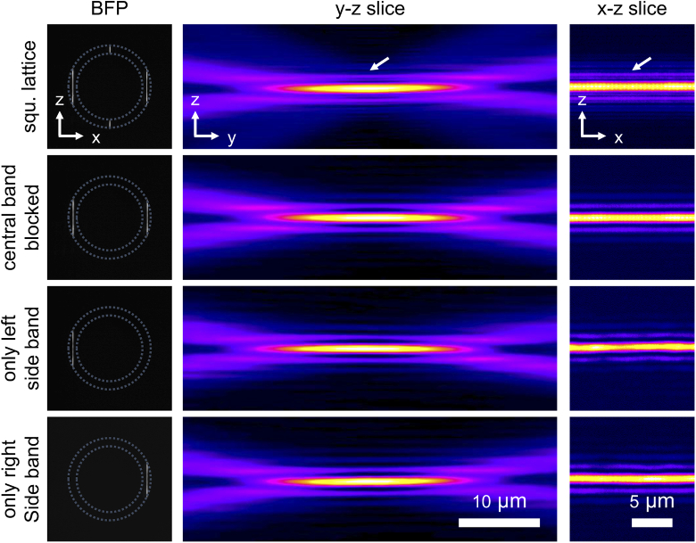
We performed measurements on square lattices before and after blocking the central and side bands. Although similar results were found for other measurements, only one data set is shown for each annulus. Table 3 in this appendix summarizes the measurements with multiple annuli. It can also be seen that when the central band is blocked, and only a single side band is allowed, the light-sheet remains similar to the original dithered square lattice light-sheet (see also Fig. 9).
Fig. 9.
Light-sheets generated by blocking the central band, or by allowing only a single side band. Images of the back focal plane (BFP) of the illumination objective, and YZ, and XZ slices of the light-sheets are shown. Sidelobes are present in all conditions, except for the one with all bands (square lattice), where they appear more prominent. The annulus used here possesses an outer and inner NA of 0.55 and 0.44, respectively.
Table 3. Summary of all experimental data shown in Fig. 9. Dependence of square lattice properties on back-pupil illumination bands.
| NA 0.35, na 0.28 | Dithered square lattice | Central band blocked | Only left or right side band transmitted |
|---|---|---|---|
| Thickness (µm) | 1.3 ± 0.01 | 1.3 ± 0.01 | |
| Axial FWHM (µm) | 42.6 ± 0.48 | 43.1 ± 0.47 | |
| Confocal parameter (µm) | 47.0 ± 0.84 | 50.1 ± 2.43 | |
| NA 0.55, na 0.40 | Dithered square lattice | Central band blocked | Only left or right side band transmitted |
| Thickness (µm) | 0.9 ± 0.01 | 0.9 ± 0.01 | |
| Axial FWHM (µm) | 16.9 ± 0.18 | 17.5 ± 0.63 | |
| Confocal parameter (µm) | 18.6 ± 0.26 | 17.2 ± 0.60 | |
| NA 0.55, na 0.44 | Dithered square lattice | Central band blocked | Only left or right side band transmitted |
| Thickness (µm) | 0.9 ± 0.02 | 0.8 ± 0.01 | 0.8 ± 0.01 |
| Axial FWHM (µm) | 17.3 ± 0.24 | 18.1 ± 0.21 | 18.3 ± 0.75 |
| Confocal parameter (µm) | 19.0 ± 0.68 | 18.5 ± 0.62 | 18.8 ± 0.89 |
| NA 0.55, na 0.48 | Dithered square lattice | Central band blocked | Only left or right side band transmitted |
| Thickness (µm) | 1.2 ± 0.01 | 1.2 ± 0.01 | |
| Axial FWHM (µm) | 30.9 ± 1.36 | 26.9 ± 0.90 | |
| Confocal parameter (µm) | 33.2 ± 0.22 | 32.2 ± 0.37 | |
| NA 0.55, na 0.52 | Dithered square lattice | Central band blocked | Only left or right side band transmitted |
| Thickness (µm) | 1.5 ± 0.01 | 1.5 ± 0.01 | |
| Axial FWHM (µm) | 44.5 ± 2.46 | 44.2 ± 2.45 | |
| Confocal parameter (µm) | 49.4 ± 1.99 | 50.0 ± 2.56 | |
| NA 0.70, na 0.56 | Dithered square lattice | Central band blocked | Only left or right side band transmitted |
| Thickness (µm) | 0.8 ± 0.02 | 0.7 ± 0.02 | |
| Axial FWHM (µm) | 10.3 ± 0.16 | 10.8 ± 0.14 | |
| Confocal parameter (µm) | 11.2 ± 0.20 | 11.1 ± 0.25 | |
| NA 0.70, na 0.60 | Dithered square lattice | Central band blocked | Only left or right side band transmitted |
| Thickness (µm) | 0.8 ± 0.01 | 0.8 ± 0.01 | |
| Axial FWHM (µm) | 14.3 ± 0.11 | 14.7 ± 0.28 | |
| Confocal parameter (µm) | 16.7 ± 0.24 | 16.9 ± 0.23 | |
| NA 0.70, na 0.67 | Dithered square lattice | Central band blocked | Only left or right side band transmitted |
| Thickness (µm) | 1.3 ± 0.01 | 1.4 ± 0.03 | |
| Axial FWHM (µm) | 29.4 ± 0.44 | 30.7 ± 3.01 | |
| Confocal parameter (µm) | 34.9 ± 0.93 | 37.0 ± 1.80 |
Appendix F. Choice of SLM parameters for lattice generation
Fig. 10.
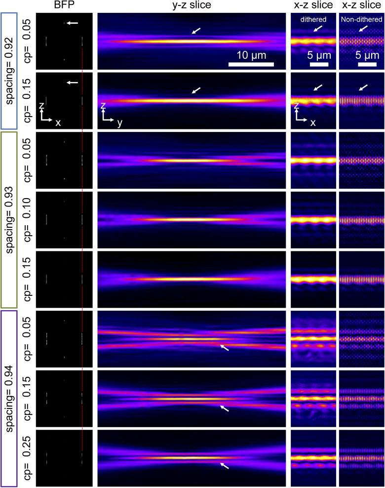
Effect of cropping factor and spacing on lattice light-sheet generation. The combination of three spacing numbers and different cropping factors are shown. From left to right, the line beams on the Back focal plane (BFP) of the illumination objective, YZ and XZ slices are shown. The XZ slice of the non-dithered lattice is shown in the rightmost column. Note that the scaling in the XZ slice is different from the YZ in order to show clearer the lattice pattern and the sidelobes. In the BFP, white arrows point to the short line beams and how they are affected by the cropping factor (cp). The red dashed line indicates the shift of the long line beams depending on the spacing. In the YZ and XZ slices, white arrows point to the appearance and disappearance of the sidelobes. The NA and na of the annulus used in this example are 0.55 and 0.52, respectively.
The SLM phase hologram is essential for generating lattice light-sheets, and it is affected by the size of the annulus, the wavelength, and the magnification of the optical path. Nonetheless, to obtain a proper lattice, several parameters need to be carefully adjusted, of which the cropping factor and the spacing are the two most important. The cropping factor is a threshold that removes low-intensity values from the calculation for the SLM phase hologram, and in practice, affects the sidelobes of the final lattice light-sheet. As shown in Fig. 10, indicated with the white arrows, as the cropping factor increases, the energy density in the central band in the back focal plane and the sidelobes in the front focal plane decreases. As a result, the lattice becomes more Gaussian-like. The spacing parameter influences the frequency of the coherent modulation present in a static lattice light-sheet by determining the position and distance between the side bands in the back focal plane. In general, and as can be seen by the dashed fiduciary lines in the figure below, as the spacing factor increases, the side bands move closer to the center of the back focal plane. Commonly, the spacing parameter is chosen such that the side bands are located in the middle of the outer and inner annulus rings. However, some flexibility remains in their fine spacing, and this also influences the propagation length of the light-sheet owing to changes in the length of the side bands (See also Appendix A for a discussion of this effect). Table 4 in this appendix provides a quantitative summary of these results. For comparison, we also provide measurements for two Gaussian light-sheets with similar thicknesses. Here, a ∼1.5 µm thick Gaussian light-sheet has a propagation length of ∼62-64 µm, which is very similar to the longest dithered square lattice (spacing = 0.92, cropping factor = 0.05) shown in this appendix. Of note, we did not include the dithered square lattice light-sheet with a spacing of 0.94 because it behaves more like a hexagonal lattice light-sheet and thus requires a distinct quantification strategy.
Table 4. The thickness and confocal parameters for dithered square lattice light-sheets using different spacing and cropping factors. The numbers for the lattice light-sheet with spacing = 0.94 are not shown because it does not behave like a square lattice. Two Gaussian light-sheets that have similar properties are shown for comparison. n=6 for the lattice light-sheet measurements and n = 24 for the Gaussian light-sheet measurements used to compute average and standard deviation of the different parameters. cp = cropping factor.
| Dithered Square lattice light-sheet | Gaussian | ||||||
|---|---|---|---|---|---|---|---|
| Spacing = 0.92 | Spacing = 0.93 | Effective NA = 0.142 | Effective NA = 0.128 | ||||
| cp=0.05 | cp=0.15 | cp=0.05 | cp=0.10 | cp=0.15 | |||
| Thickness (µm) | 1.5 ± 0.02 | 1.8 ± 0.02 | 1.3 ± 0.02 | 1.5 ± 0.02 | 1.5 ± 0.02 | 1.5 ± 0.04 | 1.6 ± 0.03 |
| Propagation length (µm) | 48.0 ± 2.38 | 53.5 ± 1.22 | 40.2 ± 0.86 | 44.3 ± 0.85 | 46.7 ± 0.63 | 53.4 ± 1.53 | 53.9 ± 2.85 |
| Confocal Parameter (µm) | 63.8 ± 2.20 | 64.1 ± 2.33 | 40.4 ± 0.99 | 47.2 ± 1.67 | 54.4 ± 2.19 | 62.4 ± 4.05 | 69.8 ± 3.72 |
Appendix G. Gaussian versus flat top light-sheets
We compared our experimentally measured Gaussian light-sheets to simulations of light-sheets generated from Gaussian and flat top input beams in the pupil plane (see also Fig. 11).
Fig. 11.
Our experimentally measured Gaussian light-sheets compared to simulated Gaussian and flat-top light-sheets. A flat-top light-sheet has a constant intensity profile in the back focal plane. Here we can see the characteristics of simulated Gaussian and flat-top light-sheets are very similar, and they both fit closely to the experimental Gaussian data. Simulated flat-top is also performed by the publicly available code from Remacha et al. [17].
Appendix H. Experimental PSFs for Gaussian and square lattice light sheets
To compare the lateral and axial resolutions obtained for Gaussian and square lattice light-sheets, we imaged 100-nm fluorescent beads embedded in 2% agarose. We chose four square lattice light-sheets with confocal parameters of ∼12, 19, 32, and 40 µm, and then we picked four Gaussian light-sheets with similar confocal parameters. The maximum intensity projections (MIPs) of bead images are shown in Fig. 12. The resolutions are summarized in Table 1. Resolution measurements were performed by using MetroloJ plugin in ImageJ [25]. Of note, three square lattice light-sheets (0.55, 044; 0.55, 0.48; 0.40, 0.32) we chose were also frequently used in the original LLSM paper.
Fig. 12.
YX and YZ MIPs of bead 3D image stack illuminated by square lattice and Gaussian light-sheets with a propagation length of (A) 12, (B), 19, (C) 32, and (D) 40 µm.
Appendix I. Estimates of point spread and optical transfer functions from experimental light-sheet measurements
To estimate the point-spread function (PSF) and the optical transfer function (OTF) we multiplied a simulated widefield PSF with the experimentally measured light-sheets. We decided to do it this way because experimentally measured 3D PSFs from beads are often compromised by noise and suffer from background accumulation. In contrast, a simulated PSF is free of any noise. The light-sheet measurements themselves are of high SNR, so we assume that the resulting “semi-simulated” light-sheet PSF has a very high SNR level and thus can faithfully demonstrate the limits of the OTF of each light-sheet system (i.e., in a real-world application, the OTF roll off to the noise level would very likely occur earlier than in our semi-simulated OTFs). The widefield PSF is generated by the “PSF Generator” plugin in ImageJ [26] using the Born & Wolf 3D Optical Model, and assuming the following parameters: = 1.33, = 520 nm, =1.1. Pixel sizes and Z-step were set to 81.25 nm to match our experimental results. The PSFs for the Gaussian light-sheet and the square lattice light-sheet are the product of the widefield PSF with an = 0.31 Gaussian light-sheet and a square lattice with an = 0.70, = 0.56 annulus, respectively. This particular square lattice was chosen because it is the thinnest square lattice (∼0.8 µm) we have created experimentally, and we selected a corresponding Gaussian light-sheet. The optical transfer function (OTF) is calculated by computing the 3D Fourier Transform of the PSF with MATLAB. As can be seen in Fig. 13, the OTF for the Gaussian and square lattice light-sheet have a similar sized support, whereas the decay of the Gaussian light-sheet OTF in the kz direction appears more linear. The lattice light-sheet OTF has two weak sidelobes, corresponding to the interference of the central bands of the lattice pattern. However, their peak strength is below 3.7% in this ideal, virtually noise free case. Furthermore, there are large gaps between the main body of the OTF and the two sidelobes. As such, we believe that these portions of the OTF unlikely contribute with useful information in a real imaging experiment.
Fig. 13.
PSFs and OTFs of wide-field, Gaussian light-sheet microscopy, and square lattice light-sheet microscopy. A Gamma correction of 0.5 was applied to all PSF and OTF images to increase the contrast of weak features. Profiles along the dashed lines across the center of OTFs (without the Gamma correction) show clearly the support of the OTFs. Scale bar in the PSFs is 1 µm.
Appendix J. Propagation length of lattice light-sheets and choice of annulus
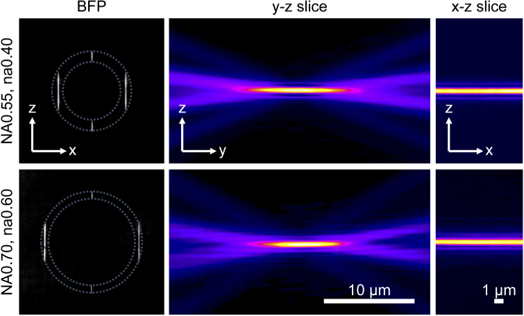
As shown in Fig. 14, we found that similar square lattice light-sheets (in the sense of having similar waist and propagation length) can be generated using different annuli. For example, the thickness and the confocal parameter of a square lattice light-sheet is 0.9 ± 0.01 µm and 18.6 ± 0.26 µm, respectively, for an NA=0.55, na=0.40 annulus. Likewise, a similar square lattice light-sheet with a thickness and propagation length of 1.0 ± 0.04 µm and 16.1 ± 0.26 µm, respectively, can be generated from an NA=0.70, na=0.60 annulus. Surprisingly, the two square lattices have comparable thickness and confocal parameter, even though they are produced from two different rings (of note, corresponding Bessel beams would have different size central lobe thicknesses). Both light-sheets share a similar effective NA ( = 0.28 for NA=0.55, na=0.40, and = 0.26 for NA = 0.70, na = 0.60 annulus, respectively), and as such, should have similar properties.
Fig. 14.
Comparison of similar square lattice light-sheet generated with different annuli. Images of the back focal plane of the illumination objective, YZ slices, and XZ slices of the light-sheets.
This result can be explained in more detail with the theory presented in Appendix A. Computing the depth of focus of the sidebands with Eq. (5) of Appendix A, we obtain 15.6 µm and 16.6 µm for annuli with NA = 0.55, na = 0.4, and NA = 0.7, na = 0.6, respectively. Likewise, using Eq. (6) in Appendix A, we find that these light-sheets have thicknesses of 0.88 µm and 0.94 µm, respectively. Thus, a similar thickness and propagation length is theoretically expected. Practically, we attribute small differences between theory and experiment as likely arising from the challenge of positioning the sidebands perfectly within the annulus (center of NA and na), and we anticipate that this becomes more significant when the sideband is mapped closer to the periphery of Ewald’s sphere.
Funding
Cancer Prevention and Research Institute of Texas10.13039/100004917 (RR160057); National Institutes of Health10.13039/100000002 (5P30CA142543, R33CA235254, R35GM133522, 1R01MH120131, 1R34NS121873).
Disclosures
K.M.D. and R.F. have an investment interest in Discovery Imaging Systems, LLC. B.-J. C. declares no conflicts of interest.
References
- 1.Power R. M., Huisken J., “A guide to light-sheet fluorescence microscopy for multiscale imaging,” Nat. Methods 14(4), 360–373 (2017). 10.1038/nmeth.4224 [DOI] [PubMed] [Google Scholar]
- 2.Huisken J., Swoger J., Bene F. D., Wittbrodt J., Stelzer E. H. K., “Optical Sectioning Deep Inside Live Embryos by Selective Plane Illumination Microscopy,” Science 305(5686), 1007–1009 (2004). 10.1126/science.1100035 [DOI] [PubMed] [Google Scholar]
- 3.Gao L., Shao L., Chen B.-C., Betzig E., “3D live fluorescence imaging of cellular dynamics using Bessel beam plane illumination microscopy,” Nat. Protoc. 9(5), 1083–1101 (2014). 10.1038/nprot.2014.087 [DOI] [PubMed] [Google Scholar]
- 4.Chen B., Chakraborty T., Daetwyler S., Manton J. D., Dean K., Fiolka R., “Extended depth of focus multiphoton microscopy via incoherent pulse splitting,” Biomed. Opt. Express 11(7), 3830 (2020). 10.1364/BOE.393931 [DOI] [PMC free article] [PubMed] [Google Scholar]
- 5.Vettenburg T., Dalgarno H. I. C., Nylk J., Coll-Lladó C., Ferrier D. E. K., Čižmár T., Gunn-Moore F. J., Dholakia K., “Light-sheet microscopy using an Airy beam,” Nat. Methods 11(5), 541–544 (2014). 10.1038/nmeth.2922 [DOI] [PubMed] [Google Scholar]
- 6.Planchon T. A., Gao L., Milkie D. E., Davidson M. W., Galbraith J. A., Galbraith C. G., Betzig E., “Rapid three-dimensional isotropic imaging of living cells using Bessel beam plane illumination,” Nat. Methods 8(5), 417–423 (2011). 10.1038/nmeth.1586 [DOI] [PMC free article] [PubMed] [Google Scholar]
- 7.Fahrbach F. O., Rohrbach A., “A line scanned light-sheet microscope with phase shaped self-reconstructing beams,” Opt. Express 18(23), 24229 (2010). 10.1364/OE.18.024229 [DOI] [PubMed] [Google Scholar]
- 8.Fahrbach F. O., Simon P., Rohrbach A., “Microscopy with self-reconstructing beams,” Nat. Photonics 4(11), 780–785 (2010). 10.1038/nphoton.2010.204 [DOI] [Google Scholar]
- 9.Dean K. M., Roudot P., Welf E. S., Danuser G., Fiolka R., “Deconvolution-free Subcellular Imaging with Axially Swept Light Sheet Microscopy,” Biophys. J. 108(12), 2807–2815 (2015). 10.1016/j.bpj.2015.05.013 [DOI] [PMC free article] [PubMed] [Google Scholar]
- 10.Welf E. S., Driscoll M. K., Dean K. M., Schäfer C., Chu J., Davidson M. W., Lin M. Z., Danuser G., Fiolka R., “Quantitative Multiscale Cell Imaging in Controlled 3D Microenvironments,” Dev. Cell 36(4), 462–475 (2016). 10.1016/j.devcel.2016.01.022 [DOI] [PMC free article] [PubMed] [Google Scholar]
- 11.Chen B.-C., Legant W. R., Wang K., Shao L., Milkie D. E., Davidson M. W., Janetopoulos C., Wu X. S., Hammer J. A., Liu Z., English B. P., Mimori-Kiyosue Y., Romero D. P., Ritter A. T., Lippincott-Schwartz J., Fritz-Laylin L., Mullins R. D., Mitchell D. M., Bembenek J. N., Reymann A.-C., Böhme R., Grill S. W., Wang J. T., Seydoux G., Tulu U. S., Kiehart D. P., Betzig E., “Lattice light-sheet microscopy: Imaging molecules to embryos at high spatiotemporal resolution,” Science 346(6208), 1257998 (2014). 10.1126/science.1257998 [DOI] [PMC free article] [PubMed] [Google Scholar]
- 12.Chang B.-J., Kittisopikul M., Dean K. M., Roudot P., Welf E. S., Fiolka R., “Universal light-sheet generation with field synthesis,” Nat. Methods 16(3), 235–238 (2019). 10.1038/s41592-019-0327-9 [DOI] [PMC free article] [PubMed] [Google Scholar]
- 13.Gao R., Asano S. M., Upadhyayula S., Pisarev I., Milkie D. E., Liu T.-L., Singh V., Graves A., Huynh G. H., Zhao Y., Bogovic J., Colonell J., Ott C. M., Zugates C., Tappan S., Rodriguez A., Mosaliganti K. R., Sheu S.-H., Pasolli H. A., Pang S., Xu C. S., Megason S. G., Hess H., Lippincott-Schwartz J., Hantman A., Rubin G. M., Kirchhausen T., Saalfeld S., Aso Y., Boyden E. S., Betzig E., “Cortical column and whole-brain imaging with molecular contrast and nanoscale resolution,” Science 363(6424), eaau8302 (2019). 10.1126/science.aau8302 [DOI] [PMC free article] [PubMed] [Google Scholar]
- 14.Liu T.-L., Upadhyayula S., Milkie D. E., Singh V., Wang K., Swinburne I. A., Mosaliganti K. R., Collins Z. M., Hiscock T. W., Shea J., Kohrman A. Q., Medwig T. N., Dambournet D., Forster R., Cunniff B., Ruan Y., Yashiro H., Scholpp S., Meyerowitz E. M., Hockemeyer D., Drubin D. G., Martin B. L., Matus D. Q., Koyama M., Megason S. G., Kirchhausen T., Betzig E., “Observing the cell in its native state: Imaging subcellular dynamics in multicellular organisms,” Science 360(6386), eaaq1392 (2018). 10.1126/science.aaq1392 [DOI] [PMC free article] [PubMed] [Google Scholar]
- 15.Fritz-Laylin L. K., Riel-Mehan M., Chen B.-C., Lord S. J., Goddard T. D., Ferrin T. E., Nicholson-Dykstra S. M., Higgs H., Johnson G. T., Betzig E., Mullins R. D., “Actin-based protrusions of migrating neutrophils are intrinsically lamellar and facilitate direction changes,” eLife 6, e26990 (2017). 10.7554/eLife.26990 [DOI] [PMC free article] [PubMed] [Google Scholar]
- 16.Legant W. R., Shao L., Grimm J. B., Brown T. A., Milkie D. E., Avants B. B., Lavis L. D., Betzig E., “High-density three-dimensional localization microscopy across large volumes,” Nat. Methods 13(4), 359–365 (2016). 10.1038/nmeth.3797 [DOI] [PMC free article] [PubMed] [Google Scholar]
- 17.Remacha E., Friedrich L., Vermot J., Fahrbach F. O., “How to define and optimize axial resolution in light-sheet microscopy: a simulation-based approach,” Biomed. Opt. Express 11(1), 8 (2020). 10.1364/BOE.11.000008 [DOI] [PMC free article] [PubMed] [Google Scholar]
- 18.Huisken J., Stainier D. Y. R., “Even fluorescence excitation by multidirectional selective plane illumination microscopy (mSPIM),” Opt. Lett. 32(17), 2608 (2007). 10.1364/OL.32.002608 [DOI] [PubMed] [Google Scholar]
- 19.Born M., Wolf E., Bhatia A. B., “Principles of optics : electromagnetic theory of propagation, interference, and diffraction of light,” Seventh (expanded) anniversary edition, 60th anniversary edition. ed. (Cambridge University Press, Cambridge, 2019), p. 1 online resource. [Google Scholar]
- 20.Valm A. M., Cohen S., Legant W. R., Melunis J., Hershberg U., Wait E., Cohen A. R., Davidson M. W., Betzig E., Lippincott-Schwartz J., “Applying systems-level spectral imaging and analysis to reveal the organelle interactome,” Nature 546(7656), 162–167 (2017). 10.1038/nature22369 [DOI] [PMC free article] [PubMed] [Google Scholar]
- 21.Veltman D. M., Williams T. D., Bloomfield G., Chen B.-C., Betzig E., Insall R. H., Kay R. R., “A plasma membrane template for macropinocytic cups,” eLife 5, e20085 (2016). 10.7554/eLife.20085 [DOI] [PMC free article] [PubMed] [Google Scholar]
- 22.Nagorni M., Hell S. W., “Coherent use of opposing lenses for axial resolution increase in fluorescence microscopy I Comparative study of concepts,” J. Opt. Soc. Am. A 18(1), 36 (2001). 10.1364/JOSAA.18.000036 [DOI] [PubMed] [Google Scholar]
- 23.Chang B.-J., Fiolka R., “Light-sheet engineering using the Field Synthesis theorem,” JPhys Photonics 2(1), 014001 (2019). 10.1088/2515-7647/ab5028 [DOI] [Google Scholar]
- 24.McCutchen C. W., “Generalized Aperture and the Three-Dimensional Diffraction Image,” J. Opt. Soc. Am. 54(2), 240 (1964). 10.1364/JOSA.54.000240 [DOI] [Google Scholar]
- 25.Matthews C., Cordelières F. P., “MetroloJ: an ImageJ plugin to help monitor microscopes’ health,” in ImageJ User & Developer Conference, (2010). [Google Scholar]
- 26.Sage D., Donati L., Soulez F., Fortun D., Schmit G., Seitz A., Guiet R., Vonesch C., Unser M., “DeconvolutionLab2: An open-source software for deconvolution microscopy,” Methods 115, 28–41 (2017). 10.1016/j.ymeth.2016.12.015 [DOI] [PubMed] [Google Scholar]


