. 2000 Feb 1;14(3):301-12.

c-Kit triggers dual phosphorylations, which couple activation and degradation of the essential melanocyte factor Mi

Affiliations

c-Kit triggers dual phosphorylations, which couple activation and degradation of the essential melanocyte factor Mi

M Wu et al. Genes Dev. .

Abstract

Microphthalmia (Mi) is a bHLHZip transcription factor that is essential for melanocyte development and postnatal function. It is thought to regulate both differentiated features of melanocytes such as pigmentation as well as proliferation/survival, based on phenotypes of mutant mouse alleles. Mi activity is controlled by at least two signaling pathways. Melanocyte-stimulating hormone (MSH) promotes transcription of the Mi gene through cAMP elevation, resulting in sustained Mi up-regulation over many hours. c-Kit signaling up-regulates Mi function through MAP kinase phosphorylation of Mi, thereby recruiting the p300 transcriptional coactivator. The current study reveals that c-Kit signaling triggers two phosphorylation events on Mi, which up-regulate transactivation potential yet simultaneously _target Mi for ubiquitin-dependent proteolysis. The specific activation/degradation signals derive from MAPK/ERK _targeting of serine 73, whereas serine 409 serves as a substrate for p90 Rsk-1. An unphosphorylatable double mutant at these two residues is at once profoundly stable and transcriptionally inert. These c-Kit-induced phosphorylations couple transactivation to proteasome-mediated degradation. c-Kit signaling thus triggers short-lived Mi activation and net Mi degradation, in contrast to the profoundly increased Mi expression after MSH signaling, potentially explaining the functional diversity of this transcription factor in regulating proliferation, survival, and differentiation in melanocytes.


Figures

Figure 1
Figure 1
c-Kit activation produces MAPK-dependent Mi degradation. (A) Human melanoma cells (501 Mel) were stimulated at the c-Kit receptor with recombinant Steel factor (Sl) in the presence or absence of the MEK inhibitor drug PD98059 and whole cell extracts probed with monoclonal antibody against Mi (doublet) or α-tubulin-loading control at the time points indicated. (B) c-Kit stimulation was also carried out in the presence of cycloheximide (CHX) to reveal Mi degradation kinetics.
Figure 2
Figure 2
c-Kit-induced Mi degradation is blocked by proteasome inhibition. Presence of a Mi dephosphorylating activity. Mi degradation kinetics were followed using cycloheximide (CHX). (A) Human melanoma cells (501 Mel) were stimulated by recombinant Steel factor (SI) in the presence of cycloheximide (Sl + CHX) and with and without the proteasome inhibitor MG132. α-Tubulin was probed as internal loading control. (B) Lactacystin was used as proteasome inhibitor on SI-stimulated human melanoma cells. (C) Primary human melanocytes were stimulated with recombinant SI in the presence of cycloheximide (Sl + CHX) in the absence or presence of Mg132. Reappearance of the lower migrating Mi isoform at later time points was reversed by treatment of cells with the phosphatase inhibitor okadaic acid in the human melanoma MeWo. (D) Mouse melanoma cells (B16) were treated with TPA to activate MAPK as these cells lack functional c-Kit receptor. Mi levels were compared in the absence or presence of Mg132 at the time points indicated. (E) Mouse melanocytes were treated with TPA as above and Mi levels assessed by Western blotting.
Figure 3
Figure 3
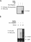
Mi is covalently ubiquitinated in a c-Kit responsive fashion. (A) Plasmids encoding Mi or His-tagged ubiquitin were transfected transiently into baby hamster kidney cells (BHK) and cell lysates were made 24 hr later. His-tagged protein (and associated protein) was affinity purified on nickel resin, proteins eluted and resolved by SDS-PAGE, and Western blotted for Mi. (B) HA-tagged ubiquitin plasmid was transfected into human melanoma cells (as indicated). Twenty-four hours later cells were stimulated with recombinant Steel factor (Sl, as indicated) and extracted 0.5 hr later. Lysates were immunoprecipitated with anti-Mi antibody followed by Western blotting for HA (ubiquitin). Endogenous Mi covalently associates with ubiquitin in a SI-inducible fashion.
Figure 4
Figure 4
Ser-73 is not essential to c-Kit-induced Mi degradation. Adenoviruses encoding wild-type Mi or ser-73 mutated to alanine (S73A) were used to infect human melanoma cells (501 Mel), which were subsequently stimulated with Steel factor (SI) in the presence or absence of cycloheximide and Mg132 proteasome inhibitor. Nuclear extracts from these cells were used in gel shift assays and the Mi-specific DNA-binding activity was identified by supershifting with anti-HA antibody addition (as indicated). Both wild-type and S73A mutant Mi DNA-binding activities diminish after SI stimulation.
Figure 5
Figure 5
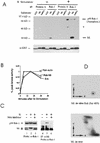
Involvement of Rsk-1 in phosphorylation of ser-409. (A) In vitro kinase assays were carried out using immunoprecipitated Rsk-1 (or protein A control) from unstimulated or Steel factor-stimulated (SI) melanoma cells. Kinase substrates consisted of GST (control), GST fusion to carboxy-terminal fragment of wild-type Mi (“Mi”), or GST fusion to carboxy-terminal fragment of Mi mutated at ser-409 to alanine (“S409A”). GST blot is shown at the bottom. Rsk-1 autophosphorylation is indicated and a weakly coprecipitating second Rsk substrate is indicated by asterisk. (B) Kinetics of Rsk and MAPK/ERK activation after SI stimulation. Kinase assays as described in A were quantitated by PhosphorImager and plotted as percent of maximal activity, indicating that Rsk activation lags ERK activation slightly. (C) Rsk-1 associates with Mi upon SI stimulation. Human melanoma cells were stimulated with SI (or unstimulated) in the presence or absence of MEK inhibitor drug (PD98059). Cell lysates were either probed directly for p90–Rsk-1 or Mi (lanes 13). MEK inhibition prevents a Steel-induced mobility shift in both Mi and a subtle mobility shift in Rsk-1. Mi was also immunoprecipitated from cell lysates followed by Western blotting for Rsk-1. Rsk-1 associated with Mi after Steel stimulation only if MAP kinase was not inhibited. (D) Two-dimensional phosphotryptic mapping indicates presence of phosphoserine 409 within endogenous cellular Mi. The two-dimensional mobility of phosphoserine 409 was determined by analyzing in vitro phosphorylated recombinant Mi (as in Fig. 5A). 32P-labeled melanoma cells were stimulated by SI and extracted; endogenous Mi was immunoprecipitated and subjected to tryptic digestion and two-dimensional analysis (simultaneously run with in vitro phosphorylated recombinant Mi). A spot was observed (dotted circles), which displays superimposable migration relative to the phosphoserine 409 spot generated by Rsk-1 phosphorylation of ser-409 in vitro.
Figure 6
Figure 6
Simultaneous mutation of ser-73 and ser-409 protects Mi from c-Kit-induced proteolysis. (A) Plasmids encoding wild-type Mi (“Mi”) or individual mutations at ser-73 or ser-409 or the double mutant at both sites (S73/409A) were transfected into NIH 3T3 cells. Twenty-four hours later MAPK was activated by TPA in the presence of cycloheximide (CHX), and Mi degradation was followed using Western blot analysis. Tubulin control is indicated. (B) The same constructs as in A were introduced into 501 Mel human melanoma cells, followed by assessment of Mi degradation after Steel stimulation (SI).
Figure 6
Figure 6
Simultaneous mutation of ser-73 and ser-409 protects Mi from c-Kit-induced proteolysis. (A) Plasmids encoding wild-type Mi (“Mi”) or individual mutations at ser-73 or ser-409 or the double mutant at both sites (S73/409A) were transfected into NIH 3T3 cells. Twenty-four hours later MAPK was activated by TPA in the presence of cycloheximide (CHX), and Mi degradation was followed using Western blot analysis. Tubulin control is indicated. (B) The same constructs as in A were introduced into 501 Mel human melanoma cells, followed by assessment of Mi degradation after Steel stimulation (SI).
Figure 7
Figure 7
Double mutant Mi, resistant to MAPK and Rsk, is transcriptionally inactive despite enhanced stability and intact DNA binding. (A) Transactivation assays were performed using wild-type Mi and single or double mutants at ser-73 and ser-409. NIH 3T3 or 501 Mel melanoma cells were cotransfected with pEBB (empty vector) or indicated Mi constructs and luciferase reporter under control of the tyrosinase promoter. Data are normalized to cotransfected sea pansy luciferase plasmid. (B) Gel shifts of BHK cells transfected with the same Mi constructs as in A after MAPK activation with TPA and cycloheximide (CHX). Mi-specific DNA-binding activity is identified using supershifting antibody.

Similar articles

Cited by

References

    1. Andrews RG, Briddell RA, Appelbaum FR, McNiece IK. Stimulation of hematopoiesis in vivo by stem cell factor. Curr Opin Hematol. 1994;1:187–196. - PubMed
    1. Armstrong JA, Emerson BM. Transcription of chromatin: These are complex times. Curr Opin Genet Dev. 1998;8:165–172. - PubMed
    1. Bentley NJ, Eisen T, Goding CR. Melanocyte-specific expression of the human tyrosinase promoter: Activation by the microphthalmia gene product and role of the initiator. Mol Cell Biol. 1994;14:7996–8006. - PMC - PubMed
    1. Berk AJ. Activation of RNA polymerase II transcription. Curr Opin Cell Biol. 1999;11:330–335. - PubMed
    1. Bertolotto C, Bille K, Ortonne J, Ballotti R. Regulation of tyrosinase gene expression by cAMP in B16 melanoma cells involves two CATGTG motifs surrounding the TATA box: Implication of the microphthalmia gene product. J Cell Biol. 1996;134:747–755. - PMC - PubMed

Publication types

MeSH terms

  NODES
INTERN 1
twitter 2