. 2000 Oct;20(20):7602-12.
doi: 10.1128/MCB.20.20.7602-7612.2000.

Inhibition of Daxx-mediated apoptosis by heat shock protein 27

Affiliations

Inhibition of Daxx-mediated apoptosis by heat shock protein 27

S J Charette et al. Mol Cell Biol. 2000 Oct.

Abstract

Heat shock protein 27 (HSP27) confers cellular protection against a variety of cytotoxic stresses and also against physiological stresses associated with growth arrest or receptor-mediated apoptosis. Phosphorylation modulates the activity of HSP27 by causing a major change in the supramolecular organization of the protein, which shifts from oligomers to dimers. Here we show that phosphorylated dimers of HSP27 interact with Daxx, a mediator of Fas-induced apoptosis, preventing the interaction of Daxx with both Ask1 and Fas and blocking Daxx-mediated apoptosis. No such inhibition was observed with an HSP27 phosphorylation mutant that is only expressed as oligomers or when apoptosis was induced by transfection of a Daxx mutant lacking its HSP27 binding domain. HSP27 expression had no effect on Fas-induced FADD- and caspase-dependent apoptosis. However, HSP27 blocked Fas-induced translocation of Daxx from the nucleus to the cytoplasm and Fas-induced Daxx- and Ask1-dependent apoptosis. The observations revealed a new level of regulation of the Fas pathway and suggest a mechanism for the phosphorylation-dependent protective function of HSP27 during stress and differentiation.


Figures

FIG. 1
FIG. 1
HSP27 interacts with Daxx. A partial cDNA of Daxx (clone 55.1) was obtained in a two-hybrid screen of a HeLa cell cDNA library using HaHSP27 as a bait. (A) Schematic representation of the Daxx sequences contained in clone 55.1. Represented are the acidic domain (acid.), the AAD, and the FBD of human Daxx. Residue numbers at domain boundaries are indicated. (B and C) GST pull-down assays. (B) Daxx is retained on immobilized HuHSP27. Extracts from pCINHuDaxx-transfected 293 cells were incubated with immobilized GST–Fas-DD, GST-HuHSP27, or GST. Retained Daxx protein was detected by Western blotting using antibody EL-68. Also shown are the GST fusion proteins in the same gel stained with Ponceau red. For convenience, bands were aligned at the same level. The input track represents 6.7% of the total extract. (C) HuHSP27 is retained on immobilized Daxx sequences contained in clone 55.1. Extracts from pCINHu27WT-transfected 293 cells were incubated with immobilized GST-HuHSP27, GST–55.1, or GST. Retained HuHSP27 protein was detected by Western blotting using an anti-HuHSP27 antibody. Also shown are the GST fusion proteins in the same gel stained with Ponceau red. For convenience, bands were aligned at the same level. The asterisk indicates a GST-HuHSP27 degradation product recognized by anti-Hu. The input track represents 7.5% of the total extract. (D and E) HSP27 interacts with the Fas binding domain of Daxx. (D) Two-hybrid interaction assay was performed with yeast after transfection of LexA-HuHSP27 or the negative controls LexA-lamin C and LexA-ras (val 12) with pGADGH-55.1, GAL4-AAD, or GAL4-FBD. The interaction was quantified by measuring β-galactosidase activities, which are shown in arbitrary units. (E) 293 cells were transfected with pCINmyc-FBD or with pCINmyc-FBD and pSVHa27WT. Extracts (6.1% of the total) were analyzed by Western blotting (WB) with anti-Myc antibody 9E10 to detect Myc-FBD protein or with anti-HaHSP27 antibody L2R3 to detect HaHSP27 expression (input). The extracts were also immunoprecipitated (IP) with L2R3 and then probed with 9E10 to detect coimmunoprecipitated Myc-FBD.
FIG. 2
FIG. 2
HSP27 phosphorylation modulates the interaction with Daxx. Extracts from 293 cells transfected with pSVHa27EE (A) or pSVHa27AA (B) were incubated with immobilized GST, GST–55.1, or GST-HuHSP27. Retained HSP27 proteins were detected by Western blotting using antibody L2R3. Also shown are the GST fusion proteins in the same gel stained with Ponceau red. For convenience, bands were aligned at the same level. The input tracks represent 5% of the total extracts.
FIG. 3
FIG. 3
HSP27 competes with Fas (A and B) and Ask1 (C and D) for interacting with Daxx. 293 cells were transfected with either pCIN-Daxx, pcDNA3-HA-Ask1, pCINHu27WT, pSVHa27WT, pSVHa27AA, or pSVHa27EE. Extracts from Daxx- (A and B) or HA-Ask1 (C and D)-expressing cells were incubated with immobilized GST–Fas-DD or GST–55.1, respectively, either alone (lanes 2) or after being mixed with extracts from cells expressing either species of HSP27 (lanes 3 to 5) as indicated in the input lanes (HuHSP27 or HaHSP27-WT, -AA, or -EE). Retained Daxx and Ask1 were detected by Western blotting using anti-Daxx EL-68 and anti-HA 12CA5 antibodies, respectively. Ponceau red-stained GST fusion proteins are shown on the bottom of each panel. The input lanes in panels A to D represent 0.6, 0.3, 7.5, and 7.5% of the total extracts, respectively.
FIG. 4
FIG. 4
Coexpression of Daxx and Ask1 induces apoptosis in 293 cells. 293 cells were cotransfected with various combinations of pCIN-Daxx, pCIN-DaxxC, pCIN-AAD, pcDNA3-HA-Ask1, and pCIN-Fas, as indicated. pCMVβ was also cotransfected as a marker to identify transfected cells. Twenty-four hours later, the percentages of β-galactosidase-positive cells with apoptotic morphology were calculated after correcting for apoptosis in cells transfected with the marker gene only.
FIG. 5
FIG. 5
HSP27 blocks apoptosis induced by coexpression of Daxx and Ask1. Apoptosis was induced by the cotransfection of the appropriate plasmids to yield the coexpression of Ask1 with either Daxx (A), DaxxC (B and D), or AAD (C), with or without HuHSP27 (Hu), HaHSP27 (Ha WT), or the phosphorylation mutants HaHSP27-AA (Ha AA) and HaHSP27-EE (Ha EE). pCMVβ was also transfected in all cases to identify transfected cells. Twenty-four hours after transfection, the cells were processed to determine the number of β-galactosidase-positive cells with apoptotic morphology. When indicated (Hu SB), the cells were incubated for the last 16 h in the presence of SB203580 (5 μM). (A and B insets) Western blots showing the concentrations of Ask1, Daxx or DaxxC, and HSP27 in an aliquot of the cell lysates for each condition corresponding to the histograms. Gel tracks in the insets are in the same order as in the histograms.
FIG. 6
FIG. 6
Specificity of the Daxx and FADD arms of the Fas pathway. (A) Effect of expressing FADD-DN or inhibiting the caspases with z-VAD-fmk on apoptosis induced by overexpression of Daxx and Ask1 or Fas. (B) Effect of expression of pseudophosphorylated HSP27 (HaHSP27-EE) on apoptosis induced by overexpression of Daxx and Ask1 or FADD. 293 cells were transfected with the appropriate plasmids together with pCMVβ, and apoptosis was measured 24 h later in β-galactosidase-positive cells. When indicated, z-VAD-fmk (50 μM) was added 12 h before staining.
FIG. 7
FIG. 7
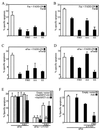
HSP27 inhibits the Daxx-sensitive arm of Fas-induced apoptosis. (A to D) Effect of expression of Daxx-FBD, HaHSP27-AA, or HaHSP27-EE, together with FADD-DN, on apoptosis induced 24 h (A and C) or 48 h (B and D) after either overexpressing Fas by transfection (A and B) or stimulating Fas with anti-Fas (C and D). Anti-Fas (100 ng/ml) was added 24 h after transfection of other plasmids. (E and F) Overexpressing Ask1 amplifies the relative importance of the Daxx pathway and of HSP27 inhibition of apoptosis induced 24 h after Fas activation. The cells were transfected with the appropriate plasmids in various combinations as indicated. Anti-Fas (100 ng/ml) was added 12 h after transfection. z-VAD-fmk (50 μM) was added 1 h before Fas activation. pCMVβ was also transfected in all cases, and apoptosis in the β-galactosidase-positive cells was measured.
FIG. 8
FIG. 8
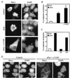
Fas-induced translocation of Daxx from the nucleus to the cytoplasm is inhibited by HSP27. (A to C) 293 cells were transfected with plasmid pCINHuDaxx, alone or together with plasmid pSVHa27AA (AA) or pSVHa27EE (EE). Twelve hours after transfection the cells were left untreated (control or con) or treated with 100 ng of anti-Fas activating antibody/ml for 12 h before fixation. Where indicated, z-VAD-fmk (50 μM) was added to the cells 30 min before Fas stimulation. Daxx was localized by immunofluorescence using anti-Daxx EL-68 (A). Nuclei were revealed by DAPI staining. Arrowheads indicate nuclei of transfected cells. The percentages of cells in which Daxx was localized in the cytoplasm (as illustrated in panel A) were determined by direct counting under the microscope. (D) Immunolocalization of endogenous Daxx. The procedure was the same as for panel A except that the cells were treated directly without transfection. Endogenous Daxx was not seen in panel A because of a much shorter exposure time (15-fold) than that used for panel D.

Similar articles

Cited by

References

    1. Algeciras-Schimnich A, Griffith T S, Lynch D H, Paya C V. Cell cycle-dependent regulation of FLIP levels and susceptibility to Fas-mediated apoptosis. J Immunol. 1999;162:5205–5211. - PubMed
    1. Arrigo A P, Landry J. Expression and function of the low-molecular-weight heat shock proteins. In: Morimoto R I, Tissières A, Georgopoulos C, editors. The biology of heat shock proteins and molecular chaperones. Cold Spring Harbor, N.Y: Cold Spring Harbor Laboratory Press; 1994. pp. 335–373.
    1. Benndorf R, Hayess K, Ryazantsev S, Wieske M, Behlke J, Lutsch G. Phosphorylation and supramolecular organization of murine small heat shock protein HSP25 abolish its actin polymerization-inhibiting activity. J Biol Chem. 1994;269:20780–20784. - PubMed
    1. Benndorf R, Kraft R, Otto A, Stahl J, Bohm H, Bielka H. Purification of the growth-related protein p25 of the Ehrlich ascites tumor and analysis of its isoforms. Biochem Int. 1988;17:225–234. - PubMed
    1. Boldin M P, Goncharov T M, Goltsev Y V, Wallach D. Involvement of MACH, a novel MORT1/FADD-interacting protease, in Fas/APO-1 and TNF receptor-induced cell death. Cell. 1996;85:803–815. - PubMed

Publication types

MeSH terms

  NODES
twitter 2