Distinct but overlapping roles of histone acetylase PCAF and of the closely related PCAF-B/GCN5 in mouse embryogenesis
- PMID: 11027331
- PMCID: PMC17195
- DOI: 10.1073/pnas.97.21.11303
Distinct but overlapping roles of histone acetylase PCAF and of the closely related PCAF-B/GCN5 in mouse embryogenesis
Abstract
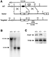
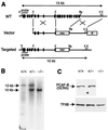
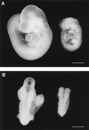
PCAF plays a role in transcriptional activation, cell-cycle arrest, and cell differentiation in cultured cells. PCAF contributes to transcriptional activation by acetylating chromatin and transcription factors through its intrinsic histone acetylase activity. In this report, we present evidence for the in vivo function of PCAF and the closely related PCAF-B/GCN5. Mice lacking PCAF are developmentally normal without a distinct phenotype. In PCAF null-zygous mice, protein levels of PCAF-B/GCN5 are drastically elevated in lung and liver, where PCAF is abundantly expressed in wild-type mice, suggesting that PCAF-B/GCN5 functionally compensates for PCAF. In contrast, animals lacking PCAF-B/GCN5 die between days 9.5 and 11.5 of gestation. Normally, PCAF-B/GCN5 mRNA is expressed at high levels already by day 8, whereas PCAF mRNA is first detected on day 12.5, which may explain, in part, the distinct knockout phenotypes. These results provide evidence that PCAF and PCAF-B/GCN5 play distinct but functionally overlapping roles in embryogenesis.
Figures


Similar articles
-
Loss of Gcn5l2 leads to increased apoptosis and mesodermal defects during mouse development.Nat Genet. 2000 Oct;26(2):229-32. doi: 10.1038/79973. Nat Genet. 2000. PMID: 11017084
-
GCN5 and p300 share essential functions during early embryogenesis.Dev Dyn. 2005 Aug;233(4):1337-47. doi: 10.1002/dvdy.20445. Dev Dyn. 2005. PMID: 15937931
-
Distinct GCN5/PCAF-containing complexes function as co-activators and are involved in transcription factor and global histone acetylation.Oncogene. 2007 Aug 13;26(37):5341-57. doi: 10.1038/sj.onc.1210604. Oncogene. 2007. PMID: 17694077 Review.
-
GCN5: a supervisor in all-inclusive control of vertebrate cell cycle progression through transcription regulation of various cell cycle-related genes.Gene. 2005 Feb 28;347(1):83-97. doi: 10.1016/j.gene.2004.12.007. Gene. 2005. PMID: 15715965
-
[Histone-like TAFs within the PCAF histone acetylase complex].Tanpakushitsu Kakusan Koso. 1998 Nov;43(14):2079-86. Tanpakushitsu Kakusan Koso. 1998. PMID: 9838849 Review. Japanese. No abstract available.
Cited by
-
KATapulting toward Pluripotency and Cancer.J Mol Biol. 2017 Jun 30;429(13):1958-1977. doi: 10.1016/j.jmb.2016.09.023. Epub 2016 Oct 6. J Mol Biol. 2017. PMID: 27720985 Free PMC article. Review.
-
Crx activates opsin transcription by recruiting HAT-containing co-activators and promoting histone acetylation.Hum Mol Genet. 2007 Oct 15;16(20):2433-52. doi: 10.1093/hmg/ddm200. Epub 2007 Jul 26. Hum Mol Genet. 2007. PMID: 17656371 Free PMC article.
-
The histone acetyltransferase GCN5 affects the inflorescence meristem and stamen development in Arabidopsis.Planta. 2009 Nov;230(6):1207-21. doi: 10.1007/s00425-009-1012-5. Epub 2009 Sep 22. Planta. 2009. PMID: 19771450
-
Pair of unusual GCN5 histone acetyltransferases and ADA2 homologues in the protozoan parasite Toxoplasma gondii.Eukaryot Cell. 2006 Jan;5(1):62-76. doi: 10.1128/EC.5.1.62-76.2006. Eukaryot Cell. 2006. PMID: 16400169 Free PMC article.
-
KAT2B Gene Polymorphisms Are Associated with Body Measure Traits in Four Chinese Cattle Breeds.Animals (Basel). 2022 Aug 1;12(15):1954. doi: 10.3390/ani12151954. Animals (Basel). 2022. PMID: 35953943 Free PMC article.
References
-
- Yang X J, Ogryzko V V, Nishikawa J, Howard B H, Nakatani Y. Nature (London) 1996;382:319–324. - PubMed
-
- Kuo M H, Allis C D. BioEssays. 1998;20:615–626. - PubMed
-
- Struhl K. Genes Dev. 1998;12:599–606. - PubMed
-
- Workman J, Kingston R E. Annu Rev Biochem. 1998;67:545–579. - PubMed
-
- Schiltz R L, Nakatani Y. Biochim Biophys Acta. 2000;1470:M37–M53. - PubMed
MeSH terms
Substances
LinkOut - more resources
Full Text Sources
Molecular Biology Databases

