. 2000 Dec;20(24):9247-61.
doi: 10.1128/MCB.20.24.9247-9261.2000.

Inhibitory regulation of Rac activation, membrane ruffling, and cell migration by the G protein-coupled sphingosine-1-phosphate receptor EDG5 but not EDG1 or EDG3

Affiliations

Inhibitory regulation of Rac activation, membrane ruffling, and cell migration by the G protein-coupled sphingosine-1-phosphate receptor EDG5 but not EDG1 or EDG3

H Okamoto et al. Mol Cell Biol. 2000 Dec.

Abstract

Sphingosine-1-phosphate (S1P) is a bioactive lysophospholipid that induces a variety of biological responses in diverse cell types. Many, if not all, of these responses are mediated by members of the EDG (endothelial differentiation gene) family G protein-coupled receptors EDG1, EDG3, and EDG5 (AGR16). Among prominent activities of S1P is the regulation of cell motility; S1P stimulates or inhibits cell motility depending on cell types. In the present study, we provide evidence for EDG subtype-specific, contrasting regulation of cell motility and cellular Rac activity. In CHO cells expressing EDG1 or EDG3 (EDG1 cells or EDG3 cells, respectively) S1P as well as insulin-like growth factor I (IGF I) induced chemotaxis and membrane ruffling in phosphoinositide (PI) 3-kinase- and Rac-dependent manners. Both S1P and IGF I induced a biphasic increase in the amount of the GTP-bound active form of Rac. In CHO cells expressing EDG5 (EDG5 cells), IGF I similarly stimulated cell migration; however, in contrast to what was found for EDG1 and EDG3 cells, S1P did not stimulate migration but totally abolished IGF I-directed chemotaxis and membrane ruffling, in a manner dependent on a concentration gradient of S1P. In EDG5 cells, S1P stimulated PI 3-kinase activity as it did in EDG1 cells but inhibited the basal Rac activity and totally abolished IGF I-induced Rac activation, which involved stimulation of Rac-GTPase-activating protein activity rather than inhibition of Rac-guanine nucleotide exchange activity. S1P induced comparable increases in the amounts of GTP-RhoA in EDG3 and EDG5 cells. Neither S1P nor IGF I increased the amount of GTP-bound Cdc42. However, expression of N(17)-Cdc42, but not N(19)-RhoA, suppressed S1P- and IGF I-directed chemotaxis, suggesting a requirement for basal Cdc42 activity for chemotaxis. Taken together, the present results demonstrate that EDG5 is the first example of a hitherto-unrecognized type of receptors that negatively regulate Rac activity, thereby inhibiting cell migration and membrane ruffling.


Figures

FIG. 1
FIG. 1
(A) EDG5 mediates inhibition by S1P of cell migration, whereas EDG1 and EDG3 mediate chemotaxis toward S1P. Migration across the porous filter of CHO cells expressing either EDG1, EDG3, or EDG5 and nontransfected CHO cells was determined in the presence of S1P alone at various concentrations (top) and of S1P plus IGF I (100 ng/ml) (bottom) in the lower wells of the Boyden chamber. OD, optical density. (B) EDG5-mediated inhibition of chemotaxis is dependent on a S1P concentration gradient. Migration of EDG5 cells was determined in the presence or absence of S1P (0.1 μM) and IGF I (100 ng/ml) in the upper and lower chambers. All data are means ± standard errors of three determinations and are representative of three independent experiments with similar results.
FIG. 1
FIG. 1
(A) EDG5 mediates inhibition by S1P of cell migration, whereas EDG1 and EDG3 mediate chemotaxis toward S1P. Migration across the porous filter of CHO cells expressing either EDG1, EDG3, or EDG5 and nontransfected CHO cells was determined in the presence of S1P alone at various concentrations (top) and of S1P plus IGF I (100 ng/ml) (bottom) in the lower wells of the Boyden chamber. OD, optical density. (B) EDG5-mediated inhibition of chemotaxis is dependent on a S1P concentration gradient. Migration of EDG5 cells was determined in the presence or absence of S1P (0.1 μM) and IGF I (100 ng/ml) in the upper and lower chambers. All data are means ± standard errors of three determinations and are representative of three independent experiments with similar results.
FIG. 2
FIG. 2
(A) EDG5 mediates formation of stress fibers and inhibition of IGF I-induced membrane ruffling in response to S1P stimulation, whereas EDG1 mediates membrane ruffling. EDG1 and EDG5 cells were nonstimulated (a and b) or stimulated with either IGF I (100 ng/ml) (c and d), S1P (0.1 μM) (e and f), or IGF I plus S1P (g and h) for 30 min and then fixed and stained for F-actin with TRITC-phalloidin. (B) EDG5 does not mediate inhibition of V12-Rac-induced membrane ruffling in response to S1P stimulation. EDG5 cells were transiently transfected with an expression vector for myc-tagged V12-Rac 48 h prior to stimulation with S1P (0.1 μM) for 30 min. The cells were then fixed and stained for visualizing F-actin and myc-tagged V12-Rac protein with TRITC-phalloidin (a and c) and 9E10 antimyc antibody (b and d), respectively. Arrows, cells expressing myc-tagged V12-Rac. The results shown are representative of three experiments with similar results.
FIG. 3
FIG. 3
Chemotaxis induced by S1P via EDG1 and by IGFI is dependent on Rac and Cdc42 but not Rho. (A) EDG1 cells were transduced with adenoviral vectors encoding either LacZ, N17-Ras, or N17-Rac and 48 h later were assayed for chemotaxis toward S1P (0.1 μM) and IGF I (100 ng/ml). Parallel dishes were processed for immunodetection of N17-Ras (anti-Ras) and N17-Rac (anti-Rac). OD, optical density. (B) EDG1 cells were cotransfected with a vector encoding N17-Cdc42, myc-N19-Rho, or myc-N17-Rac or the empty vector and the β-galactosidase (LacZ) expression vector 48 h before the migration assay. LacZ-expressing migrating cells were counted as described in Materials and Methods. The expression of the dominant-negative GTPases (arrowheads) and endogenous wild-type GTPases (arrows) was detected by Western blotting. The data in panels A and B are means ± standard errors of three determinations. (C) EDG1 cells were transduced with adenoviruses encoding LacZ or N17-Rac or pretreated with LY294002 (LY; 30 μM) or wortmannin (WMN, 0.1 μM) for 20 min and then stimulated with S1P (0.1 μM) for 30 min. The cells were fixed and stained for visualizing F-actin with TRITC-phalloidin. The results are representative of three experiments with similar results.
FIG. 4
FIG. 4
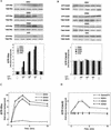
EDG1 and EDG3 mediate activation of Rac, whereas EDG5 mediates inhibition of IGF I-stimulation of Rac. (A) EDG cells and CHO cells were stimulated with various concentrations of S1P or 100 ng of IGF I/ml for 1 min, and the amount of GTP-loaded Rac (GTP-Rac) was determined as described in Materials and Methods. A portion (1/20) of the cell lysate was subjected to Western analysis for evaluating the amount of total Rac in each sample. (B) EDG1 and EDG5 cells were stimulated with IGF I (100 ng/ml) plus various concentrations of S1P and analyzed as described for panel A. (C) Time-dependent increase in the amount of GTP-Rac in IGF I-stimulated EDG5 cells in the presence and absence of S1P. The cells were pretreated with S1P (0.1 μM) for 10 min or left unpretreated and then stimulated with IGF I (100 ng/ml) for the indicated time periods. (D) Time-dependent increase in the amount of GTP-Rac in S1P (0.1 μM)-stimulated EDG1, EDG3, and EDG5 cells. (E) Time-dependent decrease in the amount of GTP-Rac in S1P-pretreated, IGF I-stimulated EDG5 cells. EDG5 cells were treated with S1P (0.1 μM) for the indicated time periods. IGF I (100 ng/ml) was present for the last 1 min of the incubation. The data in panels A to E are means ± standard errors of three determinations. (F) EDG5 cells were transiently transfected with expression vectors for myc-tagged wild-type (wt) Rac or V12-Rac and 48 h later were stimulated with S1P (0.1 μM) for 5 min or were not stimulated and assayed for the amounts of GTP-bound forms of Rac and V12-Rac. The results are representative of two or three experiments with similar results. IB, immunoblot.
FIG. 4
FIG. 4
EDG1 and EDG3 mediate activation of Rac, whereas EDG5 mediates inhibition of IGF I-stimulation of Rac. (A) EDG cells and CHO cells were stimulated with various concentrations of S1P or 100 ng of IGF I/ml for 1 min, and the amount of GTP-loaded Rac (GTP-Rac) was determined as described in Materials and Methods. A portion (1/20) of the cell lysate was subjected to Western analysis for evaluating the amount of total Rac in each sample. (B) EDG1 and EDG5 cells were stimulated with IGF I (100 ng/ml) plus various concentrations of S1P and analyzed as described for panel A. (C) Time-dependent increase in the amount of GTP-Rac in IGF I-stimulated EDG5 cells in the presence and absence of S1P. The cells were pretreated with S1P (0.1 μM) for 10 min or left unpretreated and then stimulated with IGF I (100 ng/ml) for the indicated time periods. (D) Time-dependent increase in the amount of GTP-Rac in S1P (0.1 μM)-stimulated EDG1, EDG3, and EDG5 cells. (E) Time-dependent decrease in the amount of GTP-Rac in S1P-pretreated, IGF I-stimulated EDG5 cells. EDG5 cells were treated with S1P (0.1 μM) for the indicated time periods. IGF I (100 ng/ml) was present for the last 1 min of the incubation. The data in panels A to E are means ± standard errors of three determinations. (F) EDG5 cells were transiently transfected with expression vectors for myc-tagged wild-type (wt) Rac or V12-Rac and 48 h later were stimulated with S1P (0.1 μM) for 5 min or were not stimulated and assayed for the amounts of GTP-bound forms of Rac and V12-Rac. The results are representative of two or three experiments with similar results. IB, immunoblot.
FIG. 5
FIG. 5
EDG5 mediates inhibition of IGF I-induced p65PAK activation. (A) CHO cells and EDG cells were pretreated with S1P (0.1 μM) for 5 min or left unpretreated and then stimulated with S1P (0.1 μM) or IGF I (100 ng/ml) for a further 5 min and assayed for p65PAK activity as described in Materials and Methods. Autoradiographs and anti-myc Western blots are shown at the top. MBP, myelin basic protein. (B) EDG5 cells were transiently transfected with the empty vector or expression vectors for N17-Rac and V12-Rac and 48 h later were treated with S1P (0.1 μM) or IGF I (100 ng/ml) for 5 min or were left untreated. All data are means ± standard errors of three determinations. The results are representative of two experiments with similar results.
FIG. 5
FIG. 5
EDG5 mediates inhibition of IGF I-induced p65PAK activation. (A) CHO cells and EDG cells were pretreated with S1P (0.1 μM) for 5 min or left unpretreated and then stimulated with S1P (0.1 μM) or IGF I (100 ng/ml) for a further 5 min and assayed for p65PAK activity as described in Materials and Methods. Autoradiographs and anti-myc Western blots are shown at the top. MBP, myelin basic protein. (B) EDG5 cells were transiently transfected with the empty vector or expression vectors for N17-Rac and V12-Rac and 48 h later were treated with S1P (0.1 μM) or IGF I (100 ng/ml) for 5 min or were left untreated. All data are means ± standard errors of three determinations. The results are representative of two experiments with similar results.
FIG. 6
FIG. 6
EDG5 and EDG3 mediate activation of Rho, but EDG1, EDG3, and EDG5 do not mediate activation of Cdc42. (A and B) EDG cells and CHO cells were stimulated with S1P at various concentrations or IGF I (100 ng/ml) for 3 min. (C and D) EDG cells and Swiss 3T3 cells were stimulated with S1P (0.1 μM) or bradykinin (0.1 μM), respectively, for the indicated times. The amounts of GTP-bound Rho and Cdc42 were determined as described in Materials and Methods. A portion (1/20) of the cell lysate was subjected to Western analysis for evaluating the amounts of total Rho and Cdc42 in each sample. The data are means ± standard errors of three determinations. The results are representative of two or three experiments with similar results.
FIG. 7
FIG. 7
Cyclic AMP, protein kinase A, ERK, p38MAPK, Rho kinase, PKC, and Gi protein are not involved in S1P inhibition of IGF I-induced chemotaxis and Rac activation. (A) Forskolin, dibutyryl cyclic AMP (dbcAMP), and PI 3-kinase inhibitors reduces chemotaxis toward IGF I. (B) PI 3-kinase inhibitors, but not forskolin, dbcAMP, or PTX, reduce IGF I-induced Rac activation. (C) Various protein kinase inhibitors and PTX do not block S1P inhibition of chemotaxis toward IGF I. EDG5 cells were pretreated with PTX (100 ng/ml), forskolin (FSK) (1 or 10 μM), dbcAMP (0.1 or 1 mM), LY294002 (LY) (30 μM), wortmannin (WMN) (0.1 μM), PD98059 (PD) (30 μM), SB203580 (SB) (10 μM), HA1077 (HA) (10 μM), or GF109203X (GF) (5 μM) for 20 min except for PTX, where the treatment time was 24 h, or with S1P (0.1 μM) for 10 min and then stimulated with IGF I (100 ng/ml). In the migration assay, all drugs except PTX were present throughout the 4-h incubation period. The data are means ± standard errors of three determinations. The results are representative of two experiments with similar results. OD, optical density.
FIG. 8
FIG. 8
EDG5 does not mediate inhibition of IGF I-induced PI 3-kinase activation. CHO cells and EDG cells were stimulated with S1P (0.1 μM) and/or IGF I (100 ng/ml) for 5 min and assayed for PI 3-kinase activity as described in Materials and Methods. Autoradiographs of thin-layer chromatography plates and quantitated results are shown. The data are means ± standard errors of three determinations. The results are representative of two experiments with similar results. ∗, statistically significant (P < 0.05) compared to the value for nonstimulated cells analyzed as described for Table 1.
FIG. 9
FIG. 9
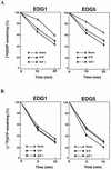
EDG5 mediates stimulation of Rac-GAP activity but not inhibition of Rac-GEF activity. (A and B) EDG1 and EDG5 cells were stimulated with S1P (0.1 μM) or IGF I (100 ng/ml) for 1 min (GEF assay) or 10 min (GAP assay) or were not stimulated. (C, left) EDG5 cells were treated with S1P (0.1 μM) for the indicated times. IGF I (100 ng/ml) was present for the last 1 min of the incubation when indicated. (Right) EDG5 cells were stimulated with various concentrations of S1P with or without IGF I (100 ng/ml) for 10 min. Cell lysate was prepared. Rac-GEF activity in cell lysate was assayed using [3H]GDP-loaded recombinant Rac protein as described in Materials and Methods. Similarly, Rac-GAP activity in cell lysate was determined using [γ-33P]GTP-loaded Rac. The values at 5 min in the Rac-GAP assay are shown. All values are means ± standard errors of three determinations. The results are representative of two or three experiments with similar results. ∗ (C), statistically significant (P < 0.05) compared to the value for nonstimulated cells analyzed as described for Table 1.
FIG. 9
FIG. 9
EDG5 mediates stimulation of Rac-GAP activity but not inhibition of Rac-GEF activity. (A and B) EDG1 and EDG5 cells were stimulated with S1P (0.1 μM) or IGF I (100 ng/ml) for 1 min (GEF assay) or 10 min (GAP assay) or were not stimulated. (C, left) EDG5 cells were treated with S1P (0.1 μM) for the indicated times. IGF I (100 ng/ml) was present for the last 1 min of the incubation when indicated. (Right) EDG5 cells were stimulated with various concentrations of S1P with or without IGF I (100 ng/ml) for 10 min. Cell lysate was prepared. Rac-GEF activity in cell lysate was assayed using [3H]GDP-loaded recombinant Rac protein as described in Materials and Methods. Similarly, Rac-GAP activity in cell lysate was determined using [γ-33P]GTP-loaded Rac. The values at 5 min in the Rac-GAP assay are shown. All values are means ± standard errors of three determinations. The results are representative of two or three experiments with similar results. ∗ (C), statistically significant (P < 0.05) compared to the value for nonstimulated cells analyzed as described for Table 1.

Similar articles

Cited by

References

    1. Adam L, Vadlamudi R, Kondapaka S B, Chernoff J, Mendelsohn J, Kumar R. Heregulin regulates cytoskeletal reorganization and cell migration through the p21-activated kinase-1 via phosphatidylinositol-3 kinase. J Biol Chem. 1998;273:28238–28246. - PubMed
    1. Akasaki T, Koga H, Sunimoto H. Phosphoinositide 3-kinase-dependent and -independent activation of the small GTPase Rac2 in human neutrophils. J Biol Chem. 1999;274:18055–18059. - PubMed
    1. Allen W E, Zicha D, Ridley A J, Jones G E. A role for Cdc42 in macrophage chemotaxis. J Cell Biol. 1998;141:1147–1157. - PMC - PubMed
    1. An S, Zheng Y, Bleu T. Sphingosine 1-phosphate-induced cell proliferation, survival, and related signaling events mediated by G protein-coupled receptors Edg3 and Edg5. J Biol Chem. 2000;275:288–296. - PubMed
    1. An S. Molecular identification and characterization of G protein-coupled receptors for lysophosphatidic acid and sphingosine 1-phosphate. Ann N Y Acad Sci. 2000;905:25–33. - PubMed

Publication types

MeSH terms

Substances

  NODES
Project 1
twitter 2