. 2001 Mar;21(6):2144-53.
doi: 10.1128/MCB.21.6.2144-2153.2001.

Dual inactivation of RB and p53 pathways in RAS-induced melanomas

Affiliations

Dual inactivation of RB and p53 pathways in RAS-induced melanomas

N Bardeesy et al. Mol Cell Biol. 2001 Mar.

Abstract

The frequent loss of both INK4a and ARF in melanoma raises the question of which INK4a-ARF gene product functions to suppress melanoma genesis in vivo. Moreover, the high incidence of INK4a-ARF inactivation in transformed melanocytes, along with the lack of p53 mutation, implies a cell type-specific role for INK4a-ARF that may not be complemented by other lesions of the RB and p53 pathways. A mouse model of cutaneous melanoma has been generated previously through the combined effects of INK4a(Delta2/3) deficiency (null for INK4a and ARF) and melanocyte-specific expression of activated RAS (tyrosinase-driven H-RAS(V12G), Tyr-RAS). In this study, we made use of this Tyr-RAS allele to determine whether activated RAS can cooperate with p53 loss in melanoma genesis, whether such melanomas are biologically comparable to those arising in INK4a(Delta2/3-/-) mice, and whether tumor-associated mutations emerge in the p16(INK4a)-RB pathway in such melanomas. Here, we report that p53 inactivation can cooperate with activated RAS to promote the development of cutaneous melanomas that are clinically indistinguishable from those arisen on the INK4a(Delta2/3) null background. Genomewide analysis of RAS-induced p53 mutant melanomas by comparative genomic hybridization and candidate gene surveys revealed alterations of key components governing RB-regulated G(1)/S transition, including c-Myc, cyclin D1, cdc25a, and p21(CIP1). Consistent with the profile of c-Myc dysregulation, the reintroduction of p16(INK4a) profoundly reduced the growth of Tyr-RAS INK4a(Delta2/3-/-) tumor cells but had no effect on tumor cells derived from Tyr-RAS p53(-/-) melanomas. Together, these data validate a role for p53 inactivation in melanomagenesis and suggest that both the RB and p53 pathways function to suppress melanocyte transformation in vivo in the mouse.


Figures

FIG. 1
FIG. 1
p53 deficiency and oncogenic RAS expression cooperate to induce melanoma. (A) Summary of tumor incidence in Tyr-RAS mice in relation to p53 status. (B) Part a, photograph of a nonpigmented cutaneous melanoma arising on the flank of animal 3. Part b, hematoxylin-and-eosin-stained tumor from animal 3 displaying nuclear pleomorphism and hyperchromasia. Part c, TRP1 immunopositivity demonstrating the melanocytic origin of the tumor. Histology and immunohistochemistry were performed as previously described (8). (C) LOH of p53 in primary melanoma specimens arising in Tyr-RAS p53+/− mice. DNA was isolated from melanomas arising in Tyr-RAS p53+/− mice (mice 1 and 2) was analyzed by multiplex PCR using primers specific for the wild-type and mutant p53 alleles. Allelotyping of normal DNA from mice of all three genotypes (p53+/+, p53+/−, and p53−/−) are presented as controls. The bands corresponding to the wild-type (WT) and knockout (KO) p53 alleles are indicated. (D) Immunoblot analysis of p53 in lysates from tumor cell lines that were either untreated (minus sign) or exposed to UV radiation at 100 J/m2 (plus sign). Cells were harvested 6 h following irradiation. Tumor A, a melanoma cell line arising in a Tyr-RAS INK4aΔ2/3−/− mouse, retains p53 function, while no p53 is induced in melanoma cell lines from Tyr-RAS p53+/− mice (mice 1 and 2). Tumor 4 is derived from a Tyr-RAS p53−/− mouse. (E) Immunoblot analyses of cell lysates from early-passage melanoma cell lines probed with specific antisera show that Tyr-RAS p53−/− melanomas retain expression of p15INK4b, p16INK4a, and p19ARF. (F) Coimmunoprecipitation analysis of melanoma cell lysates using an anti-p16INK4a antibody demonstrates that the p16INK4a expressed in the Tyr-RAS p53−/− melanomas is capable of binding to CDK4. At the top is an immunoblot analysis of melanoma cell lysates probed with antibodies to CDK4 and p16INK4A. At the bottom is an immunoblot of complexes immunoprecipitated (IP) with an anti-p16INK4a antibody and probed with antibodies to CDK4 and p16INK4a.
FIG. 1
FIG. 1
p53 deficiency and oncogenic RAS expression cooperate to induce melanoma. (A) Summary of tumor incidence in Tyr-RAS mice in relation to p53 status. (B) Part a, photograph of a nonpigmented cutaneous melanoma arising on the flank of animal 3. Part b, hematoxylin-and-eosin-stained tumor from animal 3 displaying nuclear pleomorphism and hyperchromasia. Part c, TRP1 immunopositivity demonstrating the melanocytic origin of the tumor. Histology and immunohistochemistry were performed as previously described (8). (C) LOH of p53 in primary melanoma specimens arising in Tyr-RAS p53+/− mice. DNA was isolated from melanomas arising in Tyr-RAS p53+/− mice (mice 1 and 2) was analyzed by multiplex PCR using primers specific for the wild-type and mutant p53 alleles. Allelotyping of normal DNA from mice of all three genotypes (p53+/+, p53+/−, and p53−/−) are presented as controls. The bands corresponding to the wild-type (WT) and knockout (KO) p53 alleles are indicated. (D) Immunoblot analysis of p53 in lysates from tumor cell lines that were either untreated (minus sign) or exposed to UV radiation at 100 J/m2 (plus sign). Cells were harvested 6 h following irradiation. Tumor A, a melanoma cell line arising in a Tyr-RAS INK4aΔ2/3−/− mouse, retains p53 function, while no p53 is induced in melanoma cell lines from Tyr-RAS p53+/− mice (mice 1 and 2). Tumor 4 is derived from a Tyr-RAS p53−/− mouse. (E) Immunoblot analyses of cell lysates from early-passage melanoma cell lines probed with specific antisera show that Tyr-RAS p53−/− melanomas retain expression of p15INK4b, p16INK4a, and p19ARF. (F) Coimmunoprecipitation analysis of melanoma cell lysates using an anti-p16INK4a antibody demonstrates that the p16INK4a expressed in the Tyr-RAS p53−/− melanomas is capable of binding to CDK4. At the top is an immunoblot analysis of melanoma cell lysates probed with antibodies to CDK4 and p16INK4A. At the bottom is an immunoblot of complexes immunoprecipitated (IP) with an anti-p16INK4a antibody and probed with antibodies to CDK4 and p16INK4a.
FIG. 2
FIG. 2
Chromosomal locations of DNA sequence copy number alterations detected by CGH in RAS-induced melanomas from 9 p53 mutant mice (red) and 19 INK4a-ARF mutant mice (blue). Gains are indicated by lines to the right of the chromosome ideograms, and losses are indicated by lines to the left. Amplifications are indicated by thick lines. A highly amplified focal region at chromosome 12A3 was detected in one Tyr-RAS p53−/− melanoma and one Tyr-RAS INK4a−/− melanoma. The N-myc gene is located in the proximity of these amplicons but was present in normal copy number (data not shown).
FIG. 3
FIG. 3
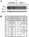
Tyr-RAS p53−/− melanomas show elevated c-Myc expression and frequent amplification of the c-Myc locus. (A) Top, immunoblot showing that c-Myc expression is strongly elevated in p53 mutant melanoma cells relative to melanocytes (M) and INK4-ARF mutant melanomas (A to G). Note the expression of the ∼46-kDa short c-Myc isoform (Myc-S) in tumors 2 and 6. The migration of the molecular size markers is indicated to the right. Bottom, immunoblot showing expression of tubulin as a loading control. (B) Summary of genomic alterations at the c-Myc locus in p53 mutant melanomas. Hybridization analysis of melanoma DNA using c-Myc and control probes was used to determine the c-Myc gene copy number (see Materials and Methods). Note that tumor 6 harbored an amplification of c-Myc which was not detected by CGH. c-Myc expression levels (see panel A) are summarized in the rightmost column (Mod, moderate increase in expression). The expression of Myc-S is also indicated. Chr, chromosome.
FIG. 4
FIG. 4
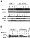
Expression analysis of G1/S regulators in RAS-induced melanomas. (A) Top, immunoblot analysis of cyclin D1 levels in melanoma cell lysates from INK4a-ARF−/− and p53−/− tumors. Below is an immunoblot probed with α-tubulin as a loading control. Bottom, immunoblot of p21CIP1, Cdc25a, and CDK4 levels. (B) Western blot analysis of RB phosphorylation status in INK4aΔ2/3-and p53-null melanomas. Tumor 7 was grown in 0% fetal calf serum (FCS; rightmost lane) to show that RB is responsive by shifting to a hypophosphorylated state.
FIG. 5
FIG. 5
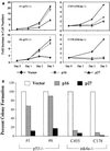
Effects of exogenous p16INK4a and p27KIP1 on the growth of Tyr-RAS melanoma cells on an INK4aΔ2/3-null or p53 mutant background. (A) Melanoma cell populations transduced with the indicated retroviruses were selected with puromycin for 2 days and then assayed for proliferation rates (see Materials and Methods). Data from two representative cell lines from each genotype are shown. Error bars indicate ranges of variability in duplicate samples assayed. (B) The relative colony-forming ability of the transduced cell lines was determined following low-density seeding (see Materials and Methods). The graph plots the number of colonies as a ratio compared to the number of colonies seen for transduction with the empty vector.
FIG. 6
FIG. 6
c-Myc and G1/S transition. The schematics show sequential phosphorylation of RB during the G1/S transition. RB phosphorylation is initiated by the activity of CDK4-cyclin D complexes and maintained by that of CDK2-cyclin E complexes. p16INK4a negatively regulates the activity of CDK4, and p27KIP1 inhibits the activity of cyclin E-CDK2, although it is also required for assembly of the active CDK4-cyclin D complex. c-Myc can activate the expression of cyclins D1 and D2 (6, 19) and CDK4 (18), leading to type cyclin sequestration of p27KIP1 from CDK2 complexes. CDK2 activity is also promoted by the abilities of c-Myc to repress the expression of p27KIP1 (29, 56), to downregulate p27KIP1 indirectly via upregulation of Cul1 (the SCF complex responsible for its degradation by ubiquitination [36]), and to induce expression of the CDK2 activator Cdc25a (14).

Similar articles

Cited by

References

    1. Akslen L A, Monstad S E, Larsen B, Straume O, Ogreid D. Frequent mutations of the p53 gene in cutaneous melanoma of the nodular type. Int J Cancer. 1998;79:91–95. - PubMed
    1. Albino A P, Vidal M J, McNutt N S, Shea C R, Prieto V G, Nanus D M, Palmer J M, Hayward N K. Mutation and expression of the p53 gene in human malignant melanoma. Melanoma Res. 1994;4:35–45. - PubMed
    1. Alevizopoulos K, Vlach J, Hennecke S, Amati B. Cyclin E and c-Myc promote cell proliferation in the presence of p16INK4a and of hypophosphorylated retinoblastoma family proteins. EMBO J. 1997;16:5322–5333. - PMC - PubMed
    1. Bastian B C, LeBoit P E, Hamm H, Brocker E B, Pinkel D. Chromosomal gains and losses in primary cutaneous melanomas detected by comparative genomic hybridization. Cancer Res. 1998;58:2170–2175. - PubMed
    1. Berns K, Hijmans E M, Bernards R. Repression of c-Myc responsive genes in cycling cells causes G1 arrest through reduction of cyclin E/CDK2 kinase activity. Oncogene. 1997;15:1347–1356. - PubMed

Publication types

MeSH terms

  NODES
Note 2
twitter 2