. 2001 Feb 19;152(4):729-40.
doi: 10.1083/jcb.152.4.729.

The karyopherin Kap142p/Msn5p mediates nuclear import and nuclear export of different cargo proteins

Affiliations

The karyopherin Kap142p/Msn5p mediates nuclear import and nuclear export of different cargo proteins

K Yoshida et al. J Cell Biol. .

Abstract

We have identified a novel pathway for protein import into the nucleus. Although the product of Saccharomyces cerevisiae gene MSN5 was previously shown to function as a karyopherin (Kap) for nuclear export of various proteins, we discovered a nuclear import pathway mediated by Msn5p (also referred to as Kap142p). We have purified from yeast cytosol a complex containing Kap142p and the trimeric replication protein A (RPA), which is required for multiple aspects of DNA metabolism, including DNA replication, DNA repair, and recombination. In wild-type cells, RPA was localized primarily to the nucleus but, in a KAP142 deletion strain, RPA was mislocalized to the cytoplasm and the strain was highly sensitive to bleomycin (BLM). BLM causes DNA double-strand breaks and, in S. cerevisiae, the DNA damage is repaired predominantly by RPA-dependent homologous recombination. Therefore, our results indicate that in wild-type cells a critical portion of RPA was imported into the nucleus by Kap142p. Like several other import-related Kap-substrate complexes, the endogenous RPA-Kap142p complex was dissociated by RanGTP, but not by RanGDP. All three RPA genes are essential for viability, whereas KAP142 is not. Perhaps explaining this disparity, we observed an interaction between RPA and Kap95p in a strain lacking Kap142p. This interaction could provide a mechanism for import of RPA into the nucleus and cell viability in the absence of Kap142p. Together with published results (Kaffman, A., N.M. Rank, E.M. O'Neill, L.S. Huang, and E.K. O'Shea. 1998. Nature. 396:482-486; Blondel, M., P.M. Alepuz, L.S. Huang, S. Shaham, G. Ammerer, and M. Peter. 1999. Genes Dev. 13:2284-2300; DeVit, M.J., and M. Johnston. 1999. Curr. Biol. 9:1231-1241; Mahanty, S.K., Y. Wang, F.W. Farley, and E.A. Elion. 1999. Cell. 98:501-512) our data indicate that the karyopherin Kap142p is able to mediate nuclear import of one set of proteins and nuclear export of a different set of proteins.


Figures

Figure 1
Figure 1
Kap142p-PrA localizes both to the cytoplasm and the nucleus. Haploid cells whose endogenous Kap142p was tagged with PrA were examined by indirect immunofluorescence of the PrA tag (left), DNA was stained with DAPI (middle), and whole cells were visualized by Nomarski optics (right).
Figure 5
Figure 5
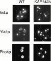
Deletion of KAP142 leads to mislocalization of Pho4p-GFP, but not to a general transport defect. Wild-type (WT) haploid strain (left) and KAP142Δ strain (right) harboring plasmid-encoded GFP-tagged hsLa (top), Yla1p (middle), and Pho4p (bottom) were visualized by direct fluorescence.
Figure 2
Figure 2
(a) Immunoisolation of cytosolic proteins interacting with Kap142p-PrA. The cytosol from a haploid KAP142-PrA strain was incubated with IgG-Sepharose. The last wash fraction and fractions eluted with a stepwise MgCl2 gradient were separated by SDS-PAGE and stained with Coomassie blue. Kap142p-PrA eluted between 1,000 and 4,500 mM MgCl2. Major bands migrating at ∼70, 37, and 14 kD were identified by mass spectrometry as Rpa1p, Rpa2p, and Rpa3p, respectively. The band migrating to ∼20 kD was identified as Ybr137wp. Relative molecular mass standards are indicated on the left. (b) Kap142p-PrA is insoluble at 4.5 M MgCl2 but soluble in SDS-PAGE sample buffer. The cytosol of a haploid KAP142-PrA strain was incubated with IgG-Sepharose. The proteins bound to the IgG-Sepharose were eluted by buffer (lane 1) and eluted with SDS sample buffer (lane 2). The proteins were separated by SDS-PAGE and then stained with Coomassie blue. Kap142p-PrA appeared as doublet bands (double arrows). h.c., heavy chain; l.c., light chain.
Figure 2
Figure 2
(a) Immunoisolation of cytosolic proteins interacting with Kap142p-PrA. The cytosol from a haploid KAP142-PrA strain was incubated with IgG-Sepharose. The last wash fraction and fractions eluted with a stepwise MgCl2 gradient were separated by SDS-PAGE and stained with Coomassie blue. Kap142p-PrA eluted between 1,000 and 4,500 mM MgCl2. Major bands migrating at ∼70, 37, and 14 kD were identified by mass spectrometry as Rpa1p, Rpa2p, and Rpa3p, respectively. The band migrating to ∼20 kD was identified as Ybr137wp. Relative molecular mass standards are indicated on the left. (b) Kap142p-PrA is insoluble at 4.5 M MgCl2 but soluble in SDS-PAGE sample buffer. The cytosol of a haploid KAP142-PrA strain was incubated with IgG-Sepharose. The proteins bound to the IgG-Sepharose were eluted by buffer (lane 1) and eluted with SDS sample buffer (lane 2). The proteins were separated by SDS-PAGE and then stained with Coomassie blue. Kap142p-PrA appeared as doublet bands (double arrows). h.c., heavy chain; l.c., light chain.
Figure 3
Figure 3
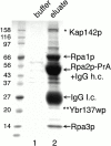
Immunoisolation of cytosolic proteins interacting with Rpa2p-PrA. Cytosol of an RPA2-PrA haploid strain was incubated with IgG-Sepharose and bound proteins were eluted and analyzed as in the legend to Fig. 2 a. The proteins bound to the IgG-Sepharose were eluted with buffer (lane 1) and eluted with SDS sample buffer (lane 2). The proteins were separated by SDS-PAGE and then stained with Coomassie blue. The bands migrating to ∼150 kD (*) and ∼20 kD (**) contained Kap142p and Ybr137wp by mass spectrometric analysis, respectively. h.c., heavy chain; l.c., light chain.
Figure 4
Figure 4
Deletion of KAP142 leads to temperature-sensitive phenotype and mislocalization of Rpa1p-PrA and Rpa2p-PrA. (a) Strains, as indicated, were streaked on yeast extract/peptone/dextrose plates and incubated at 30°C (left) and at 35°C (right). (b) Wild-type (WT) or KAP142Δ strain whose endogenous Rpa1p or Rpa2p was tagged with PrA was grown at 30°C and examined by indirect immunofluorescence of the PrA tag (left). DNA was stained with DAPI (middle) and whole cells were visualized by Nomarski optics (right).
Figure 4
Figure 4
Deletion of KAP142 leads to temperature-sensitive phenotype and mislocalization of Rpa1p-PrA and Rpa2p-PrA. (a) Strains, as indicated, were streaked on yeast extract/peptone/dextrose plates and incubated at 30°C (left) and at 35°C (right). (b) Wild-type (WT) or KAP142Δ strain whose endogenous Rpa1p or Rpa2p was tagged with PrA was grown at 30°C and examined by indirect immunofluorescence of the PrA tag (left). DNA was stained with DAPI (middle) and whole cells were visualized by Nomarski optics (right).
Figure 7
Figure 7
Deletion of KAP142 leads to impairment of DNA DSB repair. (a) Drop test on YEDA medium in the absence or presence of BLM (1 μg/ml), showing BLM sensitivity of wild-type (WT), KAP104Δ, KAP108Δ, KAP114Δ, KAP122Δ, KAP123Δ, KAP142Δ, and YBR137WΔ strains. (b) Relative quantification of survival rate in the absence and presence of various concentrations of BLM. The number of surviving wild-type colonies at each concentration was normalized to 100%. (c) Drop test on YEDA medium in the absence or presence of 4NQO (1 μM), showing 4NQO sensitivity of wild-type and KAP142Δ strains.
Figure 7
Figure 7
Deletion of KAP142 leads to impairment of DNA DSB repair. (a) Drop test on YEDA medium in the absence or presence of BLM (1 μg/ml), showing BLM sensitivity of wild-type (WT), KAP104Δ, KAP108Δ, KAP114Δ, KAP122Δ, KAP123Δ, KAP142Δ, and YBR137WΔ strains. (b) Relative quantification of survival rate in the absence and presence of various concentrations of BLM. The number of surviving wild-type colonies at each concentration was normalized to 100%. (c) Drop test on YEDA medium in the absence or presence of 4NQO (1 μM), showing 4NQO sensitivity of wild-type and KAP142Δ strains.
Figure 6
Figure 6
Dissociation of endogenous Rpa1p from Kap142p-PrA by RanGTP, but not RanGDP. The cytosol of a haploid KAP142-PrA strain was incubated with IgG-Sepharose. After washing, the proteins bound to IgG-Sepharose were incubated with buffer alone (lane 1), RanGDP (lane 2), or RanGTP (lane 3). After incubation, released proteins (unbound) were collected (top) and the remaining proteins (bound) were eluted with 50 mM (middle) and 250 mM MgCl2 (bottom). Proteins were separated by SDS-PAGE and immunoblotted with anti-Rpa1p antibody.
Figure 8
Figure 8
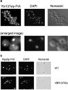
(a) Ybr137wp-PrA was localized throughout the cell and in a cytoplasmic focus. Haploid cells whose endogenous Ybr137wp was tagged with PrA were examined by indirect immunofluorescence of the PrA tag (left), DNA was stained with DAPI (middle), and whole cells were visualized by Nomarski optics (right). Images in the bottom row were enlarged using Adobe Photoshop®. (b) Deletion of YBR137W does not lead to mislocalization of Rpa2p-PrA. Wild-type (WT) haploid strain or YBR137WΔ strain (bottom) whose endogenous Rpa2p was tagged with PrA was examined by indirect immunofluorescence of the PrA tag (left), DNA was stained with DAPI (middle), and whole cells were visualized by Nomarski optics (right).
Figure 9
Figure 9
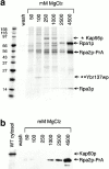
In the absence of Kap142p, RPA interacts with Kap95p and Ybr137wp. (a) Rpa2p-PrA was expressed in a KAP142Δ strain. Rpa2p-PrA and associated proteins were isolated as in the legend to Fig. 2. The bands migrating at ∼90 kD (*) and ∼20 kD (**) were identified by mass spectrometry as Kap95p and Ybr137wp, respectively. (b) Cytosol from wild-type (WT) strain and Rpa2p-PrA–associated proteins from Fig. 9 a were immunoblotted with anti-Kap60p antibody.

Similar articles

Cited by

References

    1. Aitchison J.D., Rout M.P., Marelli M., Blobel G., Wozniak R.W. Two novel related yeast nucleoporins Nup170p and Nup157pcomplementation with the vertebrate homologue Nup155p and functional interactions with the yeast nuclear pore-membrane protein Pom152p. J Cell Biol. 1995;131:1133–1148. - PMC - PubMed
    1. Aitchison J.D., Blobel G., Rout M.P. Kap104pa karyopherin involved in the nuclear transport of messenger RNA binding proteins. Science. 1996;274:624–627. - PubMed
    1. Akada R., Kallal L., Johnson D.I., Kurjan J. Genetic relationships between the G protein βγ complex, Ste5p, Ste20p and Cdc42pinvestigation of effector roles in the yeast pheromone response pathway. Genetics. 1996;143:103–117. - PMC - PubMed
    1. Albertini M., Pemberton L.F., Rosenblum J.S., Blobel G. A novel nuclear import pathway for the transcription factor TFIIS. J. Cell Biol. 1998;143:1447–1455. - PMC - PubMed
    1. Alepuz P.M., Matheos D., Cunningham K.W., Estruch F. The Saccharomyces cerevisiae RanGTP-binding protein Msn5p is involved in different signal transduction pathways. Genetics. 1999;153:1219–1231. - PMC - PubMed

Publication types

MeSH terms

  NODES
twitter 2