. 2001 May 1;20(9):2304-14.
doi: 10.1093/emboj/20.9.2304.

SMNrp is an essential pre-mRNA splicing factor required for the formation of the mature spliceosome

Affiliations

SMNrp is an essential pre-mRNA splicing factor required for the formation of the mature spliceosome

G Meister et al. EMBO J. .

Abstract

SMNrp, also termed SPF30, has recently been identified in spliceosomes assembled in vitro. We have functionally characterized this protein and show that it is an essential splicing factor. We show that SMNrp is a 17S U2 snRNP-associated protein that appears in the pre-spliceosome (complex A) and the mature spliceosome (complex B) during splicing. Immunodepletion of SMNrp from nuclear extract inhibits the first step of pre-mRNA splicing by preventing the formation of complex B. Re-addition of recombinant SMNrp to immunodepleted extract reconstitutes both spliceosome formation and splicing. Mutations in two domains of SMNrp, although similarly deleterious for splicing, differed in their consequences on U2 snRNP binding, suggesting that SMNrp may also engage in interactions with splicing factors other than the U2 snRNP. In agreement with this, we present evidence for an additional interaction between SMNrp and the [U4/U6.U5] tri-snRNP. A candidate that may mediate this interaction, namely the U4/U6-90 kDa protein, has been identified. We suggest that SMNrp, as a U2 snRNP-associated protein, facilitates the recruitment of the [U4/U6.U5] tri-snRNP to the pre-spliceosome.


Figures
None
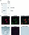
Fig. 1. SMNrp localizes in nuclear domains implicated in transcription and splicing. (A) Detection of SMNrp in HeLa nuclear extracts (NE), whole-cell extracts (TE) and Xenopus laevis oocyte extracts (OE) by western blotting using anti-SMNrp antibodies. The anti-SMNrp antibody also efficiently detects recombinant GST-tagged SMNrp (lane 4), but fails to recognize the homologous SMN protein (lane 5). (B) Confocal immunofluorescence microscopy of COS-1 cells with anti-SMNrp antibodies (1), and with anti-SMN antibody 7B10 (2). Panel 3, superimposed images 1 and 2; panel 4, a phase contrast of the same cells; panel 5, a control immunofluorescence with anti-SMNrp antibodies pre-adsorbed with recombinant SMNrp; panel 6, the phase contrast of the same cells. (C) Immunolocalization of SMNrp in the nucleus of an ultrathin section of cryofixed HTC cells by electron microscopy. Most label is associated with perichromatin fibrils (some indicated by small arrows); interchromatin granule clusters (area surrounded and indicated by large arrows) and condensed chromatin (‘C’) remain largely unlabelled. In the lower part of the image, some protein can be seen in the cytoplasm sometimes near the nuclear membrane (NE). Bar, 0.5 µm.
None
Fig. 2. SMNrp is a 17S U2 snRNP-associated protein. (A) Nuclear extract was passed over an anti-m3G/m7G-column (H-20) (lane 2) or a control column (lane 3). Bound proteins were eluted with m7G-nucleoside and analysed by western blotting. Lane 1 shows 5% of the nuclear extract applied on to the columns. SMNrp and SmB were detected using anti-SMNrp antiserum and Y12 monoclonal antibody, respectively. (B) Sedimentation analysis of SMNrp in nuclear extract by linear 15–45% sucrose gradient centrifugation. Proteins from each fraction were analysed by western blotting using anti-SMNrp antibodies (upper panel). The lower panel shows silver staining of the RNAs from the gradient fractions. (C) SMNrp is associated with 17S U2 snRNP. 17S peak fractions were immunoprecipitated with either anti-SMNrp antiserum (lane 4) or an unrelated control antibody (lane 3). RNAs from the immunoprecipitates (lanes 3 and 4) and 25% of the corresponding supernatants (lanes 1 and 2) were 3′-labelled with [32P]pCp and analysed by denaturing RNA gel electrophoresis.
None
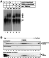
Fig. 3. SMNrp is a component of splicing complexes A and B. Radiolabelled pAd48 pre-mRNA was incubated for the indicated time points with nuclear extract active in splicing and separated by native gel electrophoresis. The gel was subsequently blotted on to a nitrocellulose membrane and splicing complexes were visualized by autoradiography (lanes 1–3). Splicing complexes H, A, B and C are indicated. The unstable complex C is only visible after extended exposure and is hence indicated in brackets. SMNrp was visualized by probing the same blot with anti-SMNrp antibodies (lanes 4–6). The band marked with an asterisk indicates an SMNrp-specific complex of unknown identity that does not contain radiolabelled pre-mRNA.
None
Fig. 4. SMNrp is an essential pre-mRNA splicing factor. (A) Affinity-purified anti-SMNrp antibodies inhibit pre-mRNA splicing in vitro. The splicing reactions were pre-incubated for 30 min with 5 µg of control antibody (lane 2), or 1, 3 or 5 µg of affinity-purified anti-SMNrp antibody (lanes 3–5). Thereafter, 32P-labelled pAd48 pre-mRNA was added and incubated for 1 h. RNA was separated on a denaturing RNA gel and visualized by autoradiography. Lane 6 shows the splicing reaction in the absence of any antibody; lane 1, the pre-mRNA transcript. (B) Analysis of splicing in Xenopus laevis oocytes. pAd48 pre-mRNA was micro-injected into the nucleus of oocytes that were pre-injected with either 200 ng of control antibody (lane 2) or anti-SMNrp antibody (lane 3). Nuclei from injected oocytes were isolated 60 min later and the RNA analysed as described in (A). (C) Western blot analysis of SMNrp (upper panel) and SMN (lower panel) in splicing extracts that were either mock-depleted (lanes 1 and 2) or immuno depleted with anti-SMNrp antibodies (lanes 3 and 4). (D) Recombinant SMNrp reconstitutes pre-mRNA splicing in immunodepleted nuclear extract. Nuclear extracts that had been either immunodepleted (lanes 2, 4 and 6–8), mock-depleted (lanes 3 and 5) or not treated (lane 1) were used for splicing of pAd48 pre-mRNA in vitro. Reactions were supple mented with either 1 µg (lanes 4 and 6) or 5 µg (lane 7) of recombinant SMNrp or with 5 µg of BSA (lane 8). The lower part of the autoradio graphy is also shown in a longer exposure to visualize better the spliced product of the reactions.
None
Fig. 5. Spliceosome assembly is arrested in SMNrp-depleted nuclear extract at the level of complex A. (A) pAd48 pre-mRNA was incubated with mock-depleted (lanes 1–3) or SMNrp-depleted (lanes 4–6) nuclear extract and analysed by native gel electrophoresis. Reactions shown in lanes 5 and 6 were complemented with 1, 5 and 3 µg of recombinant SMNrp, respectively. (B) Recombinant SMNrp is incorporated into 17S U2 snRNP in SMNrp-depleted nuclear extract. Nuclear extract that was either mock-depleted (upper panel), SMNrp-depleted (middle panel) or SMNrp-depleted and supplemented with recombinant His-tagged SMNrp (lower panel) was separated by gradient centrifugation and analysed by western blotting using anti-SMNrp antibody.
None
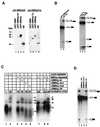
Fig. 6. The N-terminus and the Tudor domain of SMNrp mediate essential functions in spliceosome assembly. (A) Western blot analysis of affinity-purified antibodies directed against the N-terminus (lanes 1–4) and Tudor domain (lanes 5–8) of SMNrp. Recombinant GST-tagged SMN (lanes 1 and 5), SMNrp (lanes 3 and 7), SMNrp lacking the N-terminus (SMNrpΔN) (lane 2), Tudor domain of SMNrp (SMNrp-Tu) (lane 6) and nuclear extract (lanes 4 and 8) were tested to define the specificity of the antibodies. (B) Antibodies directed against the N-terminus and Tudor domain of SMNrp interfere with pre-mRNA splicing in vitro. Nuclear extract was incubated with either no antibody (lane 5) or antibodies directed against the N-terminus (lane 1), Tudor domain (lane 3) or an unrelated antibody (lanes 2 and 4). Splicing was analysed as described in Figure 4. The lower part of the autoradiography is also shown in a longer exposure (lanes 3–5). (C) Recombinant SMNrp harbouring amino acid substitutions in the Tudor domain or lacking the N-terminus fails to reconstitute spliceosome assembly in vitro. Mock-depleted (lanes 1 and 2) and SMNrp-depleted (lanes 3–9) nuclear extracts were incubated with 32P-labelled pAd48 pre-mRNA for the indicated time points in the presence of buffer (lanes 1–3 and 7), recombinant wild-type SMNrp (lanes 4 and 9), SMNrpmu1 (lane 5), SMNrpmu2 (lane 6) or SMNrpΔN (lane 8). The spliceosomal complexes were separated by native gel electrophoresis and visualized by autoradiography. (D) SMNrpΔN and SMNrpmu1 exhibit a trans-dominant-negative phenotype on pre-mRNA splicing. 32P-labelled pAd48 pre-mRNA was incubated for 60 min with nuclear extract in the presence of either SMNrp (lane 4), SMNrpmu1 (lane 3), SMNrpΔN (lane 2) (5 µg each) or buffer (lane 1). Splicing was analysed as described in Figure 4.
None
Fig. 7. Association of SMNrp mutants with 17S U2 snRNP. Sucrose gradient centrifugation of SMNrp-depleted extract supplemented with either buffer (B), 0.5 µg of recombinant proteins SMNrp (C), SMNrpmu1 (D) or SMNrpΔN (E). The mock-treated extract is shown in gradient (A). Gradient samples were separated by SDS–PAGE and SMNrp visualized by western blotting using an anti-SMNrp antibody.
None
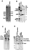
Fig. 8. Recombinant SMNrp binds to U2 snRNP and the [U4/U6⋅U5] tri-snRNP in vitro. (A) GST–SMNrp or GST tag alone was bound to glutathione–Sepharose and incubated with nuclear extract active in splicing. RNAs from the nuclear extract (lane 1), the GST–SMNrp column (lane 2) and the control column (lane 3) were separated by denaturing RNA gel electrophoresis and visualized by ethidium bromide staining. (B) Protein profile of the same fractions as in (A). Lane 2 shows proteins of U snRNPs affinity purified with H-20 monoclonal antibody.
None
Fig. 9. Binding of SMNrp to the [U4/U6⋅U5] tri-snRNP occurs via a direct interaction with the U4/U6-90 kDa protein. (A) GST–SMNrp binds specifically to U4/U6 and U2 snRNAs in nuclear extract in which the [U4/U6⋅U5] tri-snRNP had been dissociated. RNAs from the nuclear extract (lane 1), the GST–control (lane 2) and the GST–SMNrp binding reaction (lane 3) were separated and visualized as described in Figure 8. (B) Coomassie-stained protein gel of the binding experiment shown in (A). GST–SMNrp and its degradation products are marked with asterisks, U2 snRNP-specific proteins as well as Sm proteins with filled circles, and the U4/U6-specific 60 and 90 kDa proteins with arrowheads (lane 2). Lane 3 shows the protein profile of the GST–control column. (C) SMNrp binds specifically to in vitro translated U4/U6-90 kDa protein. Binding of immobilized GST–SMNrp (lanes 1–3) and GST (lane 4) to in vitro translated and 35S-labelled U4/U6-60 kDa protein (lane 1), 90 kDa protein (lane 2) or a mixture of both (lanes 3 and 4) is shown. Lanes 5 and 6 show 25% of the translated proteins used in the binding assay. (D) Direct binding of SMNrp to the U4/U6-90 kDa protein. Coomassie-stained protein gel showing binding of His-tagged recombinant U4/U6-90 kDa protein to immobilized GST–SMNrp (lane 2) or to GST tag (lane 3). Recombinant proteins used in the binding assay were separated in lanes 5–7.

Similar articles

Cited by

References

    1. Achsel T., Brahms,H., Kastner,B., Bachi,A., Wilm,M. and Lührmann,R. (1999) A doughnut-shaped heteromer of human Sm-like proteins binds to the 3′-end of U6 snRNA, thereby facilitating U4/U6 duplex formation in vitro. EMBO J., 18, 5789–5802. - PMC - PubMed
    1. Behrens S.E. and Lührmann,R. (1991) Immunoaffinity purification of a U4/U6⋅U5 tri-snRNP from human cells. Genes Dev., 5, 1439–1452. - PubMed
    1. Behrens S.E., Gallison,F., Legrain,P. and Lührmann,R. (1993a) Evidence that the 60-kDa protein of 17S U2 small nuclear ribonucleoprotein is immunologically and functionally related to the yeast PRP9 splicing factor and is required for the efficient formation of prespliceosome. Proc. Natl Acad. Sci. USA, 90, 8229–8233. - PMC - PubMed
    1. Behrens S.E., Tyc,K., Kastner,B., Reichelt,J. and Lührmann,R. (1993b) Small nuclear ribonucleoprotein (RNP) U2 contains numerous additional proteins and has a bipartite RNP structure under splicing conditions. Mol. Cell. Biol., 13, 307–319. - PMC - PubMed
    1. Bringmann P., Rinke,J., Appel,B., Reuter,R. and Lührmann,R. (1983) Purification of snRNPs U1, U2, U4, U5 and U6 with 2,2,7-trimethylguanosine-specific antibody and definition of their constituent proteins reacting with anti-Sm and anti-(U1)RNP antisera. EMBO J., 2, 1129–1135. - PMC - PubMed

Publication types

MeSH terms

Substances

  NODES
Association 3
twitter 2