. 2002 Feb 19;99(4):2175-80.
doi: 10.1073/pnas.042035699.

Pim serine/threonine kinases regulate the stability of Socs-1 protein

Affiliations

Pim serine/threonine kinases regulate the stability of Socs-1 protein

X Peter Chen et al. Proc Natl Acad Sci U S A. .

Abstract

Studies of SOCS-1-deficient mice have implicated Socs-1 in the suppression of JAK-STAT (Janus tyrosine kinase-signal transducers and activators of transcription) signaling and T cell development. It has been suggested that the levels of Socs-1 protein may be regulated through the proteasome pathway. Here we show that Socs-1 interacts with members of the Pim family of serine/threonine kinases in thymocytes. Coexpression of the Pim kinases with Socs-1 results in phosphorylation and stabilization of the Socs-1 protein. The protein levels of Socs-1 are significantly reduced in the Pim-1(-/-), Pim-2(-/-) mice as compared with wild-type mice. Similar to Socs-1(-/-) mice, thymocytes from Pim-1(-/-), Pim-2(-/-) mice showed prolonged Stat6 phosphorylation upon IL-4 stimulation. These data suggest that the Pim kinases may regulate cytokine-induced JAK-STAT signaling through modulation of Socs-1 protein levels.


Figures
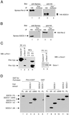
Figure 1
Figure 1
Association of Pim-2 with Socs-1 in vivo and in vitro. (A) Immunoprecipitates of Socs-1 contain Pim-2. The 293T cells were cotransfected with plasmids carrying Xpress-tagged Pim-2 and HA-tagged SOCS-1. Lysates of transfectants were immunoprecipitated with a polyclonal anti-HA Ab (lanes 1 and 3) or normal rabbit serum as a negative control (lanes 2 and 4). Both immunoprecipitates were split, loaded onto SDS gel, and immunoblotted with a monoclonal anti-Xpress (lanes 1 and 2) or anti-HA (lanes 3 and 4) Abs, respectively. (B) Socs-1 is present in Pim-2 immunoprecipitates. Same as A, except that plasmids carrying Xpress-tagged SOCS-1 and HA-tagged Pim-2 were used for transfection. (C) Endogenous association of Socs-1 and Pim-1. Total thymocytes were isolated from wild-type BALB/c mice, stimulated with PMA and ionomycin for 4 h, and lysed. Lysates were immunoprecipitated by using either preimmune serum (lanes 1 and 5) or an affinity-purified rabbit α-Socs-1 antibody (lanes 2 and 6). The immunoprecipitates were analyzed by immunoblotting first with a goat α-Socs-1 antibody (C20) (Right) and then with a monoclonal α-Pim-1 antibody (Left). As a control, 50 μg of lysates from unstimulated and stimulated thymocytes was loaded in lanes 3 and 4. (D) The N terminus of Socs-1 is required for binding to Pim-2. Constructs of Xpress-tagged SOCS-1, full-length (FL) (lanes 1 and 6), ΔN (deleting amino acids 1–79) (lane 2), ΔC (deleting amino acids 167–212) (lane 3), or ΔSH2 (deleting amino acids 80–166) (lane 4) were transiently expressed in 293T cells. Xpress-tagged SOCS-2 was used as a control (lane 5). Whole-cell lysates of the transfected 293T cells were incubated with bacterial-expressed Pim-2-GST fusion protein (lanes 1–5) or GST alone (lane 6) bound to glutathione agarose beads (see Materials and Methods). Proteins bound to the beads were analyzed by immunoblot with anti-Xpress Ab. To ensure equal input, small aliquots of whole-cell lysates were subjected to Western blot analysis with anti-Xpress Ab (lanes 7–12).
Figure 2
Figure 2
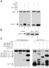
Phosphorylation of Socs-1 by Pim-2. (A) Coexpression of Socs-1 and Pim-2 results in mobility shift of Socs-1. Plasmids expressing SOCS-1 tagged with Xpress were transfected alone (lanes 1, 4, and 5) or together with Pim-2 (lane 2) or kinase-inactive Pim-2 (K61M) (lane 3) into 293T cells. An equal amount of a plasmid with the LacZ gene was included in each transfection as an internal control. Whole-cell lysates were then analyzed by immunoblot with anti-Xpress Ab. In lanes 4 and 5, DMSO control (lane 4) or 10 μM LLnL (lane 5) was added to the cells 24 h after transfection, and cells were harvested after another 24 h incubation. The expression levels of wild-type and mutant Pim-2 were comparable (data not shown). (B) The mobility shift of Socs-1 can be reversed by phosphatase. Lysate of 293T cells transfected with SOCS-1 and Pim-2 was incubated with an increasing amount of λ-phosphatase at 30°C for 90 min and analyzed by immunoblot. (C) N-terminal truncation of Socs-1 abolishes phosphorylation of Socs-1 by Pim-2. Pim-2 and various constructs of Socs-1 were expressed in bacteria as GST fusion proteins. The GST-Pim-2 fusion protein was incubated with full-length (lane 2), N-terminal truncated (lane 3), or C-terminal truncated (lane 4) Socs-1 for in vitro kinase assay (see Materials and Methods). GST alone (lane 1) was used as a negative control. A small aliquot of each sample from the kinase assay was analyzed by Western blot with anti-GST Ab to ensure that the amounts of proteins loaded in each lane were comparable (lanes 5–8)
Figure 3
Figure 3
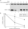
Phosphorylation by Pim kinases protects Socs-1 from degradation. (A) Phosphorylated form of Socs-1 decays more slowly than unphosphorylated form. The 293T cells were transfected with Xpress-tagged SOCS-1 alone (lanes 1–3) or together with Pim-2 (lanes 4–6). A plasmid carrying the LacZ gene was used as a control. Cycloheximide (100 μg/ml) was added to the media 36 h after transfection to block new protein synthesis. Cells were harvested at 0-, 4.5-, and 9-h time points, and total lysates were analyzed by immunoblot with anti-Xpress antibody. (B) The blot in A was scanned and quantitated by using the software nih image 1.6.2. The results from three independent experiments are plotted such that the protein level at 0-h time point is 100%.
Figure 4
Figure 4
Phosphorylation of Socs-1 decreases its binding to Elongin BC. (A) Phosphorylated Socs-1 does not bind Elongin BC as well as unphosphorylated Socs-1 in coimmunoprecipitation experiments. The 293T cells were transfected with plasmids carrying Xpress-tagged SOCS-1 alone (lanes 1 and 2) or together with plasmids expressing HA-tagged Elongin B and Elongin C (lanes 3–8), in the absence (lanes 3 and 4) or presence of Pim-2 (lanes 5 and 6) or Pim-3 (lanes 7 and 8). Total cell lysates were immunoprecipitated with anti-HA antibody to pull down Elongin BC. The immunoprecipitates (IP) were then analyzed by Western blot by using anti-Xpress antibody to detect proteins that bind to Elongin BC (lanes 2, 4, 6, and 8). For comparison, whole-cell lysates (WCL) (lanes 1, 3, 5, and 7) were analyzed on the same blot. (B) GST-Elongin C fusion protein associates preferentially with the faster-migrating band of Socs-1. The 293T cells were transfected with plasmids carrying Xpress-tagged SOCS-1 either in the absence (lanes 1–3) or presence (lanes 4–6) of Pim-1. Total cell lysates from the transfectants were incubated with bacterial expressed GST (lanes 2 and 5) or GST-Elongin C (lanes 3 and 6) immobilized on glutathione beads. The beads were washed four times before being subjected to SDS/PAGE analysis (lanes 3 and 4). Lanes 1 and 4 were small aliquots of whole-cell lysates (WCL) before incubation with glutathione beads.
Figure 5
Figure 5
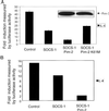
Pim-2 enhances Socs-1 inhibition of IL-4-induced Stat6 activation. (A) Pim-2 potentiates Socs-1 inhibition of JAK-STAT activation in 293T cells. The 293T cells were transfected with 2 μg of p(Iɛ-IL4RE)4-Luc reporter, 1 μg of pSV40-LacZ, and 0.6 μg of human Stat6 expression vector by calcium phosphate precipitation. Plasmid DNA (0.005 μg) carrying SOCS-1 and 2 μg of plasmid DNA carrying either wild-type Pim-2 or kinase-inactive Pim-2 were used. The total amount of transfected DNA was kept constant by addition of vector DNA as described (see Materials and Methods). Shown on the y axis is the ratio of luciferase activity between treated and untreated cells. Values reflect means of three independent experiments. (Inset) The expression levels of wild-type Pim-2 and kinase-inactive Pim2, respectively. (B) Pim-2 potentiates Socs-1 inhibition of JAK-STAT activation in NIH 3T3 cells. NIH 3T3 cells were transfected with 2 μg of p(Iɛ-IL4RE)4-Luc reporter, 1 μg of pSV40-LacZ as described in Materials and Methods. Used were 0.02 μg of plasmid DNA carrying SOCS-1 and 10 μg of Pim-2 plasmid DNA. Luciferase assay was performed essentially as described in A.
Figure 6
Figure 6
IL-4 induced Stat6 activation is prolonged in thymocytes from Pim-1−/−, Pim-2−/− mice. (A) Thymocytes from Pim-1−/−, Pim-2−/− mice and wild-type littermates were stimulated with α-CD3 (PharMingen) (1 μg/ml) plus IL-4 (10 ng/ml) and harvested at the indicated time points. Cells were then washed and lysed in 1× Nonidet P-40 lysis buffer containing inhibitors of proteases and phosphatases as described (28). Immunoprecipitates were obtained by using anti-Stat6 antibody M20 (Santa Cruz Biotechnology) and blotted with anti-phosphor-tyrosine antibody 4G10 (Upstate Biotechnology). (B) The same blots as in A were stripped and reblotted with anti-Stat6 antibody M20. (C) The levels of endogenous Socs-1 were reduced in the Pim-1−/−, Pim-2−/− mice. Thymocytes were isolated from wild-type (lanes 1 and 2) or Pim-1−/−, Pim-2−/− (lanes 3 and 4) mice, and cultured in the presence of PMA and ionomycin for 4 h. Cells were harvested and lysed, and the protein concentration was determined. Equal amounts of lysates were subjected to immunoprecipitation analysis by using preimmune serum (lanes 1 and 3) or an α-Socs-1 antibody (lanes 2 and 4) as described in Fig. 1C.

Similar articles

Cited by

References

    1. Leonard W J, O'Shea J J. Annu Rev Immunol. 1998;16:293–322. - PubMed
    1. Endo T A, Masuhara M, Yokouchi M, Suzuki R, Sakamoto H, Mitsui K, Matsumoto A, Tanimura S, Ohtsubo M, Misawa H, et al. Nature (London) 1997;387:921–924. - PubMed
    1. Naka T, Narazaki M, Hirata M, Matsumoto T, Minamoto S, Aono A, Nishimoto N, Kajita T, Taga T, Yoshizaki K, et al. Nature (London) 1997;387:924–929. - PubMed
    1. Starr R, Willson T A, Viney E M, Murray L J, Rayner J R, Jenkins B J, Gonda T J, Alexander W S, Metcalf D, Nicola N A, Hilton D J. Nature (London) 1997;387:917–921. - PubMed
    1. Chen X P, Losman J A, Rothman P. Immunity. 2000;13:287–290. - PubMed

Publication types

MeSH terms

Substances

  NODES
Association 4
INTERN 1
twitter 2