tcBid promotes Ca(2+) signal propagation to the mitochondria: control of Ca(2+) permeation through the outer mitochondrial membrane
- PMID: 11980717
- PMCID: PMC125984
- DOI: 10.1093/emboj/21.9.2198
tcBid promotes Ca(2+) signal propagation to the mitochondria: control of Ca(2+) permeation through the outer mitochondrial membrane
Abstract
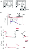
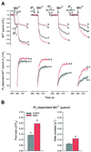
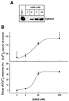
Calcium spikes established by IP(3) receptor-mediated Ca(2+) release from the endoplasmic reticulum (ER) are transmitted effectively to the mitochondria, utilizing local Ca(2+) interactions between closely associated subdomains of the ER and mitochondria. Since the outer mitochondrial membrane (OMM) has been thought to be freely permeable to Ca(2+), investigations have focused on IP(3)-driven Ca(2+) transport through the inner mitochondrial membrane (IMM). Here we demonstrate that selective permeabilization of the OMM by tcBid, a proapoptotic protein, results in an increase in the magnitude of the IP(3)-induced mitochondrial [Ca(2+)] signal. This effect of tcBid was due to promotion of activation of Ca(2+) uptake sites in the IMM and, in turn, to facilitation of mitochondrial Ca(2+) uptake. In contrast, tcBid failed to control the delivery of sustained and global Ca(2+) signals to the mitochondria. Thus, our data support a novel model that Ca(2+) permeability of the OMM at the ER- mitochondrial interface is an important determinant of local Ca(2+) signalling. Facilitation of Ca(2+) delivery to the mitochondria by tcBid may also support recruitment of mitochondria to the cell death machinery.
Figures


Similar articles
-
Apoptosis driven by IP(3)-linked mitochondrial calcium signals.EMBO J. 1999 Nov 15;18(22):6349-61. doi: 10.1093/emboj/18.22.6349. EMBO J. 1999. PMID: 10562547 Free PMC article.
-
Old players in a new role: mitochondria-associated membranes, VDAC, and ryanodine receptors as contributors to calcium signal propagation from endoplasmic reticulum to the mitochondria.Cell Calcium. 2002 Nov-Dec;32(5-6):363-77. doi: 10.1016/s0143416002001872. Cell Calcium. 2002. PMID: 12543096 Review.
-
Sorting of calcium signals at the junctions of endoplasmic reticulum and mitochondria.Cell Calcium. 2001 Apr;29(4):249-62. doi: 10.1054/ceca.2000.0191. Cell Calcium. 2001. PMID: 11243933
-
Endoplasmic reticulum stress induces calcium-dependent permeability transition, mitochondrial outer membrane permeabilization and apoptosis.Oncogene. 2008 Jan 10;27(3):285-99. doi: 10.1038/sj.onc.1210638. Epub 2007 Aug 13. Oncogene. 2008. PMID: 17700538
-
High- and low-calcium-dependent mechanisms of mitochondrial calcium signalling.Cell Calcium. 2008 Jul;44(1):51-63. doi: 10.1016/j.ceca.2007.11.015. Epub 2008 Feb 19. Cell Calcium. 2008. PMID: 18242694 Free PMC article. Review.
Cited by
-
VDAC2-specific cellular functions and the underlying structure.Biochim Biophys Acta. 2016 Oct;1863(10):2503-14. doi: 10.1016/j.bbamcr.2016.04.020. Epub 2016 Apr 23. Biochim Biophys Acta. 2016. PMID: 27116927 Free PMC article. Review.
-
Genetic dissection of the permeability transition pore.J Bioenerg Biomembr. 2005 Jun;37(3):121-8. doi: 10.1007/s10863-005-6565-9. J Bioenerg Biomembr. 2005. PMID: 16167169 Review.
-
Mitochondrial complex II prevents hypoxic but not calcium- and proapoptotic Bcl-2 protein-induced mitochondrial membrane potential loss.J Biol Chem. 2010 Aug 20;285(34):26494-505. doi: 10.1074/jbc.M110.143164. Epub 2010 Jun 21. J Biol Chem. 2010. PMID: 20566649 Free PMC article.
-
SR/ER-mitochondrial local communication: calcium and ROS.Biochim Biophys Acta. 2009 Nov;1787(11):1352-62. doi: 10.1016/j.bbabio.2009.06.004. Epub 2009 Jun 13. Biochim Biophys Acta. 2009. PMID: 19527680 Free PMC article. Review.
-
Supralinear Dependence of the IP3 Receptor-to-Mitochondria Local Ca2+ Transfer on the Endoplasmic Reticulum Ca2+ Loading.Contact (Thousand Oaks). 2024 Feb 14;7:25152564241229273. doi: 10.1177/25152564241229273. eCollection 2024 Jan-Dec. Contact (Thousand Oaks). 2024. PMID: 38362008 Free PMC article.
References
-
- Antonsson B., Montessuit,S., Sanchez,B. and Martinou,J.C. (2001) Bax is present as a high molecular weight oligomer/complex in the mitochondrial membrane of apoptotic cells. J. Biol. Chem., 276, 11615–11623. - PubMed
-
- Benz R. and Brdiczka,D. (1992) The cation-selective substate of the mitochondrial outer membrane pore: single-channel conductance and influence on intermembrane and peripheral kinases. J. Bioenerg. Biomembr., 24, 33–39. - PubMed
-
- Bernardi P. (1999) Mitochondrial transport of cations: channels, exchangers and permeability transition. Physiol. Rev., 79, 1127–1155. - PubMed
-
- Beutner G., Sharma,V.K., Giovannucci,D.R., Yule,D.I. and Sheu,S.S. (2001) Identification of a ryanodine receptor in rat heart mitochondria. J. Biol. Chem., 276, 21482–21488. - PubMed
Publication types
MeSH terms
Substances
LinkOut - more resources
Full Text Sources
Miscellaneous

