. 2002 Jun;22(12):4043-52.
doi: 10.1128/MCB.22.12.4043-4052.2002.

Chromatin disruption and histone acetylation in regulation of the human immunodeficiency virus type 1 long terminal repeat by thyroid hormone receptor

Affiliations

Chromatin disruption and histone acetylation in regulation of the human immunodeficiency virus type 1 long terminal repeat by thyroid hormone receptor

Shao-Chung Victor Hsia et al. Mol Cell Biol. 2002 Jun.

Abstract

The human immunodeficiency virus type 1 (HIV-1) long terminal repeat (LTR) controls the expression of HIV-1 viral genes and thus viral propagation and pathology. Numerous host factors participate in the regulation of the LTR promoter, including thyroid hormone (T(3)) receptor (TR). In vitro, TR can bind to the promoter region containing the NF-kappa B and Sp1 binding sites. Using the frog oocyte as a model system for chromatin assembly mimicking that in somatic cells, we demonstrated that TR alone and TR/RXR (9-cis retinoic acid receptor) can bind to the LTR in vivo independently of T(3). Consistent with their ability to bind the LTR, both TR and TR/RXR can regulate LTR activity in vivo. In addition, our analysis of the plasmid minichromosome shows that T(3)-bound TR disrupts the normal nucleosomal array structure. Chromatin immunoprecipitation assays with anti-acetylated-histone antibodies revealed that unliganded TR and TR/RXR reduce the local histone acetylation levels at the HIV-1 LTR while T(3) treatment reverses this reduction. We further demonstrated that unliganded TR recruits corepressors and at least one histone deacetylase. These results suggest that chromatin remodeling, including histone acetylation and chromatin disruption, is important for T(3) regulation of the HIV-1 LTR in vivo.


Figures

FIG. 1.
FIG. 1.
Schematic representation of the LTR promoter plasmids. pHL10 contained the HIV-1 LTR promoter region from −107 to +81 in front of a 300-bp CAT reporter fragment in pBluescript KS(−) vector. The promoter fragment included two NF-κB sites, three Sp1 sites, and a TATA box followed by a regulatory TAR sequence. Two TREs are located in the regions containing the NF-κB and Sp1 binding sites (NF-κB TRE and Sp1 TRE). The Sp1 TRE is a stronger TRE than the NF-κB TRE (17).
FIG. 2.
FIG. 2.
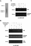
A ChIP assay demonstrates the binding of TR or TR/RXR to the LTR promoter in vivo. (A) The LTR minichromosome was sheared to an average of 0.5 kb by sonication during the ChIP assay. Oocytes were injected with the LTR plasmid, treated with formaldehyde to cross-link the DNA and protein in the minichromosome, and sonicated. The cross-links were then reversed as in the ChIP assay. The DNA was purified and subjected to Southern blot hybridization with a random-primed, 32P-labeled plasmid probe. DNA size markers are on the left. (B) ChIP assay shows that TR binds to LTR constitutively in vivo. The oocytes injected with the LTR plasmid and indicated mRNAs were treated with formaldehyde and sonicated after treatment with T3 and/or TSA. The sonicated LTR minichromosome was immunoprecipitated with an anti-TRβ antibody (63), and the precipitated DNA was analyzed by qualitative PCR. Note that TR bound to the LTR in the presence or absence of RXR and independently of T3 and/or TSA treatment. Little LTR DNA was immunoprecipitated by the anti-TR antibody when no TR mRNA was injected into the oocytes (controls, lanes 1 and 2), and equal amounts of DNA were present in all samples prior to immunoprecipitation (LTR input control). In addition, only a very weak signal independent of TR expression was detected for the ampicillin resistance gene (Amp control), indicating that the sonication sheared the minichromosome sufficiently to separate the LTR from the ampicillin resistance gene in the plasmid and that the antibody was specific in precipitating TR-bound DNA. (C) ChIP assay indicates that RXR binds to the LTR only as a TR/RXR heterodimer. The experiments were done as described for panel B except for the use of anti-RXR antibody (63). Note that the anti-RXR antibody precipitated the LTR only when TR was also present (lanes 2 and 3) but independently of T3. Again, equal amounts of DNA were present prior to immunoprecipitation (LTR input control).
FIG. 3.
FIG. 3.
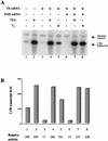
Both TR and TR/RXR repress the LTR promoter in the absence of T3 in a histone deacetylase-dependent manner. (A) Oocytes were injected with either TR or TR/RXR mRNAs followed by injection of the LTR plasmid. After overnight treatment, the promoter activity was analyzed by primer extension with a CAT1 primer (LTR transcript) and histone H4 primer (internal control). (B) Quantification of the data in panel A by PhosphorImager (Molecular Dynamics, Sunnyvale, Calif.). Note that unliganded TR and TR/RXR repressed the promoter similarly (about 6-fold, lanes 3 and 6). The addition of either T3 or TSA reversed the inhibition and further activation, resulting in an overall change of 9- to 15-fold in promoter activity.
FIG. 4.
FIG. 4.
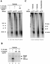
Liganded TR disrupts chromatin at the LTR through direct binding to the LTR but independently of transcriptional elongation. (A) An MNase digestion assay reveals that liganded TR disrupts the ordered nucleosome array on the LTR plasmid. Oocytes were injected and treated as described for Fig. 3 except for the presence or absence of 100 μg of α-amanitin per ml. After overnight incubation, the oocytes were harvested for the MNase digestion assay with increasing amounts of MNase (0.16, 0.8, and 4 U). The digested DNA was purified and analyzed by Southern blot analysis with a labeled LTR probe. Note that in the absence of T3 (lanes 1 to 3) or TR/RXR (not shown), an ordered nucleosomal array was present on the LTR plasmid, as indicated by the presence of the mono-, di-, and trinucleosome bands, etc. In the presence of T3 and TR/RXR, this ordered structure was disrupted, as indicated by the presence of a smear instead of discrete oligonucleosomal bands (lanes 4 to 6). Blocking transcriptional elongation with α-amanitin had no effect on the T3-induced chromatin disruption (lanes 7 to 9), suggesting that the disruption is an intrinsic property of liganded TR bound at the LTR. (B) Transcriptional activation of the LTR is inhibited by the elongation inhibitor α-amanitin. Oocytes were injected and treated as described above and were then processed for primer extension analysis. Note that TR/RXR activated the LTR in the presence of T3, and α-amanitin blocked transcription from the LTR. (C) Chromatin disruption requires the DNA binding domain of TR. mRNA encoding TR or a mutant TR lacking the DNA binding domain (TRΔDBD) (37) was injected into oocytes with RXR mRNA followed by the LTR plasmid injection. After overnight incubation in the presence or absence of T3, the plasmid minichromosome was isolated and analyzed as described for panel A. Note that disruption of the ordered nucleosomal array was observed with the liganded TR/RXR (compare lanes 4 to 6 to lanes 1 to 3) but not with the liganded TRΔDBD/RXR (lanes 7 to 9), indicating that the binding of TR to the LTR in the presence of T3 is necessary for chromatin disruption.
FIG. 5.
FIG. 5.
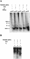
Transcription activation of the HIV-1 LTR by T3 but not TSA leads to chromatin disruption. (A) An MNase digestion assay shows that TSA treatment does not affect the nucleosomal array on the LTR plasmid. The oocytes were injected and treated as described for Fig. 4, except for TSA treatment where indicated. They were then processed for the MNase digestion assay. Again, T3 and TR/RXR together led to chromatin disruption (compare lanes 13 to 15 to lanes 1 to 3). In contrast, TR/RXR alone (lanes 7 to 9), TSA alone (lanes 4 to 6), and TSA plus TR/RXR (lanes 10 to 12) failed to alter the ordered nucleosomal array structure. (B) DNA topology analysis demonstrates that T3 but not TSA induces gross alterations of the structure of the LTR minichromosome. Oocytes were injected and treated as described for panel A, and LTR plasmid DNA was isolated for the supercoiling assay. After electrophoresis on a chloroquine-containing gel to separate the DNA with different numbers of negative superhelical turns (the higher the number of negative superhelical turns, the slower the DNA migration on the gel), the DNA was detected by Southern blot hybridization with a labeled LTR probe. Note that the average number of negative superhelical turns (star) was reduced by two to three when both TR/RXR and T3 were present (compare lanes 3 and 1). In contrast, TSA had little effect on the number of superhelical turns on the plasmid (compare lanes 2 and 1).
FIG. 6.
FIG. 6.
The ChIP assay shows that the transcription regulation by TRs leads to alterations of histone acetylation at the LTR promoter. Oocytes were injected and treated as described for Fig. 3 and were then harvested for ChIP assay with an antibody against acetylated histone H4 or histone H3 acetylated at position K9. Note that the acetylation levels of both H4 and H3 at the LTR were decreased in the presence of unliganded TR or TR/RXR (compare lanes 6 and 3, respectively, to lane 1). The addition of TSA or T3 treatment prevented the deacetylation of H4 by unliganded TR/RXR (lanes 4 and 5 for TR/RXR and 7 and 8 for TR). (H3 ChIP was not done for TR alone, as similar results were expected based on the data in the figure.) Equal amounts of DNA were present prior to immunoprecipitation (LTR input control).
FIG. 7.
FIG. 7.
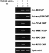
Unliganded TR/RXR recruits corepressors to the LTR in a T3-dependent manner. ChIP assays were performed as described for Fig. 6 with the indicated antibodies. As controls, anti-TR and -acetyl-H4 ChIP assays were done to show the binding of TR to the LTR and changes in histone acetylation in the samples used for the assays of the corepressors. Note that unliganded TR/RXR recruited N-CoR, SMRT, Sin3, and Rpd3 to the LTR (compare lanes 2 and 1). T3 treatment eliminated this recruitment (compare lane 3 to lanes 2 and 1). The LTR input control indicated that equal amounts of DNA were present during immunoprecipitation.

Similar articles

Cited by

References

    1. Almouzni, G., D. L. Clark, M. Mechali, and A. P. Wolffe. 1990. Chromatin assembly on replicating DNA in vitro. Nucleic Acids Res. 18:5767-5774. - PMC - PubMed
    1. Burke, L. J., and A. Baniahmad. 2000. Co-repressors 2000. FASEB J. 14:1876-1888. - PubMed
    1. Chen, H., R. J. Lin, R. L. Schiltz, D. Chakravarti, A. Nash, L. Nagy, M. L. Privalsky, Y. Nakatani, and R. M. Evans. 1997. Nuclear receptor coactivator ACTR is a novel histone acetyltransferase and forms a multimeric activation complex with P/CAF and CBP/p300. Cell 90:569-580. - PubMed
    1. Chen, J. D., and H. Li. 1998. Coactivation and corepression in transcriptional regulation by steroid/nuclear hormone receptors. Crit. Rev. Eukaryot. Gene Expr. 8:169-190. - PubMed
    1. Clark, D. J., and A. P. Wolffe. 1991. Superhelical stress and nucleosome-mediated repression of 5S RNA gene transcription in vitro. EMBO J. 10:3419-3428. - PMC - PubMed

MeSH terms

  NODES
INTERN 1
Note 9
twitter 2