Induction of sucrose utilization genes from Bifidobacterium lactis by sucrose and raffinose
- PMID: 12513973
- PMCID: PMC152442
- DOI: 10.1128/AEM.69.1.24-32.2003
Induction of sucrose utilization genes from Bifidobacterium lactis by sucrose and raffinose
Abstract
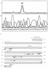
The probiotic organism Bifidobacterium lactis was isolated from a yoghurt starter culture with the aim of analyzing its use of carbohydrates for the development of prebiotics. A sucrose utilization gene cluster of B. lactis was identified by complementation of a gene library in Escherichia coli. Three genes, encoding a sucrose phosphorylase (ScrP), a GalR-LacI-type transcriptional regulator (ScrR), and a sucrose transporter (ScrT), were identified by sequence analysis. The scrP gene was expressed constitutively from its own promoter in E. coli grown in complete medium, and the strain hydrolyzed sucrose in a reaction that was dependent on the presence of phosphates. Primer extension experiments with scrP performed by using RNA isolated from B. lactis identified the transcriptional start site 102 bp upstream of the ATG start codon, immediately adjacent to a palindromic sequence resembling a regulator binding site. In B. lactis, total sucrase activity was induced by the presence of sucrose, raffinose, or oligofructose in the culture medium and was repressed by glucose. RNA analysis of the scrP, scrR, and scrT genes in B. lactis indicated that expression of these genes was influenced by transcriptional regulation and that all three genes were similarly induced by sucrose and raffinose and repressed by glucose. Analysis of the sucrase activities of deletion constructs in heterologous E. coli indicated that ScrR functions as a positive regulator.
Figures


Similar articles
-
Transcriptional regulation of the sucrase gene of Staphylococcus xylosus by the repressor ScrR.J Bacteriol. 1996 Jan;178(2):462-9. doi: 10.1128/jb.178.2.462-469.1996. J Bacteriol. 1996. PMID: 8550467 Free PMC article.
-
A functional analysis of the Bifidobacterium longum cscA and scrP genes in sucrose utilization.Appl Microbiol Biotechnol. 2006 Oct;72(5):975-81. doi: 10.1007/s00253-006-0358-x. Epub 2006 Mar 8. Appl Microbiol Biotechnol. 2006. PMID: 16523284
-
Functional characterization of sucrose phosphorylase and scrR, a regulator of sucrose metabolism in Lactobacillus reuteri.Food Microbiol. 2013 Dec;36(2):432-9. doi: 10.1016/j.fm.2013.07.011. Epub 2013 Aug 1. Food Microbiol. 2013. PMID: 24010626
-
The genes controlling sucrose utilization in Clostridium beijerinckii NCIMB 8052 constitute an operon.Microbiology (Reading). 1999 Jun;145 ( Pt 6):1461-1472. doi: 10.1099/13500872-145-6-1461. Microbiology (Reading). 1999. PMID: 10411273
-
Transcription of two adjacent carbohydrate utilization gene clusters in Bifidobacterium breve UCC2003 is controlled by LacI- and repressor open reading frame kinase (ROK)-type regulators.Appl Environ Microbiol. 2014 Jun;80(12):3604-14. doi: 10.1128/AEM.00130-14. Appl Environ Microbiol. 2014. PMID: 24705323 Free PMC article.
Cited by
-
Identification of Euglena gracilis β-1,3-glucan phosphorylase and establishment of a new glycoside hydrolase (GH) family GH149.J Biol Chem. 2018 Feb 23;293(8):2865-2876. doi: 10.1074/jbc.RA117.000936. Epub 2018 Jan 9. J Biol Chem. 2018. PMID: 29317507 Free PMC article.
-
Transcriptional control of central carbon metabolic flux in Bifidobacteria by two functionally similar, yet distinct LacI-type regulators.Sci Rep. 2019 Nov 28;9(1):17851. doi: 10.1038/s41598-019-54229-4. Sci Rep. 2019. PMID: 31780796 Free PMC article.
-
Experimental determination and characterization of the gap promoter of Bifidobacterium bifidum S17.Bioengineered. 2014;5(6):371-7. doi: 10.4161/bioe.34423. Epub 2014 Oct 30. Bioengineered. 2014. PMID: 25482086 Free PMC article.
-
Short fractions of oligofructose are preferentially metabolized by Bifidobacterium animalis DN-173 010.Appl Environ Microbiol. 2004 Apr;70(4):1923-30. doi: 10.1128/AEM.70.4.1923-1930.2004. Appl Environ Microbiol. 2004. PMID: 15066781 Free PMC article.
-
Bifidobacterium longum requires a fructokinase (Frk; ATP:D-fructose 6-phosphotransferase, EC 2.7.1.4) for fructose catabolism.J Bacteriol. 2004 Oct;186(19):6515-25. doi: 10.1128/JB.186.19.6515-6525.2004. J Bacteriol. 2004. PMID: 15375133 Free PMC article.
References
-
- Ballongue, J. 1998. Bifidobacteria and probiotic action, p. 519-587. In S. Salminen and A. Von Wright (ed.), Lactic acid bacteria. Microbiology and functional aspects. Marcel Dekker, Inc., New York, N.Y.
-
- Beg, O. U. 1995. Extraction of RNA from Gram-positive bacteria. BioTechniques 19:880-884. - PubMed
-
- Bezkorovainy, A. 1989. Classification of bifidobacteria, p. 1-28. In R. Miller-Catchpole and A. Bezkorovainy (ed.), Biochemistry and physiology of bifidobacteria. CRC Press, Boca Raton, Fla.
-
- Bradford, M. M. 1976. A rapid and sensitive method for quantitation of microgram quantities of protein utilizing the principle of protein-dye binding. Anal. Biochem. 72:248-254. - PubMed
Publication types
MeSH terms
Substances
Associated data
- Actions
- Actions
LinkOut - more resources
Full Text Sources
Other Literature Sources
Molecular Biology Databases
Miscellaneous

