Direct cAMP signaling through G-protein-coupled receptors mediates growth cone attraction induced by pituitary adenylate cyclase-activating polypeptide
- PMID: 12657686
- PMCID: PMC6742047
- DOI: 10.1523/JNEUROSCI.23-06-02274.2003
Direct cAMP signaling through G-protein-coupled receptors mediates growth cone attraction induced by pituitary adenylate cyclase-activating polypeptide
Abstract
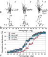
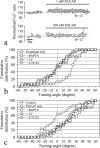
Developing axons are guided to their appropriate _targets by environmental cues through the activation of specific receptors and intracellular signaling pathways. Here we report that gradients of pituitary adenylate cyclase-activating polypeptide (PACAP), a neuropeptide widely expressed in the developing nervous system, induce marked attraction of Xenopus growth cones in vitro. PACAP exerted its chemoattractive effects through PAC1, a PACAP-selective G-protein-coupled receptor (GPRC) expressed at the growth cone. Furthermore, the attraction depended on localized cAMP signaling because it was completely blocked either by global elevation of intracellular cAMP levels using forskolin or by inhibition of protein kinase A using specific inhibitors. Moreover, local direct elevation of intracellular cAMP by focal photolysis of caged cAMP compounds was sufficient to induce growth cone attraction. On the other hand, blockade of Ca2+, phospholipase C, or phosphatidyl inositol-3 kinase signaling pathways did not affect PACAP-induced growth cone attraction. Finally, PACAP-induced attraction also involved the Rho family of small GTPases and required local protein synthesis. Taken together, our results establish cAMP signaling as an independent pathway capable of mediating growth cone attraction induced by a physiologically relevant peptide acting through GPCRs. Such a direct cAMP pathway could potentially operate in other guidance systems for the accurate wiring of the nervous system.
Figures


Similar articles
-
Pituitary adenylate cyclase-activating polypeptide receptors mediating insulin secretion in rodent pancreatic islets are coupled to adenylate cyclase but not to PLC.Endocrinology. 2002 Apr;143(4):1253-9. doi: 10.1210/endo.143.4.8739. Endocrinology. 2002. PMID: 11897681
-
Continuous activation of pituitary adenylate cyclase-activating polypeptide receptors elicits antipodal effects on cyclic AMP and inositol phospholipid signaling pathways in CATH.a cells: role of protein synthesis and protein kinases.J Neurochem. 1998 Apr;70(4):1431-40. doi: 10.1046/j.1471-4159.1998.70041431.x. J Neurochem. 1998. PMID: 9523559
-
Pituitary adenylate cyclase-activating polypeptide and vasoactive intestinal peptide inhibit dendritic growth in cultured sympathetic neurons.J Neurosci. 2002 Aug 1;22(15):6560-9. doi: 10.1523/JNEUROSCI.22-15-06560.2002. J Neurosci. 2002. PMID: 12151535 Free PMC article.
-
Pituitary adenylate cyclase-activating polypeptide (PACAP) and its receptors in the brain.Kaibogaku Zasshi. 2000 Dec;75(6):487-507. Kaibogaku Zasshi. 2000. PMID: 11197592 Review.
-
[Physiological significance of pituitary adenylate cyclase-activating polypeptide (PACAP) in the nervous system].Yakugaku Zasshi. 2002 Dec;122(12):1109-21. doi: 10.1248/yakushi.122.1109. Yakugaku Zasshi. 2002. PMID: 12510388 Review. Japanese.
Cited by
-
Dysfunctional purinergic signaling correlates with disease severity in COVID-19 patients.Front Immunol. 2022 Sep 30;13:1012027. doi: 10.3389/fimmu.2022.1012027. eCollection 2022. Front Immunol. 2022. PMID: 36248842 Free PMC article.
-
cAMP-dependent axon guidance is distinctly regulated by Epac and protein kinase A.J Neurosci. 2009 Dec 9;29(49):15434-44. doi: 10.1523/JNEUROSCI.3071-09.2009. J Neurosci. 2009. PMID: 20007468 Free PMC article.
-
Injury-associated PACAP expression in rat sensory and motor neurons is induced by endogenous BDNF.PLoS One. 2014 Jun 26;9(6):e100730. doi: 10.1371/journal.pone.0100730. eCollection 2014. PLoS One. 2014. PMID: 24968020 Free PMC article.
-
VIP and PACAP: neuropeptide modulators of CNS inflammation, injury, and repair.Br J Pharmacol. 2013 Jun;169(3):512-23. doi: 10.1111/bph.12181. Br J Pharmacol. 2013. PMID: 23517078 Free PMC article. Review.
-
Neural Explant Cultures from Xenopus laevis.J Vis Exp. 2012 Oct 15;(68):e4232. doi: 10.3791/4232. J Vis Exp. 2012. PMID: 23295240 Free PMC article.
References
-
- Arimura A. Pituitary adenylate cyclase activating polypeptide (PACAP): discovery and current status of research. Regul Pept. 1992;37:287–303. - PubMed
-
- Bolsover SR, Gilbert SH, Spector I. Intracellular cyclic AMP produces effects opposite to those of cyclic GMP and calcium on shape and motility of neuroblastoma cells. Cell Motil Cytoskeleton. 1992;22:99–116. - PubMed
-
- Campbell DS, Holt CE. Chemotropic responses of retinal growth cones mediated by rapid local protein synthesis and degradation. Neuron. 2001;32:1013–1026. - PubMed
-
- Chartrel N, Tonon MC, Vaudry H, Conlon JM. Primary structure of frog pituitary adenylate cyclase-activating polypeptide (PACAP) and effects of ovine PACAP on frog pituitary. Endocrinology. 1991;129:3367–3371. - PubMed
Publication types
MeSH terms
Substances
LinkOut - more resources
Full Text Sources
Other Literature Sources
Miscellaneous
