. 2003 May;23(9):3173-85.
doi: 10.1128/MCB.23.9.3173-3185.2003.

Role for human SIRT2 NAD-dependent deacetylase activity in control of mitotic exit in the cell cycle

Affiliations

Role for human SIRT2 NAD-dependent deacetylase activity in control of mitotic exit in the cell cycle

Sylvia C Dryden et al. Mol Cell Biol. 2003 May.

Abstract

Studies of yeast have shown that the SIR2 gene family is involved in chromatin structure, transcriptional silencing, DNA repair, and control of cellular life span. Our functional studies of human SIRT2, a homolog of the product of the yeast SIR2 gene, indicate that it plays a role in mitosis. The SIRT2 protein is a NAD-dependent deacetylase (NDAC), the abundance of which increases dramatically during mitosis and is multiply phosphorylated at the G(2)/M transition of the cell cycle. Cells stably overexpressing the wild-type SIRT2 but not missense mutants lacking NDAC activity show a marked prolongation of the mitotic phase of the cell cycle. Overexpression of the protein phosphatase CDC14B, but not its close homolog CDC14A, results in dephosphorylation of SIRT2 with a subsequent decrease in the abundance of SIRT2 protein. A CDC14B mutant defective in catalyzing dephosphorylation fails to change the phosphorylation status or abundance of SIRT2 protein. Addition of 26S proteasome inhibitors to human cells increases the abundance of SIRT2 protein, indicating that SIRT2 is _targeted for degradation by the 26S proteasome. Our data suggest that human SIRT2 is part of a phosphorylation cascade in which SIRT2 is phosphorylated late in G(2), during M, and into the period of cytokinesis. CDC14B may provoke exit from mitosis coincident with the loss of SIRT2 via ubiquitination and subsequent degradation by the 26S proteasome.


Figures
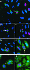
FIG. 1.
FIG. 1.
Staining of cells with SIRT2 antibody is tightly confined to cells in late G2 or M phase. Confocal microscopy was performed on Saos2 cells grown on coverslips as described in Materials and Methods. DAPI (dihydrochloride)-stained nuclei appear pink. DAPI stain is blue, but in the interests of contrast, the color was enhanced. A green fluorescent FITC-conjugated goat anti-rabbit secondary antibody was used to localize the rabbit SIRT2 peptide primary antibody. White arrows point to mitotic cells. (A) Full-field view of unsynchronized Saos2 cells. Only mitotic cells exhibit intense SIRT2 staining. (B to D) Close-up views of individual Saos2 mitotic cells. (E and F) Saos2 cells blocked at G2/M with nocodazole. The intense SIRT2 staining tightly surrounds the condensing chromatin (indicated by yellow arrows). (G) Saos2 cells blocked in metaphase with Colcemid. Again, SIRT2 is intensified around the chromatin.
FIG. 2.
FIG. 2.
There is increased phosphorylation of SIRT2 protein at G2 and in M phase. SIRT2-overexpressing cells are hyperphosphorylated. Saos2 cells (A) and a stable transfectant overexpressing SIRT2 (clone 4 in panel B) were synchronized by double-thymidine block and subsequently treated with nocodazole or Colcemid. The DNA content of synchronized cells was assayed by flow cytometry to confirm the effectiveness of cell cycle blocks. The percentage of cells with a 2N (G1 and S phase) or 4N (G2 and M phase) DNA content is shown. Cell lysates were harvested at each step during synchronization and immunoblotted with SIRT2 peptide antibody. Arrows point to the phosphorylated isoforms. The arrow with an asterisk indicates λPPase-insensitive isoforms of SIRT2. UT, untreated, unsynchronized cells; T, cells harvested after the double-thymidine block; N, cells subsequently treated with nocodazole; C, cells treated with Colcemid after thymidine treatment. Blockade in M phase (Colcemid) results in a more pronounced shift of SIRT2 to the phosphorylated isoform in Saos2 cells.
FIG. 3.
FIG. 3.
SIRT2 expression in stable transfectants of Saos2 cells. Saos2 cells stably transfected with pcDNA3.1-SIRT2 or pcDNA3.1-SIRT2 H232Y express various levels of the phosphorylated form of SIRT2. Crude lysates from individual clones were subjected to immunoblotting with the SIRT2 peptide antibody. (A) Stable transfectants expressing wild-type (wt) SIRT2. Cells ± λPPase and Na3VO4 were used as markers for phosphorylation status. Lanes: 1, untransfected Saos2 cells; 2, clone 6; 3, clone 4; 4, clone 2. Lanes 5 to 7 contained nocodazole-treated cells ± λPPase and Na3VO4. To control for protein loading, blots were also probed with actin (lower panel). (B) Stable transfectants expressing mutant SIRT2, in which histidine 232 was mutated to tyrosine. Lane 1 contained clone 4. Clones 1 and 21 (lanes 2 and 3, respectively) were used in subsequent experiments. (C) SIRT2 NDAC activity in various transfected Saos2 cell lines. Activity was assayed by monitoring the release of 3H-acetyl groups from an H4 peptide (counts per minute) under the conditions indicated. (D) The amino- and carboxy-terminal domains of the H232Y clones are intact. Nickel magnetic agarose beads were used to purify SIRT2. Crude cell lysates prior to pulldown assays are shown in lanes 1 and 2. Lysates and pulldown assays were subjected to immunoblotting with the SIRT2 peptide antibody. (E) Time course of λPPase digestion of SIRT2 stable transfectants. The hyperphosphorylated isoform appears to be the most labile, collapsing sequentially to the final hypophosphorylated SIRT2 isoform.
FIG. 3.
FIG. 3.
SIRT2 expression in stable transfectants of Saos2 cells. Saos2 cells stably transfected with pcDNA3.1-SIRT2 or pcDNA3.1-SIRT2 H232Y express various levels of the phosphorylated form of SIRT2. Crude lysates from individual clones were subjected to immunoblotting with the SIRT2 peptide antibody. (A) Stable transfectants expressing wild-type (wt) SIRT2. Cells ± λPPase and Na3VO4 were used as markers for phosphorylation status. Lanes: 1, untransfected Saos2 cells; 2, clone 6; 3, clone 4; 4, clone 2. Lanes 5 to 7 contained nocodazole-treated cells ± λPPase and Na3VO4. To control for protein loading, blots were also probed with actin (lower panel). (B) Stable transfectants expressing mutant SIRT2, in which histidine 232 was mutated to tyrosine. Lane 1 contained clone 4. Clones 1 and 21 (lanes 2 and 3, respectively) were used in subsequent experiments. (C) SIRT2 NDAC activity in various transfected Saos2 cell lines. Activity was assayed by monitoring the release of 3H-acetyl groups from an H4 peptide (counts per minute) under the conditions indicated. (D) The amino- and carboxy-terminal domains of the H232Y clones are intact. Nickel magnetic agarose beads were used to purify SIRT2. Crude cell lysates prior to pulldown assays are shown in lanes 1 and 2. Lysates and pulldown assays were subjected to immunoblotting with the SIRT2 peptide antibody. (E) Time course of λPPase digestion of SIRT2 stable transfectants. The hyperphosphorylated isoform appears to be the most labile, collapsing sequentially to the final hypophosphorylated SIRT2 isoform.
FIG. 4.
FIG. 4.
SIRT2 stable transfectants that overexpress the wild-type phosphorylated forms of SIRT2 return to G1 less effectively than cells expressing mutant SIRT2 or nonhyperphosphorylated cell lines. Cell lines were treated with nocodazole for 20 h, and then the nocodazole was washed out. Cells were collected for flow cytometry at the indicated times after the initiation of nocodazole washout. In panels A to E, graphs indicate the percentage of cells with a 2N and 4N DNA contents at each time point for each cell line. Cell lysates were harvested at each time point and immunoblotted with SIRT2 peptide antibody (results shown below each graph). UT, untreated, unsynchronized cells. These results are representative of those obtained in multiple experiments. The more hyperphosphorylated the cell line is, the slower the exit from mitosis is.
FIG. 4.
FIG. 4.
SIRT2 stable transfectants that overexpress the wild-type phosphorylated forms of SIRT2 return to G1 less effectively than cells expressing mutant SIRT2 or nonhyperphosphorylated cell lines. Cell lines were treated with nocodazole for 20 h, and then the nocodazole was washed out. Cells were collected for flow cytometry at the indicated times after the initiation of nocodazole washout. In panels A to E, graphs indicate the percentage of cells with a 2N and 4N DNA contents at each time point for each cell line. Cell lysates were harvested at each time point and immunoblotted with SIRT2 peptide antibody (results shown below each graph). UT, untreated, unsynchronized cells. These results are representative of those obtained in multiple experiments. The more hyperphosphorylated the cell line is, the slower the exit from mitosis is.
FIG.5.
FIG.5.
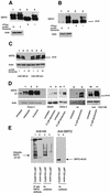
Transfection of functional Cdc14B into clone 4 causes SIRT2 protein levels to decrease and _targets SIRT2 for ubiquitination and subsequent degradation by the 26S proteasome. Clone 4 cells were transiently transfected with CDC14A, CDC14B, or the CDC14B C314S mutant at the indicated DNA concentrations. As a control, a lysate from untransfected clone 4 cells was treated with or without λPPase (lanes 3 and 4, respectively, in panels A and B). Ten micrograms of crude protein lysate was loaded in each lane and subjected to immunoblotting with the SIRT2 peptide antibody. (A) Clone 4 transfected with CDC14A. There was very little change in SIRT2 when either 6 or 12 μg of CDC14A DNA was transfected into cells (lanes 1 and 2). The arrow marked with an asterisk indicates the unphosphorylated SIRT2 isoforms. (B) Clone 4 transfected with CDC14B. When 12 μg of CDC14B was transfected into cells, there was a dramatic decrease in protein and loss of the phosphorylated SIRT2 (lane 1). (C) Clone 4 transfected with the either CDC14B wild type (wt) or a Cdc14B C314S mutant (mut) that has no phosphatase activity. With increasing amounts of CDC14B, there was a corresponding decrease in SIRT2 (lanes 1 to 3). There was no change in SIRT2 with increasing concentrations of the CDC14B C314S mutant plasmid (lanes 4 to 6). (D) The 26S proteasome inhibitors lactacystin, clasto-lactacystin β-lactone, and epoxomicin stabilize SIRT2 isoforms and prevent ubiquitin-mediated degradation. Following a 20-h incubation with the indicated inhibitors, cells were harvested and processed for Western blot analysis. Lanes: 1 and 6, untreated clone 4 lysates; 2, clone 4 treated with 10 μM lactacystin; 3, clone 4 treated with 10 μM clasto-lactacystin β-lactone; 4, untreated Saos2 lysate; 5, Saos2 treated with 0.1 μM epoxomicin; 7, clone 4 treated with 0.1 μM epoxomicin; 8 and 9, clone 4 transfected with CDC14B (wt) minus or plus 0.1 μM epoxomicin, respectively; 10 and 11, clone 4 transfected with CDC14B C314S (mut) minus or plus 0.1 μM epoxomicin, respectively. To control for protein loading, all blots were probed with actin as well. (E) SIRT2 and ubiquitin coprecipitate in both nickel magnetic agarose bead pulldown assays and protein A SIRT2 immunoprecipitations. Saos2 cells were transiently cotransfected with the HA-tagged, ubiquitin-expressing plasmid pMT123 and either pcDNA3.1-SIRT2 or the empty vector pcDNA3.1/HisC. Lysates from the transfected cells were incubated with Ni-NTA magnetic agarose beads or SIRT2 antibody bound to protein A affinity gel. Samples were processed as described above and immunoblotted with HA-ll antibody (lanes 1 to 4) or SIRT2 peptide antibody (lanes 5 and 6).

Similar articles

Cited by

References

    1. Afshar, G., and J. P. Murnane. 1999. Characterization of a human gene with sequence homology to Saccharomyces cerevisiae SIR2. Gene 234:161-168. - PubMed
    1. Avalos, J. L., I. Celic, S. Muhammad, M. S. Cosgrove, J. D. Boeke, and C. Wolberger. 2002. Structure of a Sir2 enzyme bound to an acetylated p53 peptide. Mol. Cell 10:523-535. - PubMed
    1. Bembenek, J., and H. Yu. 2001. Regulation of the anaphase-promoting complex by the dual specificity phosphatase human Cdc14a. J. Biol. Chem. 276:48237-48242. - PubMed
    1. Borra, M. T., F. J. O'Neill, M. D. Jackson, B. Marshall, E. Verdin, K. R. Foltz, and J. M. Denu. 2002. Conserved enzymatic production and biological effect of O-acetyl-ADP-ribose by silent information regulator 2-like NAD+-dependent deacetylases. J. Biol. Chem. 277:12632-12641. - PubMed
    1. Brachmann, C. G., J. M. Sherman, S. E. Devine, E. E. Cameron, L. Pillus, and J. D. Boeke. 1995. The Sir2 gene family, conserved from bacteria to humans, functions in silencing, cell cycle progression, and chromosome stability. Genes Dev. 9:2888-2902. - PubMed

Publication types

MeSH terms

  NODES
twitter 2