Characterization of extrathymic CD8 alpha beta T cells in the liver and intestine in TAP-1 deficient mice
- PMID: 12807479
- PMCID: PMC1782982
- DOI: 10.1046/j.1365-2567.2003.01654.x
Characterization of extrathymic CD8 alpha beta T cells in the liver and intestine in TAP-1 deficient mice
Abstract
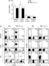
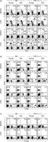
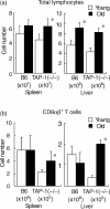
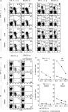
TAP-1 deficient (-/-) mice cannot transport MHC class I antigens onto the cell surface, which results in failure of the generation of CD8+ T cells in the thymus. In a series of recent studies, it has been proposed that extrathymic T cells are generated in the liver and at other extrathymic sites (e.g. the intestine). It was therefore investigated whether CD8+ extrathymic T cells require an interaction with MHC class I antigens for their differentiation in TAP-1(-/-) mice. Although CD8+ thymically derived T cells were confirmed to be absent in the spleen as well as in the thymus, CD8 alpha beta+ T cells were abundant in the livers and intestines of TAP-1(-/-) mice. These CD8+ T cells expanded in the liver as a function of age and were mainly confined to a NK1.1-CD3int population which is known to be truly of extrathymic origin. Hepatic lymphocytes, which contained CD8+ T cells and which were isolated from TAP-1(-/-) mice (H-2b), responded to neither mutated MHC class I antigens (bm1) nor allogeneic MHC class I antigens (H-2d) in in vitro mixed lymphocyte cultures. However, the results from repeated in vivo stimulations with alloantigens (H-2d) were interesting. Allogeneic cytotoxicity was induced in liver lymphocytes in TAP-1(-/-) mice, although the magnitude of cytotoxicity was lower than that of liver lymphocytes in immunized B6 mice. All allogeneic cytotoxicity disappeared with the elimination of CD8+ cells in TAP-1(-/-) mice. These results suggest that the generation and function of CD8+ extrathymic T cells are independent of the existence of the MHC class I antigens of the mouse but have a limited allorecognition ability.
Figures


Similar articles
-
Generation of CD8+ T cells specific for transporter associated with antigen processing deficient cells.Proc Natl Acad Sci U S A. 1997 Oct 14;94(21):11496-501. doi: 10.1073/pnas.94.21.11496. Proc Natl Acad Sci U S A. 1997. PMID: 9326638 Free PMC article.
-
TAP-independent selection of CD8+ intestinal intraepithelial lymphocytes.J Immunol. 1996 Jun 1;156(11):4209-16. J Immunol. 1996. PMID: 8666789
-
Existence of MHC class I-restricted alloreactive CD4+ T cells reacting with peptide transporter-deficient cells.Immunogenetics. 2001 Oct;53(8):626-33. doi: 10.1007/s00251-001-0379-7. Epub 2001 Nov 9. Immunogenetics. 2001. PMID: 11797095
-
Physiological responses of extrathymic T cells in the liver.Immunol Rev. 2000 Apr;174:135-49. doi: 10.1034/j.1600-0528.2002.017415.x. Immunol Rev. 2000. PMID: 10807513 Review.
-
Repertoire-determining role of peptide in the positive selection of CD8+ T cells.Immunol Rev. 1993 Oct;135:157-82. doi: 10.1111/j.1600-065x.1993.tb00648.x. Immunol Rev. 1993. PMID: 8282312 Review. No abstract available.
Cited by
-
Rapid migration of thymic emigrants to the colonic mucosa in ulcerative colitis patients.Clin Exp Immunol. 2010 Nov;162(2):325-36. doi: 10.1111/j.1365-2249.2010.04230.x. Epub 2010 Sep 14. Clin Exp Immunol. 2010. PMID: 20840654 Free PMC article.
-
Systemic CD8 T-cell memory response to a Salmonella pathogenicity island 2 effector is restricted to Salmonella enterica encountered in the gastrointestinal mucosa.Infect Immun. 2007 Jun;75(6):2708-16. doi: 10.1128/IAI.01905-06. Epub 2007 Apr 2. Infect Immun. 2007. PMID: 17403871 Free PMC article.
-
TAP1-/- mice present oligoclonal BV-BJ expansions following the rejection of grafts bearing self antigens.Immunology. 2006 Aug;118(4):461-71. doi: 10.1111/j.1365-2567.2006.02387.x. Immunology. 2006. PMID: 16895555 Free PMC article.
References
-
- Van Kaer L, Ashton-Rickardt PG, Ploegh HL, Tonegawa S. TAP1 mutant mice are deficient in antigen presentation, surface class I molecules, and CD4−8+ T cells. Cell. 1992;71:1205–14. - PubMed
-
- Ljunggren H-G, Glas R, Sandberg JK, Kärre K. Reactivity and specificity of CD8+ T cells in mice with defects in the MHC class I antigen-presenting pathway. Immunol Rev. 1996;151:123–48. - PubMed
MeSH terms
Substances
LinkOut - more resources
Full Text Sources
Molecular Biology Databases
Research Materials
Miscellaneous

