. 2003 Sep 15;31(18):5266-74.
doi: 10.1093/nar/gkg731.

Ligand-independent activation of estrogen receptor alpha by XBP-1

Affiliations

Ligand-independent activation of estrogen receptor alpha by XBP-1

Lihua Ding et al. Nucleic Acids Res. .

Abstract

The estrogen receptor (ER) is a member of a large superfamily of nuclear receptors that regulates the transcription of estrogen-responsive genes. Several recent studies have demonstrated that XBP-1 mRNA expression is associated with ERalpha status in breast tumors. However, the role of XBP-1 in ERalpha signaling remains to be elucidated. More recently, two forms of XBP-1 were identified due to its unconventional splicing. We refer to the spliced and unspliced forms of XBP-1 as XBP-1S and XBP-1U, respectively. Here, we report that XBP-1S and XBP-1U enhanced ERalpha-dependent transcriptional activity in a ligand-independent manner. XBP-1S had stronger activity than XBP-1U. The maximal effects of XBP-1S and XBP-1U on ERalpha transactivation were observed when they were co-expressed with full-length ERalpha. SRC-1, the p160 steroid receptor coactivator family member, synergized with XBP-1S or XBP-1U to potentiate ERalpha activity. XBP-1S and XBP-1U bound to the ERalpha both in vitro and in vivo in a ligand-independent fashion. XBP-1S and XBP-1U interacted with the ERalpha region containing the DNA-binding domain. The ERalpha-interacting regions on XBP-1S and XBP-1U have been mapped to two regions, including the N-terminal basic region leucine zipper domain (bZIP) and the C-terminal activation domain. The bZIP-deleted mutants of XBP-1S and XBP-1U completely abolished ERalpha transactivation by XBP-1S and XBP-1U. These findings suggest that XBP-1S and XBP-1U may directly modulate ERalpha signaling in both the absence and presence of estrogen and, therefore, may play important roles in the proliferation of normal and malignant estrogen-regulated tissues.


Figures
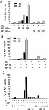
Figure 1
Figure 1
XBP-1S and XBP-1U enhance ERα-mediated transactivation in MDA-MB-435 cells. (A) Effects of XBP-1S and XBP-1U on ERα-mediated transactivation. Cells were co-transfected with 0.2 µg of ERE-LUC, 50 ng of the expression plasmid for ERα and increasing amounts of the expression vector for either XBP-1S or XBP-1U as indicated. Cells were then treated with control (0.1% ethanol) vehicle or 10 nM E2 for 24 h before luciferase assay. The LUC activity obtained on transfection of ERE-LUC and ERα without exogenous XBP-1S and XBP-1U in the absence of E2 was set as 1. Results are expressed as means ± SE for three independent experiments. (B) Effect of ERα on ERE-LUC reporter gene transcription by XBP-1S and XBP-1U. Cells were co-transfected with 0.2 µg of ERE-LUC and 0.5 µg of the expression vector for either XBP-1S or XBP-1U in both the absence and presence of the expression plasmid for ERα. Cells were then treated and analyzed as in (A). (C) Effects of antiestrogens on ERE-LUC reporter gene transcription by XBP-1S and XBP-1U. Cells were co-transfected with 0.2 µg of ERE-LUC, 50 ng of the expression plasmid for ERα and 0.5 µg of the expression vector for either XBP-1S or XBP-1U. Cells were then treated with control (0.1% ethanol) vehicle, 10 nM E2, 100 nM 4-OHT or 100 nM ICI 182,780 for 24 h before luciferase assay. Cells were analyzed as in (A).
Figure 2
Figure 2
Western blotting showing the ERα, XBP-1S and XBP-1U protein levels in 293T cells. Cells were transfected as in Table 1. Whole-cell extracts were prepared, and equivalent amounts of each extract were probed with anti-ERα antibody (H-184; Santa Cruz Biotech) or anti-XBP-1 antibody (SC-7160; Santa Cruz Biotechnology).
Figure 3
Figure 3
Both N- and C-terminal domains contribute to ERα transcriptional activity regulated by XBP-1S and XBP-1U. MDA-MB-435 cells were co-transfected with 50 ng of the expression vector for ERα, ERα ABC domain or ERα CDEF domain, 0.2 µg of ERE-LUC and 0.5 µg of the expression vector for either XBP-1S or XBP-1U as indicated. The LUC activity obtained on transfection of ERE-LUC without exogenous ERα, ERα ABC domain, ERα CDEF domain, XBP-1S and XBP-1U in the absence of E2 was set as 1. Results are expressed as means ± SE for three independent experiments.
Figure 4
Figure 4
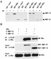
XBP-1S and XBP-1U bind to ERα in vitro and in vivo. (A) Interaction of XBP-1S and XBP-1U with ERα in vitro. GST pull-down assay was performed as described in Materials and Methods. Full-length GST–ERα fusion proteins, immobilized on beads, were mixed with in vitro translation reaction mixtures of XBP-1S or XBP-1U in the absence or presence of E2 (10–6 M) as indicated. The bound proteins were subjected to SDS–PAGE followed by autoradiography. (B) Interactions between either XBP-1S or XBP-1U and ERα in vivo. 293T cells were co-transfected with the expression vectors for either the FLAG-tagged XBP-1 or the FLAG-tagged XBP-1U and ERα as indicated. Lysates from the transfected cells were immunoprecipitated (IP) using anti-FLAG antibody (Sigma-Aldrich), and the immunoprecipitates were probed with an anti-ERα antibody (HC-20; Santa Cruz Biotech). (C) Mapping of interaction regions of XBP-1S and XBP-1U in ERα. GST pull-down assay was performed using 35S-labeled XBP-1S or XBP-1U and fusion proteins between GST and three different ERα fragments. (D) Mapping of the ERα interaction region in XBP-1S and XBP-1U. GST pull-down assay was performed using 35S-labeled ERα and fusion proteins between GST and six different XBP-1 fragments.
Figure 4
Figure 4
XBP-1S and XBP-1U bind to ERα in vitro and in vivo. (A) Interaction of XBP-1S and XBP-1U with ERα in vitro. GST pull-down assay was performed as described in Materials and Methods. Full-length GST–ERα fusion proteins, immobilized on beads, were mixed with in vitro translation reaction mixtures of XBP-1S or XBP-1U in the absence or presence of E2 (10–6 M) as indicated. The bound proteins were subjected to SDS–PAGE followed by autoradiography. (B) Interactions between either XBP-1S or XBP-1U and ERα in vivo. 293T cells were co-transfected with the expression vectors for either the FLAG-tagged XBP-1 or the FLAG-tagged XBP-1U and ERα as indicated. Lysates from the transfected cells were immunoprecipitated (IP) using anti-FLAG antibody (Sigma-Aldrich), and the immunoprecipitates were probed with an anti-ERα antibody (HC-20; Santa Cruz Biotech). (C) Mapping of interaction regions of XBP-1S and XBP-1U in ERα. GST pull-down assay was performed using 35S-labeled XBP-1S or XBP-1U and fusion proteins between GST and three different ERα fragments. (D) Mapping of the ERα interaction region in XBP-1S and XBP-1U. GST pull-down assay was performed using 35S-labeled ERα and fusion proteins between GST and six different XBP-1 fragments.
Figure 5
Figure 5
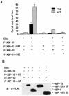
The deletion mutants of XBP-1S and XBP-1U abolished the ERα transactivation. (A) MDA-MB-435 cells were co-transfected with 0.2 µg of ERE-LUC, 50 ng of the expression plasmid for ERα and 0.5 µg of the expression vector for FLAG-tagged XBP-1S, XBP-1SΔ82, XBP-1U or XBP-1UΔ82 as indicated. Cells were then treated with control (0.1% ethanol) vehicle or 10 nM E2 for 24 h before luciferase assay. The LUC activity obtained on transfection of ERE-LUC and ERα without exogenous XBP-1 and its derivatives in the absence of E2 was set as 1. Results are expressed as means ± SE for three independent experiments. (B) Western blotting showing expression of FLAG-tagged XBP-1S, XBP-1SΔ82, XBP-1U and XBP-1UΔ82. Cells were transfected as in (A). Whole-cell extracts were prepared, and equivalent amounts of each extract were probed with anti-FLAG antibody (Sigma-Aldrich).
Figure 6
Figure 6
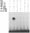
Neither XBP-1S nor XBP-1U binds to ERE. Gel shift assay was performed as described in Materials and Methods. The 32P-labeled ERE probe was incubated with the in vitro-translated ERα, XBP-1S and XBP-1U proteins as indicated, in the presence of 1 µM E2. For competition experiments, a 100-fold molar excess of unlabeled ERE was mixed with the radioactive probe. The 32P-labeled mutant ERE (EREM) probe was used as a negative control.
Figure 7
Figure 7
XBP-1 mRNA expression in breast cancer cell lines. RT–PCR from the selected cell lines and cDNA libraries was performed as described in Materials and Methods. β-Actin was used as an internal control.

Similar articles

Cited by

References

    1. Klinge C.M. (2000) Estrogen receptor interaction with co-activators and co-repressors. Steroids, 65, 227–251. - PubMed
    1. Katzenellenbogen B.S. and Katzenellenbogen,J.A. (2000) Estrogen receptor transcription and transactivation: estrogen receptor alpha and estrogen receptor beta: regulation by selective estrogen receptor modulators and importance in breast cancer. Breast Cancer Res., 2, 335–344. - PMC - PubMed
    1. Aranda A. and Pascual,A. (2001) Nuclear hormone receptors and gene expression. Physiol. Rev., 81, 1269–1304. - PubMed
    1. Krust A., Green,S., Argos,P., Kumar,V., Walter,P., Bornert,J.M. and Chambon,P. (1986) The chicken oestrogen receptor sequence: homology with v-erbA and the human oestrogen and glucocorticoid receptors. EMBO J., 5, 891–897. - PMC - PubMed
    1. Mangelsdorf D.J., Thummel,C., Beato,M., Herrlich,P., Schutz,G., Umesono,K., Blumberg,B., Kastner,P., Mark,M. and Chambon,P. (1995) The nuclear receptor superfamily: the second decade, Cell, 83, 835–839. - PMC - PubMed

Publication types

MeSH terms

  NODES
INTERN 1
twitter 2