. 2003 Nov;23(21):7926-36.
doi: 10.1128/MCB.23.21.7926-7936.2003.

BARD1 participates with BRCA1 in homology-directed repair of chromosome breaks

Affiliations

BARD1 participates with BRCA1 in homology-directed repair of chromosome breaks

Ulrica K Westermark et al. Mol Cell Biol. 2003 Nov.

Abstract

The BRCA1 tumor suppressor has been implicated in the maintenance of chromosomal stability through homology-directed repair of DNA double-strand breaks. Much of the BRCA1 in cells forms a heterodimeric complex with a structurally related protein BARD1. We report that expression of truncated mouse or human BARD1 peptides capable of interacting with Brca1 results in a homologous-repair deficiency. Repair is mildly reduced in Brca1 wild-type cells and severely reduced in cells that harbor a Brca1 splice product deleted for exon 11. Nuclear localization of the Brca1 or BARD1 peptides is not compromised, implying that the repair deficiency is caused by a more direct effect on repair. The tumor suppressor activity of BRCA1 may require the participation of BARD1 to maintain chromosome integrity through the homologous-repair pathway.


Figures

FIG. 1.
FIG. 1.
Overexpression of truncated mBard1 decreases HDR in Brca1+/+ and Brca1−/− cells. (A) Schematic drawing of wild-type murine Bard1 and an N-terminal fragment of mBard1 (mB301). The RING domains of both Bard1 and Brca1 consist of a central RING motif, which binds two zinc atoms flanked by antiparallel α-helices, which are indicated by the darker shading. Bard1, like Brca1, contains an N-terminal RING domain and two C-terminal BRCT repeats. Bard1 also has three central tandem ankyrin repeats. mB301 contains amino acids 1 to 301 of the Bard1 protein, which retains the N-terminal RING interacting domain but deletes the C-terminal domains. Both mBard1 constructs have an N-terminal FLAG epitope (not depicted). Representative Western blots of Brca1+/+ cell extracts after transient transfection of the FLAG-tagged mBard1 and mB301 expression plasmids are shown. In the left panel, an anti-BARD1 antibody was used to demonstrate endogenous mBard1expression (lane 1) compared to overexpression of a full-length 93-kDa mBard1 protein (lane 2) and a 40-kDa truncated mB301 peptide (lane 3). The amount of endogenous mBard1 protein does not appear to be affected by expression of the truncated mB301 peptide (lane 3). An anti-FLAG-M2 antibody was also used to detect exogenous mBard1 peptide expression (right panel). Lane 1 depicts cell lysates transfected with the empty vector, pCAGGS. The expression levels of the peptides were similar for both Brca1+/+ and Brca1−/− cells (see Fig. 3). (B) The gene conversion repair substrate, p59x-DR-GFP6 (34), is gene _targeted to chromosome 17 at the pim1 locus in both the Brca1+/+ and Brca1−/− cells. After in vivo expression of the I-SceI endonuclease, a chromosome DSB is introduced at the I-SceI site in SceGFP. If the break is repaired by HDR with the donor iGFP gene, a functional GFP+ gene is created. This repair event can be observed by using cellular green fluorescence and then quantitated by flow cytometry. (C) mBard1 and mB301 constructs were transiently cotransfected with an I-SceI expression vector in Brca1+/+ (open bars) or Brca1−/− (gray bars) cells. Green fluorescent cells that have undergone HDR by gene conversion can be detected by two-color fluorescence analysis 48 h after transfection. When mB301 is expressed in Brca1+/+ cells HDR, as deduced from the percentage of GFP-positive cells, was reduced 3.3-fold (P = 0.03). The Brca1−/− cells are deficient in HDR (33) due to the _targeted disruption of full-length Brca1. The ability to perform HDR was further decreased 7.3-fold when mB301 was expressed compared to the Brca1−/− cells expressing full-length mBard1 (P = 0.03) and 24-fold compared to the Brca1+/+ cells transfected with control DNA. Error bars represent the standard error from four replicate experiments. (D) Schematic drawing of wild-type murine Brca1 and an exon 11-deleted Brca1 splice product with the remaining N-terminal RING domain and C-terminal BRCT repeats. In Brca1−/− cells, exon 10 sequences are spliced directly to exon 12, deleting exon 11, which encodes amino acids 223 though 1327. A Western blot of whole-cell extracts from Brca1+/+ (+/+) and Brca1−/− (−/−) ES cells depicts expression of the 210-kDa wild-type (wt) Brca1 and the 92-kDa Brca1Δ11 spliced product with a Brca1 antibody.
FIG. 2.
FIG. 2.
Expression of truncated human BARD1 peptides that interact with Brca1 are dominant-negative for HDR. (A) Schematic drawing of full-length human BARD1 (hBARD1), an N-terminal fragment of hBARD1 (hB202), an N-terminal fragment of hBARD1 with a point mutation in a zinc-binding residue in the RING motif (hB202-C83G), and an N-terminal fragment containing a point mutation in the second BRCA1-interacting α-helix of BARD1 (hB202-L107P). All peptides have an N-terminal FLAG epitope (not shown). (B) Western analysis with a FLAG antibody with Brca1+/+ cell lysates after transient transfection of the hBARD1 peptides. Lanes: 1, control DNA; 2, full-length hBARD1; 3, hB202; 4, hB202-C83G; 5, hB202-L107P. The hB202-C83G peptide (lane 4) was consistently expressed at lower levels, and the hB202-L107P peptide (lane 5) was consistently expressed at higher levels compared to the hB202 peptide (the asterisk denotes a comparison of short and long exposures of hB202). The peptides were expressed at similar levels in Brca1+/+ and Brca1−/− cells (see Fig. 3). (C)Whole-cell extracts (WCE) of Brca1+/+ and Brca1−/− cells were obtained after transient transfection of control DNA and hBARD1 peptides. Lanes: 1 and 5, untransfected; 2 and 6, control DNA; 3 and 7, full-length hBARD1; 4 and 8, hB202. To detect an interaction with Brca1, extracts were immunoprecipitated (lanes 5 to 8) with anti-FLAG-M2 antibody (IP-FLAG). For Western analysis, anti-FLAG antibody was used to detect the FLAG-tagged hBARD1 and hB202 peptides, and anti-Brca1 antibody (GH118) was used to detect endogenous Brca1. Immune complexes generated from untransfected (lane 5) and control DNA transfected (lane 6) cell extracts revealed no detectable Brca1 protein, whereas immune complexes generated from transfection with hBARD1 (lane 7) and hB202 (lane 8) cell extracts revealed an association with Brca1 and Brca1Δ11. A cross-reacting band that was slightly larger than the Brca1Δ11 product was observed in all extracts. (D) The hBARD1 and hB202 constructs were transiently coexpressed with an I-SceI expression vector in Brca1+/+ (open bars) or Brca1−/− (gray bars) cells. HDR events were scored as GFP-positive cells 48 h after transfection as described in Fig. 1. When the N-terminal fragment of hBARD1, hB202, was expressed in Brca1+/+ cells, a 3.7-fold decrease in HDR was observed compared to expression of hBARD1 (P = 0.0012). The defect was more pronounced in Brca1−/− cells, which exhibited an 8.6-fold decrease compared to the Brca1−/− cells transfected with hBARD1 (P = 0.00018) and a 36-fold decrease compared to the Brca1+/+ control transfection. The point mutation in the RING of hB202 (hB202-C83G) showed a weaker, but significant defect in HDR in Brca1+/+ (1.8-fold, P = 0.0043) and Brca1−/− (3.3-fold, P = 0.0058) cells. The hB202-L107P point mutation that disrupts the interaction with the second Brca1 α-helix abolished the defect in HDR observed in the Brca1+/+ and Brca1−/− cells. Error bars represent the standard error. (E) Mammalian two-hybrid analysis with the GAL4 DNA-binding domain fused to B202 (BARD1 amino acids 1 to 202) and the VP-16 transactivation domain fused to BR304 (BRCA1 amino acids 1 to 304). Luciferase activity after cotransfection of the hybrid peptides and a GAL4-luciferase reporter plasmid in 293 cells was determined in triplicate experiments. Both B202 (lane 4) and B202-C83G (lane 8) mutation retain a strong interaction with BRCA1, whereas the B202-L107P mutation (lane 6) abolishes the interaction with BRCA1. Error bars represent the standard deviation of six values.
FIG. 3.
FIG. 3.
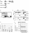
Nuclear localization of Brca1 and exogenous BARD1 peptides. Cytoplasmic (C) and nuclear (N) protein fractions were prepared from Brca1+/+ (+/+) and Brca1−/− (−/−) cells after transient transfection with control (pCAGGS), hBARD1, hB202, or hB202-L107P expression vectors. hBARD1, hB202, and hB202-L107P, as detected by anti-FLAG-M2 antibody, are predominantly nuclear in the Brca1+/+ and Brca1−/− cells (top panel). Expression or localization of endogenous Brca1 proteins, either the 210-kDa wild-type (Brca1+/+) or the 92-kDa Brca1Δ11 (Brca1−/−), was not affected by overexpression of full-length or truncated hBARD1. Brca1 was located exclusively in the nucleus. hB202-L107P showed higher expression levels than hB202, a finding consistent with Fig. 2B. The protein amount and fractionation procedure were analyzed by staining with anti-tubulin antibody (bottom panel).
FIG. 4.
FIG. 4.
Stable expression of a truncated hBARD1 results in MMC hypersensitivity. (A) Comparison of hB202 expression in Brca1+/+ cells after transient transfection with the empty vector pCAGGs (lane 1), hB202 (lane 2), and cells selected for stable hB202 expression (lane 3, clone 6B, and lane 4, clone 9B). Multiple hygromycin-resistant cell lines that contained a stable integration of the hB202 construct were analyzed for expression of the hB202 peptide by Western analysis. None of the isolated cell lines exhibited strong expression of the hB202 peptide (representative clones, lanes 3 and 4), as was typically observed for transiently transfected cells (lane 2). (B) Clonogenic survival of parental control cells (▪), two independently isolated cell lines stably expressing hB202 (6B [▴] and 9B [▾]) and Brca1−/− cells (⧫) were assessed after treatment with two doses of MMC. At the higher 0.6 μM MMC concentration, the cells that stably express low levels of hB202 were significantly more sensitive than wild-type cells (twofold; P = 0.01). At the lower 0.2 μM MMC concentration, the difference in MMC sensitivity between the cells stably expressing low levels of the hB202 compared to wild-type cell was not statistically significant (P = 0.08). The Brca1−/− cells were highly sensitive to both doses of MMC compared to the parental cells and cells that stably express low levels of hB202. The error bars represent the standard error.
FIG. 5.
FIG. 5.
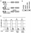
Overexpression of mutant full-length hBARD1 does not effect HDR. (A) Schematic drawing of hBARD1, hBARD1 with a RING domain mutation (hBARD1-C83G) and the hBARD1 tumor-associated point mutation (hBARD1-Q564H). The Q564H mutation is located between the ankyrin and BRCT repeats. (B) As with the hB202-C83G peptide, hBARD1-C83G was expressed at lower levels compared to hBARD1 and hBARD1-Q564H as depicted in this representative Western analysis with an anti-FLAG antibody of Brca1+/+ cell lysates after transient transfection of hBARD1 expression plasmids. (C) HDR as deduced from the number of GFP-positive cells after transient cotransfection with I-SceI and the constructs described above. The hBARD1-C83G and h BARD1-Q564H mutations did not decrease HDR in Brca1+/+ cells (open bars) or the Brca1−/− (gray bars). The error bars represent the standard error.

Similar articles

Cited by

References

    1. Ayi, T. C., J. T. Tsan, L. Y. Hwang, A. M. Bowcock, and R. Baer. 1998. Conservation of function and primary structure in the Brca1-associated ring domain (Bard1) protein. Oncogene 17:2143-2148. - PubMed
    1. Baer, R., and T. Ludwig. 2002. The BRCA1/BARD1 heterodimer, a tumor suppressor complex with ubiquitin E3 ligase activity. Curr. Opin. Genet. Dev. 12:86-91. - PubMed
    1. Bhattacharyya, A., U. S. Ear, B. H. Koller, R. R. Weichselbaum, and D. K. Bishop. 2000. The breast cancer susceptibility gene BRCA1 is required for subnuclear assembly of Rad51 and survival following treatment with the DNA cross-linking agent cisplatin. J. Biol. Chem. 275:23899-23903. - PubMed
    1. Brodie, S. G., X. Xu, W. Qiao, W. M. Li, L. Cao, and C. X. Deng. 2001. Multiple genetic changes are associated with mammary tumorigenesis in Brca1 conditional knockout mice. Oncogene 20:7514-7523. - PubMed
    1. Brzovic, P. S., J. E. Meza, M. C. King, and R. E. Klevit. 2001. BRCA1 RING domain cancer-predisposing mutations. Structural consequences and effects on protein-protein interactions. J. Biol. Chem. 276:41399-41406. - PubMed

Publication types

MeSH terms

  NODES
Association 1
Note 1
twitter 2