Comparative Study
. 2003 Dec;140(8):1351-62.
doi: 10.1038/sj.bjp.0705529. Epub 2003 Nov 24.

Anandamide initiates Ca(2+) signaling via CB2 receptor linked to phospholipase C in calf pulmonary endothelial cells

Affiliations
Comparative Study

Anandamide initiates Ca(2+) signaling via CB2 receptor linked to phospholipase C in calf pulmonary endothelial cells

Cristina Zoratti et al. Br J Pharmacol. 2003 Dec.

Abstract

The endocannabinoid anandamide has been reported to affect neuronal cells, immune cells and smooth muscle cells via either CB1 or CB2 receptors. In endothelial cells, the receptors involved in activating signal transduction are still unclear, despite the fact that anandamide is produced in this cell type. The present study was designed to explore in detail the effect of this endocannabinoid on Ca2+ signaling in single cells of a calf pulmonary endothelial cell line. Anandamide initiated a transient Ca2+ elevation that was prevented by the CB2 receptor antagonist SR144528, but not by the CB1 antagonist SR141716A. These data were confirmed by molecular identification of the bovine CB2 receptor in these endothelial cells by partial sequencing. The phospholipase C inhibitor 1-[6-[[(17beta)-3-methoxyestra-1,3,5(10)-trien-17-yl]amino]hexyl]-1H-pyrrole-2,5dione and the inositol 1,4,5-trisphosphate receptor antagonist 2-aminoethoxydiphenylborate prevented Ca2+ signaling in response to anandamide. Using an improved cameleon probe _targeted to the endoplasmic reticulum (ER), fura-2 and ratiometric-pericam, which is _targeted to the mitochondria, anandamide was found to induce Ca2+ depletion of the ER accompanied by the activation of capacitative Ca2+ entry (CCE) and a transient elevation of mitochondrial Ca2+. These data demonstrate that anandamide stimulates the endothelial cells used in this study via CB2 receptor-mediated activation of phospholipase C, formation of inositol 1,4,5-trisphosphate, Ca2+ release from the ER and subsequent activation of CCE. Moreover, the cytosolic Ca2+ elevation was accompanied by a transient Ca2+ increase in the mitochondria. Thus, in addition to its actions on smooth muscle cells, anandamide also acts as a powerful stimulus for endothelial cells.


Figures
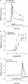
Figure 1
Figure 1
Anandamide evoked increases in [Ca2+]cyto in a calf pulmonary endothelial cell line. Panel a: Comparison of the effect of anandamide on [Ca2+]cyto in CPAE cells with that of bradykinin. As indicated, cells were stimulated with 10 μM anandamide (continuous lines and open circles, n=23) or 0.1 μM bradykinin (dotted lines and closed circles, n=25) in the presence of 2 mM extracellular Ca2+. *P<0.001 vs the effect of anandamide. Panel b: Concentration–response curves for anandamide (continuous curve and open circles each point, n=10) and its analogue methandamide (dotted curve and closed circles; n=8) in a physiological salt solution containing 2 mM CaCl2. Points represent the peak [Ca2+]cyto response at each concentration. *P<0.001 vs effect of anandamide at the same concentration. Panel c: Effect of 10 μM anandamide in cells prestimulated with 10 μM arachidonic acid (dotted lines and closed circles, n=26) or solvent (0.1% ethanol, continuous lines and open circles, n=18) as indicated by the bars.
Figure 2
Figure 2
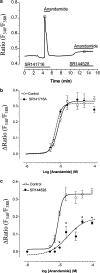
CB2 receptor antagonist SR144528 diminished the effect of anandamide on endothelial [Ca2+]cyto, but the CB1 receptor antagonist SR141716A did not. Panel a: Concentration–response curves of anandamide in the absence (continuous curve and open circles, n=10) and the presence of 1 μM SR141716 (CB1 antagonist; dotted curve and closed circles, n=7). Panel b: CPAE cells were stimulated with 10 μM anandamide in the presence of 1 μM SR141716A as indicated. After washout, the same cells were activated again with 10 μM anandamide in the presence of 1 μM SR144528 (n=7). Panel c: Concentration–response curves of anandamide in the absence (continuous curve and open circles, n=10) and presence of 1 μM SR144528 (dotted curve and closed circles, n=7). Owing to the potential effects of the solvent at higher anandamide concentrations, a maximal concentration of 100 μM anandamide was used. Points represent the peak response at the given anandamide concentration expressed as the change (Δ) in the fluorescence ratio (F340/F380). *P<0.001 vs effect of anandamide alone.
Figure 3
Figure 3
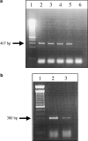
RT–PCR analysis of CB2 receptor expression. Panel a: mRNA expression of CB2 was determined by RT–PCR (407 bp) of mRNA from human monocytic leukemia cells (THP-1, lane 2), the human B lymphoblastoid cell line (DAUDI, lane 3), mouse spleen (lane 4), bovine spleen (reamplified with an additional 40 cycles; lane 5) and CPAE cells (reamplified with an additional 40 cycles; lane 6) as described in Methods. Lane 1 shows the 100-bp standard. Panel b: Based on the sequence analysis of the PCR product in bovine spleen, a new set of primers was designed for mRNA expression analysis of CB2 receptor in CPAE cells by RT-PCR (300 bp). Lane 1 shows the 100 bp standard, lane 2 is bovine spleen and lane 3 represents CPAE cells. PCR products were separated on 2% TAE–agarose.
Figure 4
Figure 4
Sequence alignment of parts of the human, mouse, bovine spleen and CPAE CB2 receptors. The Genbank accession numbers are gi 407806 for human CB2 and gi 791081 for mouse CB2; the bovine sequence has not yet been published. Bases in the bovine sequence that are identical to either or both of the human and murine sequences are indicated in bold. Bases highlighted in gray indicate regions that are conserved in the human and mouse sequences, but differ from the bovine sequence.
Figure 5
Figure 5
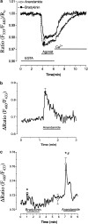
Measurements of [Ca2+]cyto during anandamide- and bradykinin-initiated intracellular Ca2+ release and CCE. Representative recordings of the effect of 10 μM anandamide (continuous lines and open circles, n=8) on intracellular Ca2+ release in the absence of extracellular Ca2+ and following the addition of 2 mM extracellular Ca2+ at the times indicated. After approximately 10 min in a Ca2+-containing solution, the same experiment was repeated with 0.1 μM bradykinin (dotted lines and closed circles, n=8). Except for the faster recovery following the addition of Ca2+ in anandamide-stimulated cells compared with that found in cells stimulated with bradykinin (*P<0.01), no differences were found. Results were identical when the order of testing the autacoids was reversed.
Figure 6
Figure 6
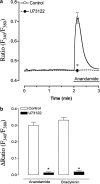
Effect of anandamide on endothelial [Ca2+]cyto was prevented by U73122, an inhibitor of phospholipase C. Panel a: Single endothelial cells were stimulated with 10 μM anandamide in the absence (continuous lines and open circles, n=32) and presence of 2 μM U73122 (dotted lines and closed circles, n=29). Points represent the means±s.e.m. *P<0.0001 vs in the absence of U73122. Panel b: Comparison of the effects of 10 μM anandamide and 0.1 μM bradykinin on endothelial [Ca2+]cyto in the absence (white columns; anadamide n=33, bradykinin n=41) and presence of 2 μM U73122 (black columns; anadamide n=29, bradykinin n=31). *P<0.0001 vs in the absence of U73122.
Figure 7
Figure 7
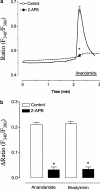
The inhibitor of inositol 1,4,5-trisphosphate receptors 2-APB prevented the anandamide-induced elevation in [Ca2+]cyto in CPAE cells. Panel a: Single endothelial cells were stimulated with 10 μM anandamide in the absence (continuous lines and open circles, n=24) and presence of 50 μM 2-APB (dotted lines and closed circles, n=29). *P < 0.0001 vs in the absence of 2-APB. Panel b: Comparison of cytosolic Ca2+ response to 10 μM anandamide and 0.1 μM bradykinin stimulation in the absence (white columns; anadamide n=40, bradykinin n=36) and presence of 2 μM U73122 (black columns; anadamide n=29, bradykinin n=34). *P<0.0001 vs in the absence of 2-APB.
Figure 8
Figure 8
Anandamide initiated Ca2+ depletion of the ER and evoked a transient mitochondrial Ca2+ signal in CPAE cells. Endothelial cells were transiently transfected with vYC4-ER (panel a) or ratiometric-pericam-mt (panel b). Panel a: As indicated by the bars, cells were stimulated with 20 μM anandamide or 0.1 μM bradykinin and the effect on the ER Ca2+ content (n=5; panel a) was monitored. Panel b: Transiently transfected endothelial cells were stimulated with 20 μM anandamide and the mitochondrial Ca2+ concentration (n=8) was followed as described in Methods. Panel c: Comparison of the effect of maximally effective concentrations of bradykinin (0.1 μM) and anandamide (20 μM) on [Ca2+]mito. As indicated by the bars, cells were stimulated with bradykinin and anandamide in the presence of extracellular Ca2+ (n=8). *P<0.01 vs basal and #P<0.01 vs the effect of bradykinin.

Similar articles

Cited by

References

    1. ASCHNER J.L., LUM H., FLETCHER P.W., MALIK A.B. Bradykinin- and thrombin-induced increases in endothelial permeability occur independently of phospholipase C but require protein kinase C activation. J. Cell. Physiol. 1997;173:387–396. - PubMed
    1. BUKOSKI R., BÁTKAI S., JÁRAI Z., WANG Y., OFFERTALER L., JACKSON W.F., KUNOS K. CB1 receptor antagonist SR141716A inhibits Ca2+-induced relaxation in CB1 receptor-deficient mice. Hypertension. 2002;39:251–257. - PMC - PubMed
    1. CHANTAIGNEAU T., FÉLÉTOU M., THOLLON C., VILLENEUVE N., VILAINE J.-P., DUHALT J., VANHOUTTE P.M. Cannabinoid CB1 receptor and endothelium-dependent hyperpolarization in guinea-pig carotid, rat mesenteric and porcine coronary arteries. Br. J. Pharmacol. 1998;123:968–974. - PMC - PubMed
    1. CHAYTOR A.T., MARTIN P.E.M., EVANS W.H., RANDALL M.D., GRIFFITH T.M. The endothelial component of cannabinoid-induced relaxations in rat mesenteric artery depends on gap junctional communication but not nitric oxide. J. Physiol. 1999;520:539–550. - PMC - PubMed
    1. DEDKOVA E.N., BLATTER L.A. Nitric oxide inhibits capacitative Ca2+ entry and enhances endoplasmic reticulum Ca2+ uptake in bovine vascular endothelial cells. J. Physiol. 2002;539:77–91. - PMC - PubMed

Publication types

MeSH terms

LinkOut - more resources

  NODES
twitter 2