Requirement of matrix metalloproteinase-9 for the transformation of human mammary epithelial cells by Stat3-C
- PMID: 15249664
- PMCID: PMC489981
- DOI: 10.1073/pnas.0404100101
Requirement of matrix metalloproteinase-9 for the transformation of human mammary epithelial cells by Stat3-C
Abstract
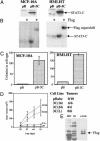
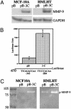
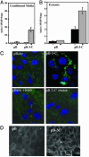
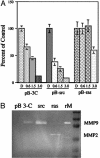
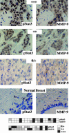
Persistently activated Stat3 is found in many different cancers, including approximately 60% of breast tumors. Here, we demonstrate that a constitutively activated Stat3 transforms immortalized human mammary epithelial cells and that this oncogenic event requires the activity of matrix metalloproteinase-9 (MMP-9). By immunohistochemical analysis, we observe a positive correlation between strong MMP-9 expression and tyrosine phosphorylated Stat3 in primary breast cancer specimens. These results demonstrate a relationship between activated Stat3 and MMP-9 in breast oncogenesis.
Figures
Similar articles
-
CCAAT/Enhancer binding protein delta (c/EBPdelta) regulation and expression in human mammary epithelial cells: I. "Loss of function" alterations in the c/EBPdelta growth inhibitory pathway in breast cancer cell lines.J Cell Biochem. 2004 Nov 1;93(4):830-43. doi: 10.1002/jcb.20223. J Cell Biochem. 2004. PMID: 15389879
-
Stable expression of constitutively-activated STAT3 in benign prostatic epithelial cells changes their phenotype to that resembling malignant cells.Mol Cancer. 2005 Jan 12;4(1):2. doi: 10.1186/1476-4598-4-2. Mol Cancer. 2005. PMID: 15647107 Free PMC article.
-
Progestins induce transcriptional activation of signal transducer and activator of transcription 3 (Stat3) via a Jak- and Src-dependent mechanism in breast cancer cells.Mol Cell Biol. 2005 Jun;25(12):4826-40. doi: 10.1128/MCB.25.12.4826-4840.2005. Mol Cell Biol. 2005. PMID: 15923602 Free PMC article.
-
The Multifaceted Role of STAT3 in Mammary Gland Involution and Breast Cancer.Int J Mol Sci. 2018 Jun 7;19(6):1695. doi: 10.3390/ijms19061695. Int J Mol Sci. 2018. PMID: 29875329 Free PMC article. Review.
-
Mammary epithelial cell transformation: insights from cell culture and mouse models.Breast Cancer Res. 2005;7(4):171-9. doi: 10.1186/bcr1275. Epub 2005 Jun 3. Breast Cancer Res. 2005. PMID: 15987472 Free PMC article. Review.
Cited by
-
Anticancer effect of salidroside on colon cancer through inhibiting JAK2/STAT3 signaling pathway.Int J Clin Exp Pathol. 2015 Jan 1;8(1):615-21. eCollection 2015. Int J Clin Exp Pathol. 2015. PMID: 25755753 Free PMC article.
-
The complementary roles of STAT3 and STAT1 in cancer biology: insights into tumor pathogenesis and therapeutic strategies.Front Immunol. 2023 Nov 8;14:1265818. doi: 10.3389/fimmu.2023.1265818. eCollection 2023. Front Immunol. 2023. PMID: 38022653 Free PMC article. Review.
-
leptin-induced growth stimulation of breast cancer cells involves recruitment of histone acetyltransferases and mediator complex to CYCLIN D1 promoter via activation of Stat3.J Biol Chem. 2007 May 4;282(18):13316-25. doi: 10.1074/jbc.M609798200. Epub 2007 Mar 7. J Biol Chem. 2007. PMID: 17344214 Free PMC article.
-
Identification of a structural motif in the tumor-suppressive protein GRIM-19 required for its antitumor activity.Am J Pathol. 2010 Aug;177(2):896-907. doi: 10.2353/ajpath.2010.091280. Epub 2010 Jul 1. Am J Pathol. 2010. PMID: 20595633 Free PMC article.
-
Inflammation: a driving force speeds cancer metastasis.Cell Cycle. 2009 Oct 15;8(20):3267-73. doi: 10.4161/cc.8.20.9699. Epub 2009 Oct 3. Cell Cycle. 2009. PMID: 19770594 Free PMC article. Review.
References
-
- Darnell, J. E., Jr. (1997) Science 277, 1630–1635. - PubMed
-
- Garcia, R., Bowman, T. L., Niu, G., Yu, H., Minton, S., Muro-Cacho, C. A., Cox, C. E., Falcone, R., Fairclough, R., Parsons, S., et al. (2001) Oncogene 20, 2499–2513. - PubMed
-
- Bromberg, J., Wrzeszczynska, M., Devgan, G., Zhao, Y., Albanese, C., Pestell, R. & Darnell, J. E. J. (1999) Cell 98, 295–303. - PubMed
-
- Dolled-Filhart, M., Camp, R. L., Kowalski, D. P., Smith, B. L. & Rimm, D. L. (2003) Clin. Cancer Res. 9, 594–600. - PubMed
Publication types
MeSH terms
Substances
Grants and funding
LinkOut - more resources
Full Text Sources
Other Literature Sources
Miscellaneous

