. 2004 Sep 27;166(7):1041-54.
doi: 10.1083/jcb.200406060.

Presenilin 1 mediates the turnover of telencephalin in hippocampal neurons via an autophagic degradative pathway

Affiliations

Presenilin 1 mediates the turnover of telencephalin in hippocampal neurons via an autophagic degradative pathway

Cary Esselens et al. J Cell Biol. .

Abstract

Presenilin 1 (PS1) interacts with telencephalin (TLN) and the amyloid precursor protein via their transmembrane domain (Annaert, W.G., C. Esselens, V. Baert, C. Boeve, G. Snellings, P. Cupers, K. Craessaerts, and B. De Strooper. 2001. Neuron. 32:579-589). Here, we demonstrate that TLN is not a substrate for gamma-secretase cleavage, but displays a prolonged half-life in PS1(-/-) hippocampal neurons. TLN accumulates in intracellular structures bearing characteristics of autophagic vacuoles including the presence of Apg12p and LC3. Importantly, the TLN accumulations are suppressed by adenoviral expression of wild-type, FAD-linked and D257A mutant PS1, indicating that this phenotype is independent from gamma-secretase activity. Cathepsin D deficiency also results in the localization of TLN to autophagic vacuoles. TLN mediates the uptake of microbeads concomitant with actin and PIP2 recruitment, indicating a phagocytic origin of TLN accumulations. Absence of endosomal/lysosomal proteins suggests that the TLN-positive vacuoles fail to fuse with endosomes/lysosomes, preventing their acidification and further degradation. Collectively, PS1 deficiency affects in a gamma-secretase-independent fashion the turnover of TLN through autophagic vacuoles, most likely by an impaired capability to fuse with lysosomes.


Figures

Figure 1.
Figure 1.
Full-length TLN (but not APP) accumulates in a distinct compartment in PS1 / hippocampal neurons. Double-immunofluorescence staining for endogenous TLN (green, B36.1) and APP (red, mAb C1 6.1) in wild-type (top) and PS1−/− (bottom) hippocampal neurons (15-d culture). The inset shows an overview of the neuron. In wild-type neurons TLN labeling is essentially localized to the somatodendritic plasmalemma in contrast to APP, which is found in intracellular compartments. In addition, PS1 deficiency causes TLN, but not APP, to cluster in large somatic accumulations. Bars, 10 μm.
Figure 2.
Figure 2.
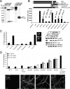
TLN lacks the characteristics of a γ-secretase substrate. (A) Western blot analysis of endogenous TLN and APP in wild-type and PS1−/− hippocampal neurons. PS1 deficiency results in the accumulation of APP-CTF. No TLN-CTF (estimated ± 7 kD) was detected, neither was it detected after SFV-induced exogenous overexpression. Also, overexpression of a TLNΔE fragment did not generate a PS1-dependent TLN intracellular domain. (B) Different constructs fused to the Gal4VP16 domain were used in a γ-secretase reporter assay. Control transfections (pIPspAdApt empty vector and pIPspAdApt-APP without Gal4VP16) show no luciferase activity. APP-Gal4VP16 and NotchΔE-Gal4VP16 activate the luciferase gene upon cleavage and are inhibited by DAPT, L658,458, or compound 32 (125 nM). No luciferase activity was detected using TLNΔE or APP in which the transmembrane domain was replaced by that of TLN (APP/TLNTMR chimaera), indicating that this region is not cleaved by γ-secretase. (C) Left: γ-secretase inhibitors do not induce the formation of TLN accumulations. Wild-type hippocampal neurons were treated with γ-secretase inhibitors (125 nM, daily from d 7–14 post-plating), fixed, and immunostained for TLN and the percentage of neurons with TLN accumulations was scored. Culturing neurons for 25 d increases the size and number (insets), but not the frequency of TLN accumulations (mean ± SEM, n = 3, 200–700 neurons per time point). Right: Western blot showing that only APP-CTF accumulates after chronic treatment with γ-secretase inhibitors. (D) Adenoviral reintroduction of human PS1 suppresses TLN accumulations. 30–35% of PS1−/− neurons display TLN accumulations, in contrast to <5% in PS1+/+ and +/− cultures as well as in cultures expressing human PS1 in a PS1−/− background. However, suppression of TLN accumulations was obtained by adenoviral introduction of human PS1. A clear dose-dependent inhibition was seen with increasing MOI of adenoviral (AV) human PS1, but also the familial Alzheimer's disease-linked PS1G384A and dominant-negative PS1D257A mutants. Control adenoviral infections with eGFP did not have a significant effect. After 9–11 d post-infection (15 d post-plating), neurons displayed significant PS1 protein expression as demonstrated by double staining for TLN and human PS1(mAb 5.2). Bars, 20 μm.
Figure 3.
Figure 3.
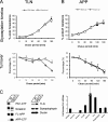
Turnover of full-length TLN is altered in PS1 / neurons. Wild-type and PS1−/− cortical neurons were transduced with SFV-TLN (A) or -APP (B), pulse labeled for 15 min with [35S]methionine, and chased for different time periods. Immunoprecipitated TLN and APP were treated with EndoH and analyzed by SDS-PAGE and phosphorimaging. The accumulation of an EndoH-resistant band indicates progressive maturation during Golgi passage; however, the ratio of EndoH-resistant to -sensitive TLN (A, top) and APP (B, top) revealed no difference (mean ± SEM, n = 3). Instead, the half-life of overexpressed TLN (A, bottom), but not full-length APP (B, bottom), is significantly prolonged in PS1−/− neurons (mean ± SEM, n = 3). (C) Wild-type and PS1−/− hippocampal neurons (12 d) were scraped, pelleted, and treated with 10 mU EndoH, followed by Western blot detection of APP, TLN, PS1, nicastrin (NCT), synaptophysin, and ductin. TLN levels are increased almost fourfold in PS1−/− neurons. No EndoH-sensitive TLN was detected in PS1−/− neurons, indicating a complete maturation. APP-CTF shows a dramatic accumulation (44-fold; mean ± SEM, n = 3).
Figure 4.
Figure 4.
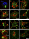
TLN accumulation occurs in a compartment distinct from classic biosynthetic and endosomal pathways. (A–F) Early compartments. No colocalization was observed between TLN (green in all panels) and nuclei (A, TOPRO-3), ER-markers BIP (B) and calnexin (C), ERGIC-53 (D), β-COP (E) or GM130 (F), and labeling cis-Golgi. (G–L) Late compartments. TLN accumulations are not accessible for exogenous biotin (G). Early and recycling endosomes (H, EEA1) and transferrin receptor (I, TFR), late endosomes (J, LBPA), and lysosomes (K, Lamp-2; L, catD) also stained negative for TLN. Vertical sections (x–z, arrowheads in I and J) clearly distinguish TLN accumulations from recycling (I, TFR) and late endosomes (J, LBPA). Bars, 10 μm.
Figure 5.
Figure 5.
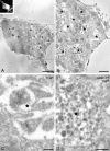
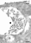
Correlative light immuno-EM analysis of TLN accumulations. (A) Insert: overview of the selected neuron as seen by immunocytochemistry. The bright signal for TLN reflects the numerous membranous accumulations seen at the ultrastructural immuno-EM level (star). (B) Higher magnification of the boxed area in A. (C) High magnification of the boxed structure seen in B. 10-nm gold particles label TLN. Note the complex composition of the internal membranes of the TLN-positive compartments. (D) Example of a TLN (15-nm gold)-containing structure obtained by the conventional cryosectioning technique. Note some heterogeneity in the internal membranes between C and D. Lamp-1 (10-nm gold) is absent from the TLN-containing compartment. Arrows point to invaginations of the cytoplasm. N, nucleus; P, plasma membrane. Bars: A, 5 μm; B, 2 μm; C, 500 nm; D, 200 nm.
Figure 6.
Figure 6.
TLN accumulations are distinct from lysosomes. A TLN (15-nm gold)-containing structure as obtained by the conventional cryosectioning technique double labeled with Lamp-1 (10-nm gold). The TLN-positive compartment is clearly distinct in size and from lysosomes (L) and lacks Lamp-1. P, plasma membrane. Bar, 200 nm.
Figure 7.
Figure 7.
TLN accumulations have characteristics of autophagic vacuoles. (A) Wild-type and PS1−/− hippocampal neurons (15 d) were incubated with 50 mM MDC, fixed, and counterstained with anti-TLN (red). Uptake of MDC was only apparent in PS1−/− neurons where it colocalized with TLN accumulations (arrowheads). Bar, 10 μm. (B and C) Double immunostaining of wild-type and PS1−/− hippocampal neurons for TLN (green) and autophagic vacuole-associated Apg12p (B) and LC3 (C). To detect both sets of pAbs, an alternative method was developed using biotinylated anti-TLN (see Materials and methods). Each experiment included controls for specificity (not depicted). In wild-type neurons, both Apg12p and LC3 (B and C, top row) were broadly distributed in cell bodies and neurites, but did not colocalize with TLN. Colocalization was only observed in multiple TLN accumulations in PS1−/− hippocampal neurons (second row in B and C, respectively). For Apg12p, coimmunostaining was also noticed in small discrete structures in the proximal dendrites (B, bottom row). Bars, 10 μm.
Figure 7.
Figure 7.
TLN accumulations have characteristics of autophagic vacuoles. (A) Wild-type and PS1−/− hippocampal neurons (15 d) were incubated with 50 mM MDC, fixed, and counterstained with anti-TLN (red). Uptake of MDC was only apparent in PS1−/− neurons where it colocalized with TLN accumulations (arrowheads). Bar, 10 μm. (B and C) Double immunostaining of wild-type and PS1−/− hippocampal neurons for TLN (green) and autophagic vacuole-associated Apg12p (B) and LC3 (C). To detect both sets of pAbs, an alternative method was developed using biotinylated anti-TLN (see Materials and methods). Each experiment included controls for specificity (not depicted). In wild-type neurons, both Apg12p and LC3 (B and C, top row) were broadly distributed in cell bodies and neurites, but did not colocalize with TLN. Colocalization was only observed in multiple TLN accumulations in PS1−/− hippocampal neurons (second row in B and C, respectively). For Apg12p, coimmunostaining was also noticed in small discrete structures in the proximal dendrites (B, bottom row). Bars, 10 μm.
Figure 8.
Figure 8.
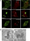
CatD / hippocampal neurons as a model system for autophagic vacuole accumulation. (A) Intracellular sorting and maturation of catD. Wild-type and PS1−/− neurons were metabolically labeled and chased for the indicated time periods. Phosphorimaging of immunoprecipitated radiolabeled catD did not reveal a difference in the ratio of proteolytically processed 46-kD intermediate vs. 52-kD precursor (mean ± SEM, n = 3). (B–D) TLN localizes to autophagic vacuoles of catD −/− hippocampal neurons. Lysotracker staining revealed sparsely distributed small-sized organelles in wild-type and PS1−/− neurons in contrast to catD−/− neurons where high numbers of large Lysotracker-positive organelles were found (B, arrowheads). Although TLN accumulations in PS1−/− were fully negative for Lysotracker, some acidic organelles tended to closely associate (C, arrowheads, middle). In catD−/− neurons, TLN was clearly detected in the large acidic organelles (C, bottom). Bars, 20 μm. (D) At the immuno-EM level, the large acidic organelles seen in catD−/− neurons represent dense autophagic vacuole-like structures (star) that label positive for TLN (10-nm gold). M, mitochondrion. Bars, 200 nm.
Figure 8.
Figure 8.
CatD / hippocampal neurons as a model system for autophagic vacuole accumulation. (A) Intracellular sorting and maturation of catD. Wild-type and PS1−/− neurons were metabolically labeled and chased for the indicated time periods. Phosphorimaging of immunoprecipitated radiolabeled catD did not reveal a difference in the ratio of proteolytically processed 46-kD intermediate vs. 52-kD precursor (mean ± SEM, n = 3). (B–D) TLN localizes to autophagic vacuoles of catD −/− hippocampal neurons. Lysotracker staining revealed sparsely distributed small-sized organelles in wild-type and PS1−/− neurons in contrast to catD−/− neurons where high numbers of large Lysotracker-positive organelles were found (B, arrowheads). Although TLN accumulations in PS1−/− were fully negative for Lysotracker, some acidic organelles tended to closely associate (C, arrowheads, middle). In catD−/− neurons, TLN was clearly detected in the large acidic organelles (C, bottom). Bars, 20 μm. (D) At the immuno-EM level, the large acidic organelles seen in catD−/− neurons represent dense autophagic vacuole-like structures (star) that label positive for TLN (10-nm gold). M, mitochondrion. Bars, 200 nm.
Figure 9.
Figure 9.
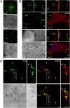
TLN mediates phagocytic uptake of microbeads in hippocampal neurons. (A) Microbeads align along neurites of hippocampal neurons as demonstrated by phase contrast (DIC). Already after 4 h, many beads stain positive for TLN (arrowheads, top). At 48 h (bottom), most TLN immunoreactivity was associated with microbeads at the expense of its typical plasma membrane staining. *, cell body. Note that microbeads were not found associated with preexisting TLN accumulations (asterisk in top panels, 4 h). (B) Microbeads (24 h) accumulate TLN and actin, as shown by phalloidin-Alexa 568. An overview of the neuron is given in each top left inset, next to two detailed areas (white boxes). The differential interference contrast (DIC) clearly demonstrates a complete colocalization of microbeads with TLN and phalloidin (arrowheads). TOPRO-3 (blue) marks the nucleus (see also Fig. S1, available at http://www.jcb.org/cgi/content/full/jcb.200406060/DC1). (C) Similar as in B, but immunostained for TLN and endogenous PIP2. The overview demonstrates the overall recruitment of PIP2 to microbeads (arrows). The parallel insets show a detailed area (white box) with PIP2 being colocalized with TLN on individual microbeads (arrowheads). Note that PIP2 occasionally colocalizes with TLN at the plasma membrane (asterisk). Bars (A–C), 10 μm.

Similar articles

Cited by

References

    1. Annaert, W., and B. De Strooper. 2002. A cell biological perspective on Alzheimer's disease. Annu. Rev. Cell Dev. Biol. 18:25–51. - PubMed
    1. Annaert, W.G., L. Levesque, K. Craessaerts, I. Dierinck, G. Snellings, D. Westaway, P.S. George-Hyslop, B. Cordell, P. Fraser, and B. De Strooper. 1999. Presenilin 1 controls γ-secretase processing of the amyloid precursor protein in pre-Golgi compartments of hippocampal neurons. J. Cell Biol. 147:277–294. - PMC - PubMed
    1. Annaert, W.G., C. Esselens, V. Baert, C. Boeve, G. Snellings, P. Cupers, K. Craessaerts, and B. De Strooper. 2001. Interaction with telencephalin and the amyloid precursor protein predicts a ring structure for presenilins. Neuron. 32:579–589. - PubMed
    1. Botelho, R.J., M. Teruel, R. Dierckman, R. Anderson, A. Wells, J.D. York, T. Meyer, and S. Grinstein. 2000. Localized biphasic changes in phosphatidylinositol-4,5-bisphosphate at sites of phagocytosis. J. Cell Biol. 151:1353–1368. - PMC - PubMed
    1. Cai, D., J.Y. Leem, J.P. Greenfield, P. Wang, B.S. Kim, R. Wang, K.O. Lopes, S.H. Kim, H. Zheng, P. Greengard, et al. 2003. Presenilin-1 regulates intracellular trafficking and cell surface delivery of β-amyloid precursor protein. J. Biol. Chem. 278:3446–3454. - PubMed

Publication types

MeSH terms

Substances

  NODES
INTERN 2
Note 4
twitter 2