Molecular genetic evidence for different clonal origins of epithelial and stromal components of phyllodes tumor of the prostate
- PMID: 15466403
- PMCID: PMC1618623
- DOI: 10.1016/S0002-9440(10)63397-4
Molecular genetic evidence for different clonal origins of epithelial and stromal components of phyllodes tumor of the prostate
Abstract
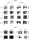
Phyllodes tumor of the prostate is a rare neoplasm, composed of epithelium-lined cysts and channels embedded in a variably cellular stroma. The pathogenetic relationship of the epithelium and stroma is unknown and whether each is a clonal neoplastic element is uncertain. We studied the clonality of phyllodes tumors from six patients who underwent either enucleation or transurethral resection as their initial treatment. This was followed by total prostatectomy in three of the patients. Laser-assisted microdissection was performed to extract epithelial and stromal components of phyllodes tumor from formalin-fixed, paraffin-embedded tissue. Polymerase chain reaction was used to amplify genomic DNA at specific loci on chromosome 7q31 (D7S522), 8p21.3-q11.1 (D8S133, D8S137), 8p22 (D8S261), 10q23 (D10S168, D10S571), 17p13 (TP53), 16q23.2 (D16S507), 12q11-12 (D12S264), 17q (D17S855), 18p11.22-p11 (D18S53), and 22q11.2 (D22S264). In each tumor, stroma and epithelium were analyzed separately. Gel electrophoresis with autoradiography was used to detect loss of heterozygosity. All tumors showed allelic loss in one or more loci of both the epithelial and stromal components. The frequency of allelic loss in the epithelial component was 2 of 5 (40%) at D7S522, 2 of 6 (33%) at D8S133, 1 of 5 (20%) at D8S137, 3 of 6 (50%) at D8S261, 4 of 4 (100%) at D10S168, 4 of 6 (67%) at TP53, 2 of 6 (33%) at D10S571, 6 of 6 (100%) at D16S507, 1 of 5 (20%) at D12S264, 1 of 6 (17%) at D17S855, 2 of 6 (33%) at D18S53, and 2 of 5 (40%) at D22S264. The frequency of allelic loss in the stromal component was 2 of 5 (40%) at D7S522, 1 of 6 (17%) at D8S133, 2 of 5 (40%) at D8S137, 3 of 6 (50%) at D8S261, 1 of 4 (25%) at D10S168, 3 of 6 (50%) at TP53, 2 of 6 (33%) at D10S571, 3 of 6 (50%) at D16S507, 1 of 5 (20%) at D12S264, 0 of 6 (0%) at D17S855, 1 of 6 (17%) at D18S53, and 0 of 5 (0%) at D22S264. The pattern of allelic loss is significantly different in both stroma and epithelium statistically; completely concordant allelic loss patterns were not seen in any tumor examined. Our data demonstrate that both epithelial and stromal components of phyllodes tumor of the prostate are clonal, supporting the hypothesis that both elements are neoplastic. While both epithelium and stroma are clonal proliferations, they appear to have different clonal origins.
Figures


Similar articles
-
Clonal divergence and genetic heterogeneity in clear cell renal cell carcinomas with sarcomatoid transformation.Cancer. 2005 Sep 15;104(6):1195-203. doi: 10.1002/cncr.21288. Cancer. 2005. PMID: 16047350
-
Evidence for transformation of fibroadenoma of the breast to malignant phyllodes tumor.Appl Immunohistochem Mol Morphol. 2009 Jul;17(4):345-50. doi: 10.1097/PAI.0b013e318194d992. Appl Immunohistochem Mol Morphol. 2009. PMID: 19276971
-
Molecular genetic evidence for the independent origin of multifocal papillary tumors in patients with papillary renal cell carcinomas.Clin Cancer Res. 2005 Oct 15;11(20):7226-33. doi: 10.1158/1078-0432.CCR-04-2597. Clin Cancer Res. 2005. PMID: 16243792
-
[Recent advances in molecular pathology of phyllodes tumor of breast].Zhonghua Bing Li Xue Za Zhi. 2011 Feb;40(2):135-7. Zhonghua Bing Li Xue Za Zhi. 2011. PMID: 21426820 Review. Chinese. No abstract available.
-
[Phyllodes tumor of the prostate: a case report].Nihon Hinyokika Gakkai Zasshi. 2002 Jan;93(1):52-7. doi: 10.5980/jpnjurol1989.93.52. Nihon Hinyokika Gakkai Zasshi. 2002. PMID: 11842541 Review. Japanese.
Cited by
-
The steroid receptor coactivator-3 is required for developing neuroendocrine tumor in the mouse prostate.Int J Biol Sci. 2014 Oct 2;10(10):1116-27. doi: 10.7150/ijbs.10236. eCollection 2014. Int J Biol Sci. 2014. PMID: 25332686 Free PMC article.
-
Microenvironmental regulators of tissue structure and function also regulate tumor induction and progression: the role of extracellular matrix and its degrading enzymes.Cold Spring Harb Symp Quant Biol. 2005;70:343-56. doi: 10.1101/sqb.2005.70.013. Cold Spring Harb Symp Quant Biol. 2005. PMID: 16869771 Free PMC article.
-
Epithelial-mesenchymal transition: a cancer researcher's conceptual friend and foe.Am J Pathol. 2009 May;174(5):1588-93. doi: 10.2353/ajpath.2009.080545. Epub 2009 Mar 26. Am J Pathol. 2009. PMID: 19342369 Free PMC article. Review.
-
Epithelial-to-mesenchymal transition in prostate cancer: paradigm or puzzle?Nat Rev Urol. 2011 Jun 21;8(8):428-39. doi: 10.1038/nrurol.2011.85. Nat Rev Urol. 2011. PMID: 21691304 Free PMC article. Review.
-
Laser capture microdissection in the genomic and proteomic era: _targeting the genetic basis of cancer.Int J Clin Exp Pathol. 2008 Mar 15;1(6):475-88. Int J Clin Exp Pathol. 2008. PMID: 18787684 Free PMC article.
References
-
- Reese JH, Lombard CM, Krone K, Stamey TA. Phyllodes type of atypical prostatic hyperplasia: a report of 3 new cases. J Urol. 1987;138:623–626. - PubMed
-
- Attah EB, Nkposong EO. Phyllodes type of atypical prostatic hyperplasia. J Urol. 1976;115:762–764. - PubMed
-
- Kevwitch MK, Walloch JL, Waters WB, Flanigan RC. Prostatic cystic epithelial-stromal tumors: a report of 2 new cases. J Urol. 1993;149:860–864. - PubMed
-
- Yokota T, Yamashita Y, Okuzono Y, Takahashi M, Fujihara S, Akizuki S, Ishihara T, Uchino F, Iwata T. Malignant cystosarcoma phyllodes of prostate. Acta Pathol Jpn. 1984;34:663–668. - PubMed
-
- Lopez-Beltran A, Gaeta JF, Huben R, Croghan GA. Malignant phyllodes tumor of prostate. Urology. 1990;35:164–167. - PubMed
Publication types
MeSH terms
Substances
LinkOut - more resources
Full Text Sources
Medical
Research Materials
Miscellaneous

