. 2004 Oct 8;16(1):23-34.
doi: 10.1016/j.molcel.2004.09.003.

Expansion and compression of a protein folding intermediate by GroEL

Affiliations

Expansion and compression of a protein folding intermediate by GroEL

Zong Lin et al. Mol Cell. .

Erratum in

  • Mol Cell. 2004 Oct 22;16(2):317

Abstract

The GroEL-GroES chaperonin system is required for the assisted folding of many essential proteins. The precise nature of this assistance remains unclear, however. Here we show that denatured RuBisCO from Rhodospirillum rubrum populates a stable, nonaggregating, and kinetically trapped monomeric state at low temperature. Productive folding of this nonnative intermediate is fully dependent on GroEL, GroES, and ATP. Reactivation of the trapped RuBisCO monomer proceeds through a series of GroEL-induced structural rearrangements, as judged by resonance energy transfer measurements between the amino- and carboxy-terminal domains of RuBisCO. A general mechanism used by GroEL to push large, recalcitrant proteins like RuBisCO toward their native states thus appears to involve two steps: partial unfolding or rearrangement of a nonnative protein upon capture by a GroEL ring, followed by spatial constriction within the GroEL-GroES cavity that favors or enforces compact, folding-competent intermediate states.


Figures
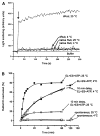
Figure 1
Figure 1. Low Temperature Suppresses High-Order Aggregation, but Not GroEL-Mediated Refolding, of Denatured RuBisCO
(A) Static light scattering of different RuBisCO samples was monitored at 90° and 340 nm. In all cases, the final protein concentration was 100 nM RuBisCO monomer. The arrow indicates the addition of either acid-urea-denatured RuBisCO (“dRub”) or native RuBisCO to standard refolding buffer equilibrated at the indicated temperature. (B) Spontaneous and GroEL-mediated refolding of RuBisCO at 4°C and 25°C. In all cases, 100 nM acid-urea-denatured RuBisCO was diluted either directly into refolding buffer (“spontaneous”) or into buffer containing 200 nM GroEL (“EL+ES+ATP”). Samples of denatured RuBisCO were also diluted into refolding buffer alone and incubated at the indicated temperature for 10 min, prior to the addition of GroEL (“10 min delay, EL+ES+ATP”). For GroEL-mediated refolding, samples were then supplemented with 400 nM GroES and 1 mM ATP to initiate folding.
Figure 2
Figure 2. GroEL Efficiently Refolds Fluorescent Derivatives of RuBisCO
(A) Ribbon diagram of the R. rubrum RuBisCO structure (9RUB [Schneider et al., 1990]) derivatized with AEDANS (a FRET donor) and fluorescein (“F”; a FRET acceptor). The fluorescent dyes were modeled into the RuBisCO structure using PyMOL (DeLano Scientific), ChemDraw, and Chem3D (CambridgeSoft). Dye orientations are for reference only and have not been energy minimized. (B) Refolding of fluorescent RuBisCO variants requires GroEL and GroES and is comparable to the refolding of wild-type, unmodified RuBisCO. Acid-urea-denatured samples of each protein (100 nM final) were mixed with 150 nM GroEL, and folding was then initiated by adding 300 nM GroES and 2 mM ATP.
Figure 3
Figure 3. The Kinetically Trapped State Populated by RuBisCO at Low Protein Concentration and Low Temperature Is Not a Low-Order Aggregate
(A) The extent of monomer-monomer contact upon formation of low-order RuBisCO aggregates was monitored by donor-side, steady-state FRET. Samples of unlabeled, donor-only (AEDANS; “58-D”)- and acceptor-only (fluorescein; “58-A”)-labeled RuBisCO were denatured in acid-urea and then mixed together in 1:1 ratios, as indicated. These concentrated, denatured samples were then diluted to 100 nM (final protein concentration) in standard refolding buffer at the indicated temperature. (B) The dependence of low-order aggregate formation on the total RuBisCO monomer concentration at 4°C is shown. The extent of FRET for each pair of donor- and acceptor-labeled monomers at each final protein concentration was determined by both steady-state (closed symbols) and time-resolved fluorescence measurements (open symbols). Three different pairs of labeled proteins were examined: (triangles) RubC58A/A454C-AEDANS (“454-D”) mixed with RubC58A/A454C-F (“454-A”); (circles) RubC58-AEDANS (“58-D”) mixed with RubC58-F (“58-A”); (squares) RubC58-AEDANS (“58-D”) RubC58A/A454C-F (“454-A”).
Figure 4
Figure 4. The Low-Temperature Kinetically Trapped RuBisCO Monomer Is Expanded and Displays a Shortened, Nonnative Distance between the N- and C-Terminal Domains
Samples of pyrene-labeled RuBisCO were denatured in acid-urea and then diluted into refolding buffer at 4°C. The anisotropy decays for 50 and 400 nM pyrene-labeled RuBisCO (“58-pyr”) are shown in (A). The global fit of this data to a rotational diffusion model containing a single rotational decay term and two lifetime components is shown. The rotational correlation times associated with overall rotational diffusion (ϕg) were 92 ± 14 ns at 50 nM and 438 ± 52 ns at 400 nM. (B) Measurement of the global rotational correlation time of the denatured, pyrene-labeled RuBisCO monomer at 4°C as a function of total protein concentration. The curve shown is present for illustration purposes only. (C and D) FRET measurements of the kinetically trapped RuBisCO monomer at 4°C. A sample of the donor-only (RubC58A/A454C-AEDANS; “454-D”)- and donor-acceptor (RubC58-F/A454C-AEDANS; “454-D/58-A”)-labeled RuBisCO were denatured in acid-urea and diluted into refolding buffer at 4°C to a final protein concentration of 100 nM. Both steady-state (C) and time-resolved (D) measurements were taken on each sample. The instrument response function (“IRF”) for the lifetime measurement is shown in (D).
Figure 5
Figure 5. Direct Binding of the Trapped RuBisCO Intermediate to a GroEL Ring Results in a Stretching of the N- to C-Terminal Domain Distance
(A) Binding of the kinetically trapped RuBisCO monomer to both wild-type GroEL and SR1 induces a significant decrease in the FRET efficiency of the labeled monomer. Samples of labeled RuBisCO were denatured in acid-urea, diluted into refolding buffer at 4°C (100 nM), and then loaded into a stopped-flow device thermally jacketed at 4°C. The kinetically trapped monomer samples were rapidly mixed 1:1 with either buffer only (“+ buffer”) or with wild-type GroEL (“+ EL”; 400 nM final concentration) or SR1 (“+ SR1”; 400 nM). The observed change in the FRET efficiency upon binding to both wild-type GroEL and SR1 required two exponential fitting components (kfast and kslow). (B) The rate of the fast change in the FRET signal (kfast) is directly proportional to the concentration of GroEL rings present for both wild-type GroEL (closed circles) and SR1 (closed squares). (C) Example of a manual mixing experiment, with GroEL and SR1 present at molar ratios of 4:1 relative to RuBisCO. The rate of the slower FRET change for both SR1 and GroEL requires two exponential fitting components (kslow,1 = 0.0094 ± 0.0005 s−1 and kslow,2 = 0.0008 ± 0.0001 s−1 for GroEL; kslow,1 = 0.015 ± 0.0035 s−1 and kslow,2 = 0.0016 ± 0.0004 s−1 for SR1), neither of which was dependent on the chaperonin concentration. The dominant component of the slow phase (∼95%) is shown at different GroEL ([B]; open circles) or SR1 ([B]; open squares) concentrations. (D) The intraprobe distance distribution between the N- and C-terminal domains of RuBisCO expands upon binding to SR1. The apparent distance distributions consistent with time-resolved FRET measurements of the native RuBisCO dimer (“native”), the kinetically trapped intermediate (“int”), and the SR1 (“+ SR1”) and wild-type GroEL (“+ EL”) binary complexes are shown. The apparent distributions were obtained by global fitting of the time-resolved FRET data to a distributed distance model (see Experimental Procedures). Note that the distance distribution measured for the native RuBisCO dimer is measured across the dimer interface between the sites on different monomers, while the other distributions are measured for intrasite distances within individual monomers. The apparent distribution recovered for the GroEL binary complex is outside the reliable distance range measurable with the AEDANS-fluorescein energy transfer pair, as indicated by the shaded area.
Figure 6
Figure 6. Binding of GroES over the Top of a GroEL Bound RuBisCO Monomer Induces a Significant and Transient Compaction of the Distance between the N- and C-Terminal Domains
A binary complex between SR1 (400 nM) and either donor-only- or donor-acceptor-labeled RuBisCO (100 nM) was prepared and loaded into a stopped-flow device thermally jacketed at 25°C. The binary complex was then rapidly mixed (1:1) with either buffer only, ATP only (2 mM; “ATP”), or ATP and GroES (2 mM ATP and 800 nM GroES; “ATP + GroES”). Using both EDANS-fluorescein (A) and fluorescein-rhodamine (B) as the FRET pair, a significant (∼4–5 Å) and rapid (t1/2 = 0.5 s) increase in FRET is observed as ATP and GroES bind and encapsulate the SR1-captured RuBisCO. Subsequently, the FRET signal shows a slow decrease (t1/2 = 175 s). The inset is an expansion of the first 20 s of each data set. The preshot before the arrow shows the FRET efficiency of the SR1-RuBisCO binary complex mixed only with buffer. The data after the arrow are the result of mixing either ATP only or ATP and GroES with the SR1 binary complex.
Figure 7
Figure 7. Three-Stage Model for GroEL-Dependent RuBisCO Refolding
Denaturation of a RuBisCO dimer results in the formation of a misfolded, kinetically trapped monomer. The misfolded monomer is highly prone to aggregation, which in its early stages is reversible. Binding of GroEL to the aggregation-prone monomer can block the progression of aggregation. Fully productive refolding of RuBisCO requires two additional stages of directed structural modification of the misfolded monomer. First, binding of the RuBisCO monomer to a GroEL ring results in expansion or stretching of the distance between the N and C-terminal domains. Additional conformational alteration of the RuBisCO intermediate, attendant with the observed N-to-C terminal expansion, is probable. Subsequent encapsulation of the RuBisCO monomer upon binding of ATP and GroES results in a compaction of the folding intermediate inside the GroEL-GroES cavity.

Similar articles

Cited by

References

    1. Baumketner A, Jewett A, Shea JE. Effects of confinement in chaperonin assisted protein folding: rate enhancement by decreasing the roughness of the folding energy landscape. J Mol Biol. 2003;332:701–713. - PubMed
    1. Beechem JM, Haas E. Simultaneous determination of intramolecular distance distributions and conformational dynamics by global analysis of energy transfer measurements. Biophys J. 1989;55:1225–1236. - PMC - PubMed
    1. Bhutani N, Udgaonkar JB. A thermodynamic coupling mechanism can explain the GroEL-mediated acceleration of the folding of barstar. J Mol Biol. 2000;297:1037–1044. - PubMed
    1. Braig K, Otwinowski Z, Hegde R, Boisvert DC, Joachimiak A, Horwich AL, Sigler PB. The crystal structure of the bacterial chaperonin GroEL at 2.8 A. Nature. 1994;371:578–586. - PubMed
    1. Brinker A, Pfeifer G, Kerner MJ, Naylor DJ, Hartl FU, Hayer-Hartl M. Dual function of protein confinement in chaperonin-assisted protein folding. Cell. 2001;107:223–233. - PubMed

Publication types

MeSH terms

LinkOut - more resources

  NODES
Note 1
twitter 2