. 2004 Dec 7;101(49):17264-9.
doi: 10.1073/pnas.0407639101. Epub 2004 Nov 24.

The V proteins of paramyxoviruses bind the IFN-inducible RNA helicase, mda-5, and inhibit its activation of the IFN-beta promoter

Affiliations

The V proteins of paramyxoviruses bind the IFN-inducible RNA helicase, mda-5, and inhibit its activation of the IFN-beta promoter

J Andrejeva et al. Proc Natl Acad Sci U S A. .

Abstract

Most paramyxoviruses circumvent the IFN response by blocking IFN signaling and limiting the production of IFN by virus-infected cells. Here we report that the highly conserved cysteine-rich C-terminal domain of the V proteins of a wide variety of paramyxoviruses binds melanoma differentiation-associated gene 5 (mda-5) product. mda-5 is an IFN-inducible host cell DExD/H box helicase that contains a caspase recruitment domain at its N terminus. Overexpression of mda-5 stimulated the basal activity of the IFN-beta promoter in reporter gene assays and significantly enhanced the activation of the IFN-beta promoter by intracellular dsRNA. Both these activities were repressed by coexpression of the V proteins of simian virus 5, human parainfluenza virus 2, mumps virus, Sendai virus, and Hendra virus. Similar results to the reporter assays were obtained by measuring IFN production. Inhibition of mda-5 by RNA interference or by dominant interfering forms of mda-5 significantly inhibited the activation of the IFN-beta promoter by dsRNA. It thus appears that mda-5 plays a central role in an intracellular signal transduction pathway that can lead to the activation of the IFN-beta promoter, and that the V proteins of paramyxoviruses interact with mda-5 to block its activity.


Figures

Fig. 1.
Fig. 1.
An IFN-inducible 150-kDa protein interacts with the SV5 V protein. (a) Vero cells were mock-infected or infected with SV5 (CPI-) at a high multiplicity of infection (10 plaque-forming units per cell) for 12 h, incubated in the presence (+) or absence (–) of rHuIFN-αA/D for 6 h, and metabolically labeled with 35[S]methionine for 1 h. Virus proteins (and associated cellular proteins) were immuno-precipitated with a pool of mAbs to the NP, P, V, M, and HN proteins. (b) Vero cells were transfected with plasmids that express either the V (lanes 1 and 2) or P (lanes 3 and 4) proteins of SV5 (CPI-). Thirty-six hours posttransfection, the cells were, or were not, treated with IFN-β for 6 h (as shown); the cells metabolically labeled with 35[S]methionine for 1 h; and the P and V proteins immunoprecipitated. (c) Confluent monolayers of 2f/SV5-V(CPI-) cells were, or were not, treated with IFN-β for 6 h before metabolic labeling with 35[S]methionine for 1 h and subjected to immunoprecipitation by using polyclonal anti-P/V antisera. Precipitated proteins were separated by electrophoresis through 4–12% (a) or 10% (b and c) polyacrylamide gels and radioactively labeled proteins visualized by PhosphorImager analysis. The 150-kDa IFN-inducible protein is highlighted with *.
Fig. 2.
Fig. 2.
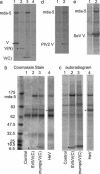
mda-5 interacts with the C terminus of the SV5 V protein and with other paramyxovirus V proteins. (a) Cells were transfected for 36 h with vectors expressing nontagged mda-5 (lane 4), together with myc-tagged versions of SV5 V(W3A) (lane 1), the N terminus of V (1–174) [V(N); lane 2], or the C terminus of V (126–222) [V(C); lane 3], or a control “empty vector” (lane 4) and immunoprecipitated with anti-myc antibody. (b and c) Coomassie stain and autoradiogram, respectively, of immunoprecipitates, by using anti-myc antibody, of cells transfected for 36 h with plasmids expressing nontagged mda-5, together with a control “empty vector” (lane 1) or plasmids expressing myctagged versions of the C terminus of SV5 [SV5/V(C); lane 2] and of mumps [mumps/V(C); lane 3] or myc-tagged HeV V (lane 4). (d and e) Autoradiograms of immunoprecipitates from cells transfected with a plasmid that express the nontagged mda-5, together with a control plasmid (lane 1), a plasmid expressing PIV 2/V(d, lane 2), or a plasmid expressing myc-tagged SeV V (e, lane 2). Cells were metabolically labeled with 35[S]methionine for 1 h before the myc-tagged proteins being immunoprecipitated with the 9E10 mAb and the V protein of hPIV-2 immunoprecipitated with the mAb Pk2 (32). *, the bands corresponding to the V proteins or derivatives. Proteins were separated on 4–12% polyacrylamide gradient gels (Invitrogen).
Fig. 3.
Fig. 3.
mda-5 is an activator of dsRNA-responsive IFN-β transcription and is a _target for inhibition by the SV5 virus V protein. Vero cells (a and c–g) or HeLaE cells (b) were cotransfected with the indicated luciferase reporter constructs; the β-galactosidase expression vector, pJATlacZ; and mammalian expression plasmids driving the overexpression of either mda-5 or fragments thereof, SV5-V, or the control “empty vector.” Transfected cells were either mock-treated or treated with dsRNA and cell extracts prepared for reporter gene assays. In each case, luciferase activity was corrected to the β-galactosidase activity to normalize for variations in the transfection efficiency. Transfection experiments were repeated at least twice, and averages and error bars are shown. Expression levels are compared with the basal level of the empty vector (=1) in each case except c and e, where the reference point is the induced level of the empty vector (=100%). (a and b) mda-5 stimulates basal and dsRNA-induced activity of the IFN-β promoter. Note that a Inset shows a magnified scale of the effects of mda-5 on basal expression. (c) mda-5 is essential for dsRNA response of the IFN-β promoter. Lanes 1–8 show the effect of dsRNA on IFN-β induction in the presence of the empty vector (lanes 1 and 2), the SV5 V protein (lanes 3 and 4), the mda-5 CARD domain (lanes 5 and 6), and mda-5 lacking the CARD domain (mda-5ΔN, lanes 7 and 8). Lanes 9–12 show the effect on induction of short dsRNAi molecules directed against mda-5 (lanes 11 and 12) compared with a control dsRNAi with two nucleotide mismatches (lanes 9 and 10). Lanes 13–16 show the same effect by using pRetroSuper-derived RNAi against mda-5 (lanes 15 and 16) compared with a control against CD2 (lanes 13 and 14). (d and e) mda-5 stimulates basal and dsRNA-induced activity of NF-κB(d) and IRF-3 (e) reporters. In the latter case, the reporter is a dimerized lexA operator sequence placed upstream of the herpes simplex virus 1 (HSV-1) thymidine kinase promoter, and transcriptional activity is conferred by a cotransfected Lex-IRF-3 fusion. (f and g) The basal (f) and dsRNA-induced (g) activation of the IFN-β promoter by mda-5 is inhibited by the SV5 V protein. Lanes 1 and 9 show the activity of the IFN-β promoter in the absence of mda-5, whereas lanes 2–8 show the effect of an increasing level of mda-5 expression plasmid (4, 8, 16, 40, 80, 320, and 468 ng). Lanes 10–13 show the effect of increasing levels of SV5 V expression plasmid (40, 80, and 160 ng) in the presence of 40 ng of mda-5 expression plasmid.
Fig. 4.
Fig. 4.
mda-5 stimulates the production of endogenous IFN, and this activity can be blocked by coexpression of the SV5 V protein. (a and b): 293 cells were transfected with control “empty vector” (5 μg), mda-5 (1.25 μg and “empty vector” 3.75 μg), or mda-5 (1.25 μg) and SV5-V (3.75 μg) expression vectors. After 24 h, cells were either mock-treated or treated with dsRNA for a further 18 h. The amount of IFN present in the culture media was estimated by either a virus reduction assay (a) or an IFN-stimulated regulatory element reporter assay (b, as described in ref. 6) in Vero cells. (c) As a above, but increasing amounts (1.25–5 μg) of the mda-5 expression vector were transfected with 3.75 μg of the SV5 V plasmid.
Fig. 5.
Fig. 5.
mda-5 activity is inhibited by the C terminus of SV5 V and is a property of other paramyxovirus V proteins. Vero cells were cotransfected with the IFN-β luciferase reporter, the β-galactosidase expression vector, pJATlacZ, and mammalian expression plasmids driving the overexpression of mda-5 or the control “empty vector,” and either the V or P proteins of SV5 or truncations of the SV5 V protein (a) or the V proteins of other paramyxoviruses (b). In each case, luciferase activity was corrected to the β-galactosidase activity to normalize for variations in the transfection efficiency. Transfection experiments were repeated at least twice, and averages and error bars are shown. Expression levels are compared with the basal level of the empty vector (=1) in each case.

Similar articles

Cited by

References

    1. Lamb, R. A. & Kolakofsky, D. (2001) in Fields' Virology, eds. Fields, B. N., Knipe, D. M., Howley, P. M. & Griffin, D. E. (Lippincott Williams & Wilkins, Philadelphia), Vol. 1, pp. 1305–1340.
    1. Wang, L.-F. & Eaton, B. (2001) Infect. Dis. Rev. 3, 52–69.
    1. Goodbourn, S., Didcock, L. & Randall, R. E. (2000) J. Gen. Virol. 81, 2341–2364. - PubMed
    1. Sen, G. C. (2001) Annu. Rev. Microbiol. 55, 255–281. - PubMed
    1. Grandvaux, N., tenOever, B. R., Servant, M. J. & Hiscott, J. (2002) Curr. Opin. Infect. Dis. 15, 259–267. - PubMed

Publication types

MeSH terms

LinkOut - more resources

  NODES
Note 1
twitter 2