. 2005 Feb;3(2):e41.
doi: 10.1371/journal.pbio.0030041. Epub 2005 Feb 8.

SIRT1 regulates HIV transcription via Tat deacetylation

Affiliations

SIRT1 regulates HIV transcription via Tat deacetylation

Sara Pagans et al. PLoS Biol. 2005 Feb.

Abstract

The human immunodeficiency virus (HIV) Tat protein is acetylated by the transcriptional coactivator p300, a necessary step in Tat-mediated transactivation. We report here that Tat is deacetylated by human sirtuin 1 (SIRT1), a nicotinamide adenine dinucleotide-dependent class III protein deacetylase in vitro and in vivo. Tat and SIRT1 coimmunoprecipitate and synergistically activate the HIV promoter. Conversely, knockdown of SIRT1 via small interfering RNAs or treatment with a novel small molecule inhibitor of the SIRT1 deacetylase activity inhibit Tat-mediated transactivation of the HIV long terminal repeat. Tat transactivation is defective in SIRT1-null mouse embryonic fibroblasts and can be rescued by expression of SIRT1. These results support a model in which cycles of Tat acetylation and deacetylation regulate HIV transcription. SIRT1 recycles Tat to its unacetylated form and acts as a transcriptional coactivator during Tat transactivation.


Figures

Figure 1
Figure 1. In Vitro Tat Deacetylation by Human SIRT Proteins
(A) Scheme of Tat deacetylation assay with immunoprecipitated SIRT1–7 proteins. Expression vectors for FLAG-tagged SIRT proteins were transfected into HEK 293 cells, immunoprecipitated, and incubated with synthetic Tat (72 amino acids) carrying an N-terminal biotin label and an acetyl group at position 50 (AcTat) in the presence of NAD+. Immunoprecipitated material was also analyzed in a radioactive (3H) histone deacetylase assay using an H3 peptide as a substrate. (B) WB analysis of deacetylation reactions with antibodies specific for acetylated lysine 50 in Tat (α-AcTat), with SA-HRP, or with α-FLAG antibodies. (C) WB of Tat deacetylation by immunoprecipitated SIRT1 in the presence or absence of NAD+, TSA, or nicotinamide (Nic).
Figure 2
Figure 2. Physical Interaction between Tat and SIRT1
(A) Immunoprecipitation (IP) and WB of FLAG-tagged Tat (Tat-FLAG) and HA-tagged SIRT1 (SIRT1-HA) after transfection of corresponding expression vectors (+) or empty vector controls (−) into HEK 293 cells. (B) The same experiments as in (A) performed with T7-tagged Tat and FLAG-tagged SIRT1, SIRT2, and SIRT6. (C) Coimmunoprecipitation of FLAG-tagged Tat with endogenous SIRT1 in HEK 293 cells transfected with the Tat expression vector or the empty vector control. IPs were performed with or without rabbit α-SIRT1 antibodies. (D) WB of recombinant SIRT1 protein after pulldown with synthetic biotinylated Tat or AcTat. Tat proteins were detected with antibodies specific for acetylated lysine 50 in the Tat ARM (α-AcTat) or SA-HRP. (E) Immunoprecipitation/WB of FLAG-tagged Tat or TatK50R and HA-tagged SIRT1. WT, wild type.
Figure 3
Figure 3. SIRT1 Is a Positive Cofactor for Tat Transactivation
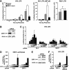
(A) Cotransfection of SIRT1 or the catalytically inactive SIRT1 mutant SIRT1H363Y with the HIV LTR luciferase construct and increasing amounts of a Tat expression vector (RSV-Tat: 0, 2, 20, and 200 ng), an HIV LTR luciferase construct containing mutated binding sites for the transcription factor NF-κB and RSV-Tat (20 ng), or with an RSV-luciferase construct (200 ng) in HeLa cells. The average of three experiments is shown (± standard error of the mean [SEM]). (B) WB analysis of HeLa cells 72 h after transfection of siRNAs directed against SIRT1 or GL3 control siRNAs. (C) Cotransfection of the HIV LTR luciferase construct with increasing amounts of CMV-Tat or CMV-TatK50R (0, 50, 100, 200, 400, and 800 ng) 48 h after transfection of double-stranded siRNAs directed against SIRT1 or GL3 control siRNAs in HeLa cells. Luciferase activity was measured 24 h after plasmid transfection and 72 h after siRNA transfection. Note that all luciferase reporter vectors used in this study are based on the pGL2 luciferase vector, which is not affected by GL3-specific siRNAs [36]. The average of three experiments is shown (± SEM). (D) The transcriptional activity of increasing amounts of the CMV-luciferase reporter (0, 50, 100, 200, 400, and 800 ng) was similar in SIRT1 knockdown or GL3-treated control cells. The average of two experiments performed in duplicate is shown (± SEM). (E) WB of endogenous SIRT1 or actin 72 h after transfection of siRNA directed against SIRT1 or mutated SIRT1 siRNA. (F) Cotransfection of the HIV LTR luciferase with increasing amounts of CMV-Tat (0, 2, 20, and 200 ng) in HeLa cells pretransfected with wild-type or mutant SIRT1 siRNA oligonucleotides as described in (C). WT, wild-type.
Figure 4
Figure 4. Impaired Tat Transcriptional Activity in Murine SIRT1−/− Cells
(A) Nuclear microinjection of HIV LTR luciferase, RSV-Tat, and a human cyclinT1-expressing construct into MEFs derived from SIRT+/+ or SIRT−/− mice. In all experiments, a fixed amount of DNA was injected by adding the empty vector control. Cells were coinjected with CMV-GFP, and the luciferase activity per GFP-positive cell was calculated. An average of two injections is shown. (B) The HIV LTR luciferase construct together with RSV-Tat and the cyclinT1-expressing construct were coinjected into SIRT−/− MEFs in the presence of increasing amounts of a plasmid expressing human SIRT1. The average of three experiments is shown (± SEM). (C) Coinjection of the human SIRT1 expression vector together with the 5xUAS luciferase construct containing five Gal4 binding sites upstream of the thymidine kinase promoter and a Gal4-VP16 expression plasmid into SIRT1−/− MEFs. The average of three experiments is shown (± SEM).
Figure 5
Figure 5. Transcriptional Activity of AcTat Depends on Deacetylation by SIRT1
(A) AcTat functions through TAR and cyclinT1 binding. Nuclear microinjection of increasing amounts of synthetic Tat or AcTat together with wild-type (wt TAR), TAR Δbulge, or TAR Δloop mutant HIV LTR luciferase constructs into HeLa cells. Cells were coinjected with CMV-GFP, and luciferase activity was calculated per GFP-positive cell. An average of three experiments is shown (± SEM). (B) AcTat transactivation requires CDK9. HeLa cells microinjected with Tat or AcTat (each 30 ng/μl) and the HIV LTR luciferase reporter were treated with increasing amounts of DRB, a known CDK9 inhibitor, for 4 h. (C) AcTat transcriptional activity is inhibited by nicotinamide, but not TSA. HeLa cells injected with HIV LTR luciferase and increasing amounts of AcTat were treated with TSA (400 nM) or nicotinamide (5 mM) for 4 h. The average of two experiments is shown. (D) SIRT1 is necessary for AcTat, but not Tat function. HeLa cells were transfected with siRNAs specific for SIRT1 or GL3 control siRNAs 48 h before microinjection of HIV LTR luciferase and Tat or AcTat (each 30 ng/μl). The average of three experiments is shown (± SEM).
Figure 6
Figure 6. Inhibition of HIV Gene Expression by a Small Molecule Inhibitor of SIRT1
(A) In vitro histone deacetylation assays including recombinant SIRT1, radioactively labeled histone H3 peptide, and indicated concentrations of splitomicin or HR73. The average (± SEM) of two experiments performed in duplicate is shown for each point. (B) Chemical structures of splitomicin and its derivative HR73. (C) Inhibition of Tat transactivation by HR73. RSV-Tat (0, 20, and 200 ng) and HIV LTR luciferase (200 ng) or RSV-luciferase (200 ng) vectors were transfected into HeLa cells. Transfected cells were treated with indicated concentrations of HR73 or DMSO for 8 h. (D) Inhibition of HIV gene expression by HR73. GFP expression in Jurkat T cells infected with HIVNL4–3 containing the GFP open reading frame in place of the viral nef gene or with an HIV-based lentiviral vector expressing GFP from the heterologous EF-1α promoter. Treatment with HR73 (1 μM in DMSO) or DMSO was performed for 36 h after overnight infection. The average (± SEM) of four experiments is shown.

Similar articles

Cited by

References

    1. Kao SY, Calman AF, Luciw PA, Peterlin BM. Anti-termination of transcription within the long terminal repeat of HIV-1 by tat gene product. Nature. 1987;330:489–493. - PubMed
    1. Toohey MG, Jones KA. In vitro formation of short RNA polymerase II transcripts that terminate within the HIV-1 and HIV-2 promoter-proximal downstream regions. Genes Dev. 1989;3:265–282. - PubMed
    1. Rosen CA, Sodroski JG, Haseltine WA. The location of cis-acting regulatory sequences in the human T cell lymphotropic virus type III (HTLV-III/LAV) long terminal repeat. Cell. 1985;41:813–823. - PubMed
    1. Feng S, Holland EC. HIV-1 tat trans-activation requires the loop sequence within tar. Nature. 1988;334:165–167. - PubMed
    1. Wei P, Garber ME, Fang SM, Fischer WH, Jones KA. A novel CDK9-associated C-type cyclin interacts directly with HIV-1 Tat and mediates its high-affinity, loop-specific binding to TAR RNA. Cell. 1998;92:451–462. - PubMed

Publication types

  NODES
Note 1
twitter 2