. 2005 Mar 8;33(5):1474-86.
doi: 10.1093/nar/gki292. Print 2005.

Translation of the F protein of hepatitis C virus is initiated at a non-AUG codon in a +1 reading frame relative to the polyprotein

Affiliations

Translation of the F protein of hepatitis C virus is initiated at a non-AUG codon in a +1 reading frame relative to the polyprotein

Martin Baril et al. Nucleic Acids Res. .

Abstract

The hepatitis C virus (HCV) genome contains an internal ribosome entry site (IRES) followed by a large open reading frame coding for a polyprotein that is cleaved into 10 proteins. An additional HCV protein, the F protein, was recently suggested to result from a +1 frameshift by a minority of ribosomes that initiated translation at the HCV AUG initiator codon of the polyprotein. In the present study, we reassessed the mechanism accounting for the synthesis of the F protein by measuring the expression in cultured cells of a luciferase reporter gene with an insertion encompassing the IRES plus the beginning of the HCV-coding region preceding the luciferase-coding sequence. The insertion was such that luciferase expression was either in the +1 reading frame relative to the HCV AUG initiator codon, mimicking the expression of the F protein, or in-frame with this AUG, mimicking the expression of the polyprotein. Introduction of a stop codon at various positions in-frame with the AUG initiator codon and substitution of this AUG with UAC inhibited luciferase expression in the 0 reading frame but not in the +1 reading frame, ruling out that the synthesis of the F protein results from a +1 frameshift. Introduction of a stop codon at various positions in the +1 reading frame identified the codon overlapping codon 26 of the polyprotein in the +1 reading frame as the translation start site for the F protein. This codon 26(+1) is either GUG or GCG in the viral variants. Expression of the F protein strongly increased when codon 26(+1) was replaced with AUG, or when its context was mutated into an optimal Kozak context, but was severely decreased in the presence of low concentrations of edeine. These observations are consistent with a Met-tRNA(i)-dependent initiation of translation at a non-AUG codon for the synthesis of the F protein.


Figures
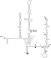
Figure 1
Figure 1
Sequence and secondary structure of the HCV IRES [based on reference (60)] and beginning of the HCV-coding sequence (nt 1–510 of genotype 1a). The initiation codon of the HCV polyprotein (codon 1, nt 342–344) is highlighted. The gray arrows point to the 5′ end (nt 1) of the IRES and to the 5′end (nt 290) of the HCV insertion in the constructs without IRES (see Figure 2). The black arrows point to the 3′ end of segments of different length of the HCV-coding sequence that were used in this study. The number preceding the arrows corresponds to the last nucleotide of these segments. The sites where a +1 ribosomal frameshift was previously proposed to occur (see the text) are in bold and marked by arrowheads. The brackets indicate codons of the HCV polyprotein with their corresponding number, which are referred to in the text. The codon overlapping codon 26 in the +1 reading frame to which we also refer in the text is in bold. It was verified, using the mfold program (61), that the predicted secondary structure of the HCV sequence was not altered when it was fused to the luciferase-coding sequence.
Figure 2
Figure 2
Description of the luciferase vectors used for the study of the expression of HCV F protein in vitro and in cultured cells. Nucleotides are numbered according to Figure 1. (A) The construct presented in this figure is pHCV-447-LUC, where a portion of the HCV-coding sequence extending to nt 447 is inserted upstream the coding sequence of the firefly luciferase reporter gene. Inserts of different length are: pHCV-387-LUC, pHCV-426-LUC, pHCV-447-LUC and pHCV-510-LUC, where the number indicates the last nucleotide of the insertion (see Figure 1). The AUG initiation codon (nt 342–344) of the HCV polyprotein is underlined. For the (0) constructs, the luciferase sequence is in-frame with this AUG initiation codon. For these (0) constructs, the expression of luciferase mimics the synthesis of the polyprotein. For the (+1) constructs, an adenine was added immediately after the BamHI restriction site (at position 455), so that only ribosomes translating the HCV sequence in the +1 reading frame relative to the HCV AUG initiator codon synthesize luciferase. With these (+1) constructs the expression of luciferase mimics the synthesis of the F protein. All the constructs were cloned by inserting a PCR product containing the investigated HCV sequence between the KpnI–BamHI sites of the pcDNA3.1-LUC vector. This HCV sequence was obtained from a plasmid that contains the 5′-UTR plus the beginning of the coding sequence of HCV genotype 1a (see Materials and Methods). In all the pHCV-LUC (0) and (+1) constructs, the HCV-coding sequence is preceded either by the complete HCV 5′-UTR (IRES constructs) or by a small segment (nt 290–341) of the HCV 5′-UTR region (cap constructs). The boxed sequence, which corresponds to a HCV consensus sequence, was mutated in pHCV-387-LUC, pHCV-426-LUC, pHCV-447-LUC and pHCV-510-LUC, creating derivatives with a stretch of 10A (10A constructs). (B) Sequences of the HCV-coding region of the derivatives of pHCV-447-LUC constructed in this study. Nucleotides that are mutated are underlined and nucleotides that are inserted and deleted are in bold and indicated by a dashed line, respectively.
Figure 3
Figure 3
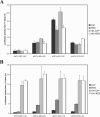
Synthesis of the F protein with constructs containing insertions of different length of the HCV-coding sequence. The synthesis of the F protein was measured with constructs containing HCV segments of different length described in Figure 2. In the pHCV-387-LUC, pHCV-426-LUC, pHCV-447-LUC and pHCV-510-LUC cap and IRES constructs, the consensus HCV sequence (AAAGAAAAAC, nt 364–373) occupies the site where a +1 frameshift was proposed to occur. In the 10A corresponding constructs, the consensus HCV sequence is replaced with a 10A stretch. (A) Synthesis of the F protein in cultured cells. Synthesis of the F protein was measured after co-transfection of 293FT cells with 3 μg of a pHCV-LUC (0) or (+1) construct and 1 μg of pcDNA3.1/Hygro(+)/lacZ, which is used to normalize for variations in transfection efficiency. (B) Synthesis of the F protein in vitro. In vitro translation experiments were carried out in 25 μl of RRL with 0.2 μg of mRNAs transcribed from the FbaI-digested pHCV-LUC constructs. Results are reported as the amount of F protein synthesized (assessed by the activity of luciferase in the +1 reading frame) relative to the amount of the polyprotein synthesized (assessed by the activity of luciferase in the 0 reading frame) and were calculated as described in the text. Each value represents the mean ± standard error of four to six independent experiments.
Figure 4
Figure 4
Synthesis of the F protein when a stop codon is introduced in the reading frame of the polyprotein. A UGA stop codon was introduced in pHCV-447-LUC, in the reading frame of the polyprotein, upstream or across the previously proposed +1 ribosomal frameshift site (15) at codon 3, 9, 10, 11 or 12. This generated pHCV-UGA3-LUC, pHCV-UGA9-LUC, pHCV-UGA10-LUC, pHCV-UGA11-LUC and pHCV-UGA12-LUC. All these constructs either contained or did not contain the HCV IRES (IRES and cap construct, respectively), except pHCV-UGA3-LUC for which the HCV IRES was not constructed since mutating nt 348–350 of the HCV RNA destabilizes a hairpin structure that is important for the IRES function (62). Assays were made in cultured cells as described in the legend to Figure 3. The synthesis of the F protein in the mutant constructs is expressed relative to that obtained with pHCV-447-LUC (+1), which is arbitrarily set at 100%. Each value represents the mean ± standard error of four to six independent experiments.
Figure 5
Figure 5
Synthesis of the polyprotein and the F protein when the AUG start codon of the polyprotein or its context is mutated. Derivatives of pHCV-447-LUC were constructed by mutating the AUG (nt 342–344) polyprotein start codon to UAC or its upstream context from ACC to UUU, generating pHCV-accUAC-LUC and pHCV-uuuAUG-LUC, respectively. Translation efficiencies are indicated on a logarithmic scale for the polyprotein and the F protein. The synthesis of the polyprotein measured with the mutant (0) constructs and that of the F protein measured with the (+1) constructs are expressed relative to the synthesis of the polyprotein with pHCV-447-LUC (0), which is arbitrarily set at 100%. Assays were made in cultured cells as described in the legend to Figure 3. Each value represents the mean ± standard error of four to six independent experiments.
Figure 6
Figure 6
Identification of the translation start site of the F protein. A stop codon was introduced in pHCV-447-LUC at codons overlapping codon 23, 24, 25 or 26 of the polyprotein in the +1 reading frame, generating pHCV-UAA23+1-LUC, pHCV-UGA24+1-LUC, pHCV-UGA25+1-LUC and pHCV-UGA26+1-LUC. Assays were made in cultured cells, as described in the legend to Figure 3. The efficiency of the synthesis of the F protein from construct pHCV-447-LUC (+1) is arbitrarily set at 100%. Each value represents the mean ± standard error of four to six independent experiments.
Figure 7
Figure 7
Confirmation of the location of the translation start site of the F protein. (A) Description of the mutants used for this confirmation. A nucleotide was added in pHCV-447-LUC (+1) immediately after codon 25(+1) or 26(+1), generating pHCV+U418-LUC and pHCV+U421-LUC, respectively, where the reading frame of luciferase that monitors the synthesis of the F protein is shifted by 1 nt relative to its reading frame in pHCV-447-LUC (+1). Nt 447 was deleted (dashed line) in pHCV+U421-LUC, generating pHCV+U421ΔU447-LUC, in which the luciferase reading frame is restored. (B) Assays were made in cultured cells as described in the legend to Figure 3. The efficiency of synthesis of the F protein from construct pHCV-447-LUC (+1) is arbitrarily set at 100%. Each value represents the mean ± standard error of four to six independent experiments.
Figure 8
Figure 8
Further details on the initiation of translation of the F protein. In pHCV-GCG26+1-LUC, the initiation codon of the F protein, which is GUG in pHCV-447-LUC, was mutated to GCG. In pHCV-AUG23-LUC, an AUG codon with an optimal Kozak context for initiation was introduced in the 0 frame at codon 23 of pHCV-447-LUC. In pHCV-AUG26+1-LUC, the GUG initiation codon of the F protein was mutated to AUG. In pHCV-accGUG26+1-LUC and pHCV-accGCG26+1-LUC, the Kozak context of codon 26(+1) was mutated to ACC, the initiator codon being GUG and GCG, respectively. The synthesis of the F protein is indicated on a logarithmic scale. Assays were made in cultured cells, as described in the legend to Figure 3. The synthesis of the F protein from pHCV-447-LUC (+1) is arbitrarily set at 100%. Each value represents the mean ± standard error of four to six independent experiments.
Figure 9
Figure 9
Inhibition of the synthesis of the polyprotein and the F protein by edeine. In vitro translation experiments were carried out in 25 μl of RRL containing 0, 0.1, 0.2 or 0.3 μM edeine, with 0.2 μg of mRNAs transcribed from the FbaI-digested pHCV-LUC IRES constructs. For each construct, the protein synthesis in the absence of edeine (0 μM) is arbitrarily set at 100%. Each value represents the mean ± standard error of three independent experiments.

Similar articles

Cited by

References

    1. Lefkowitch J.H., Schiff E.R., Davis G.L., Perrillo R.P., Lindsay K., Bodenheimer H.C., Balart L.A., Ortego T.J., Payne J., Dienstag J.L. Pathological diagnosis of chronic hepatitis C: a multicenter comparative study with chronic hepatitis B. The Hepatitis Interventional Therapy Group. Gastroenterology. 1993;104:595–603. - PubMed
    1. Saito I., Miyamura T., Ohbayashi A., Harada H., Katayama T., Kikuchi S., Watanabe Y., Koi S., Onji M., Ohta Y., et al. Hepatitis C virus infection is associated with the development of hepatocellular carcinoma. Proc. Natl Acad. Sci. USA. 1990;87:6547–6549. - PMC - PubMed
    1. Scheuer P.J., Ashrafzadeh P., Sherlock S., Brown D., Dusheiko G.M. The pathology of hepatitis C. Hepatology. 1992;15:567–571. - PubMed
    1. Choo Q.L., Richman K.H., Han J.H., Berger K., Lee C., Dong C., Gallegos C., Coit D., Medina-Selby R., Barr P.J., et al. Genetic organization and diversity of the hepatitis C virus. Proc. Natl Acad. Sci. USA. 1991;88:2451–2455. - PMC - PubMed
    1. Reynolds J.E., Kaminski A., Carroll A.R., Clarke B.E., Rowlands D.J., Jackson R.J. Internal initiation of translation of hepatitis C virus RNA: the ribosome entry site is at the authentic initiation codon. RNA. 1996;2:867–878. - PMC - PubMed

Publication types

  NODES
INTERN 5
twitter 2