. 2005 Jun 15;19(12):1444-54.
doi: 10.1101/gad.1315905. Epub 2005 Jun 2.

Specificity and mechanism of the histone methyltransferase Pr-Set7

Affiliations

Specificity and mechanism of the histone methyltransferase Pr-Set7

Bing Xiao et al. Genes Dev. .

Abstract

Methylation of lysine residues of histones is an important epigenetic mark that correlates with functionally distinct regions of chromatin. We present here the crystal structure of a ternary complex of the enzyme Pr-Set7 (also known as Set8) that methylates Lys 20 of histone H4 (H4-K20). We show that the enzyme is exclusively a mono-methylase and is therefore responsible for a signaling role quite distinct from that established by other enzymes that _target this histone residue. We provide evidence from NMR for the C-flanking domains of SET proteins becoming ordered upon addition of AdoMet cofactor and develop a model for the catalytic cycle of these enzymes. The crystal structure reveals the basis of the specificity of the enzyme for H4-K20 because a histidine residue within the substrate, close to the _target lysine, is required for completion of the active site. We also show how a highly variable component of the SET domain is responsible for many of the enzymes' interactions with its _target histone peptide and probably also how this part of the structure ensures that Pr-Set7 is nucleosome specific.


Figures

Figure 1.
Figure 1.
Ternary structure of the histone 4 K20 methyltransferase Pr-Set7. (A) A ribbons representation of Pr-Set7 colored according to its subdomains: N-flanking (magenta); core SET domain (cyan); SET-I (blue); C-flanking (beige). The H4 peptide and AdoHcy cofactor products are shown as stick representations with the peptide carbons in green and AdoHcy carbons in yellow. Key secondary structure elements have been labeled. (B) Surface representation of Pr-Set7 in the same orientation as A showing the relationship of the four subdomains and the peptide-binding groove. (C) Surface representation rotated 90° about the X-axis to show the AdoHcy-binding pocket.
Figure 2.
Figure 2.
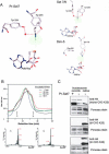
Pr-Set7 is a monomethylase. (A) Key residues in the active site of Pr-Set7 in stick representation. The hydrogen bonds that coordinate the lysine Nε atom are represented by dashed red lines. A dashed white line represents a theoretical line between the lysine Nε and the AdoHcy S atom. The transferred methyl group is also shown in a space-filling representation (in green) to give an indication of size and why it is not possible to accommodate extra bulky methyl groups at the position of the hydrogens currently forming H-bonds either with Tyr 245 or the water molecule. The upper right panel shows the equivalent view in Set7/9 (PDB 1o9s), also a mono-methylase, and the lower right panel shows the equivalent view in Dim-5 (PDB 1peg), a tri-methylase. (B) Analytical analysis of the reaction products obtained following incubation of Pr-Set7 with unmodified H4 30-mer peptide. The products were separated by HPLC and fractions analyzed by MALDI time of flight mass spectrometry, two example traces of which are shown beneath. (C) Western blots obtained after incubating Pr-Set7 with nucleosome substrates and AdoMet. The substrates are reconstituted recombinant nucleosomes (Xenopus histone) or derived from Hela cells (human histone) as indicated. In each the upper panel has been stained with the indicated antibody with a Ponceau stained control shown beneath. (D) Sequence alignment of the Pr-Set7 SET domain with two other histone 4 K20 methyltransferase proteins (Suv-20h1 [accession no. AAT00539] and NSD-1 [NP_758859]) and with SET domains of known structure (Set7/9 [NP_085151], Dim-5 [AAL35215], and Clr4 [T43745]). The sequence is colored according to domain. The secondary structure elements of Pr-Set7 are indicated above. Green and red circles indicate the Pr-Set7 active site Y-245 and Y-334 equivalent residues (dashed circles for intuited equivalent residues). Yellow circles indicate protein main-chain-peptide interactions; yellow stars indicate protein side-chain-peptide interactions.
Figure 2.
Figure 2.
Pr-Set7 is a monomethylase. (A) Key residues in the active site of Pr-Set7 in stick representation. The hydrogen bonds that coordinate the lysine Nε atom are represented by dashed red lines. A dashed white line represents a theoretical line between the lysine Nε and the AdoHcy S atom. The transferred methyl group is also shown in a space-filling representation (in green) to give an indication of size and why it is not possible to accommodate extra bulky methyl groups at the position of the hydrogens currently forming H-bonds either with Tyr 245 or the water molecule. The upper right panel shows the equivalent view in Set7/9 (PDB 1o9s), also a mono-methylase, and the lower right panel shows the equivalent view in Dim-5 (PDB 1peg), a tri-methylase. (B) Analytical analysis of the reaction products obtained following incubation of Pr-Set7 with unmodified H4 30-mer peptide. The products were separated by HPLC and fractions analyzed by MALDI time of flight mass spectrometry, two example traces of which are shown beneath. (C) Western blots obtained after incubating Pr-Set7 with nucleosome substrates and AdoMet. The substrates are reconstituted recombinant nucleosomes (Xenopus histone) or derived from Hela cells (human histone) as indicated. In each the upper panel has been stained with the indicated antibody with a Ponceau stained control shown beneath. (D) Sequence alignment of the Pr-Set7 SET domain with two other histone 4 K20 methyltransferase proteins (Suv-20h1 [accession no. AAT00539] and NSD-1 [NP_758859]) and with SET domains of known structure (Set7/9 [NP_085151], Dim-5 [AAL35215], and Clr4 [T43745]). The sequence is colored according to domain. The secondary structure elements of Pr-Set7 are indicated above. Green and red circles indicate the Pr-Set7 active site Y-245 and Y-334 equivalent residues (dashed circles for intuited equivalent residues). Yellow circles indicate protein main-chain-peptide interactions; yellow stars indicate protein side-chain-peptide interactions.
Figure 3.
Figure 3.
H4 peptide-binding interactions in Pr-Set7. A schematic representation of the interactions observed between Pr-Set7 and its H4 peptide substrate. Hydrogen bonds are shown as a red dotted line. The peptide main-chain interactions are shown above the peptide trace (green) and side-chain interactions are shown below. The _target lysine is shown in black but its interactions are omitted in this figure for clarity. The Pr-Set7 residues involved in interactions are colored according to the scheme used in Figure 1 and protein side-chain or main-chain interactions are indicated accordingly.
Figure 4.
Figure 4.
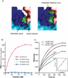
The H4 peptide substrate contributes to formation of the lysine access channel in Pr-Set7. (A) A surface representation of the lysine access channel in Pr-Set7 colored according to the scheme in Figure 1. The substrate H4-K20 side chain is shown as a space-filling representation. The left panel is a view without the His(-2) residue and to the right the histidine has been added completing the access channel shown by the dashed line. (B) Histone methyltransferase assay of Pr-Set7 incubated with increasing concentration of H4 30-mer peptide substrate. Traces are shown for wild-type substrate and peptides with substituted residues at the -2 position relative to the _target lysine as indicated. Rates are derived from the incorporation of [3H CH3] after 10 min as measured by liquid scintillation counting of peptide separated from free AdoMet by reverse phase chromatography. (C) Plots showing the binding phase from surface plasmon resonance experiments for Pr-Set7 protein injections (as indicated) onto surfaces containing either wild-type or H(-2)A peptide. The inset shows a plot of the observed on rate versus Pr-Set7 concentration.
Figure 5.
Figure 5.
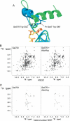
Flexibility of the C-flanking domain. (A) A ribbons representation of the structures of the C-flanking domains of Pr-Set7 (blue) and Set7/9 (green) aligned on the AdoHcy cofactor (yellow and orange, respectively). The figure shows that the benzyl rings of Trp 349 of Pr-Set7 and Trp 352 of Set7/9 occupy equivalent positions and make the same interaction with the adenine ring of AdoHcy. (B) 15N-HSQC spectra of apo-Set7/9 (left) and Set7/9 with 1.2 M equivalents of AdoHcy (right). Note there are fewer resonances in the binary spectrum. (C) {1H} 15N-heteronuclear NOE data. The plots represent the difference spectra (saturated - 0.8× control) for apo-Set7/9 (left) and Set7/9 + AdoHcy (right). (D) 1H-13CO projection of the 3D trosy-HNCO spectrum used for accurate peak counting. (E) Selected sections from the 3D trosyHNCO projection demonstrating the characteristic “doubling” of resonances for some residues. (F) 15N-HSQC spectra of selectively Trp-15N-labeled apo-Set7/9 (left) and Set7/9 with 1.2 M equivalents of AdoHcy (right). The assigned Trp resonances are indicated. Below are cross-sections through the spectra at the positions indicated.
Figure 5.
Figure 5.
Flexibility of the C-flanking domain. (A) A ribbons representation of the structures of the C-flanking domains of Pr-Set7 (blue) and Set7/9 (green) aligned on the AdoHcy cofactor (yellow and orange, respectively). The figure shows that the benzyl rings of Trp 349 of Pr-Set7 and Trp 352 of Set7/9 occupy equivalent positions and make the same interaction with the adenine ring of AdoHcy. (B) 15N-HSQC spectra of apo-Set7/9 (left) and Set7/9 with 1.2 M equivalents of AdoHcy (right). Note there are fewer resonances in the binary spectrum. (C) {1H} 15N-heteronuclear NOE data. The plots represent the difference spectra (saturated - 0.8× control) for apo-Set7/9 (left) and Set7/9 + AdoHcy (right). (D) 1H-13CO projection of the 3D trosy-HNCO spectrum used for accurate peak counting. (E) Selected sections from the 3D trosyHNCO projection demonstrating the characteristic “doubling” of resonances for some residues. (F) 15N-HSQC spectra of selectively Trp-15N-labeled apo-Set7/9 (left) and Set7/9 with 1.2 M equivalents of AdoHcy (right). The assigned Trp resonances are indicated. Below are cross-sections through the spectra at the positions indicated.
Figure 6.
Figure 6.
Processivity of SET domain enzymes. Our observations fit the following scheme: (1) The lysine loses a proton to the bulk solvent and binds to the methyltranferase. (2) Upon binding of the cofactor (AdoMet) the C-flanking domain completes the active site. (3) Methyl transfer occurs via a simple nucleophilic substitution reaction. (4) The C-flanking domain dissociates from the SET domain allowing release of the cofactor product (AdoHcy). In the case of a monomethylase, for example Pr-Set7, the methylated lysine can now dissociate. For higher-order methyltransferase another cofactor molecule could bind and the cycle repeated.
Figure 7.
Figure 7.
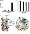
Interaction of Pr-Set7 with the nucleosome. (A) Histone methyltransferase assay showing incorporation of [3H CH3] into H4-K20 as peptide, histone octamer, or nucleosome substrate. (B) Histone methyltransferase assay following incorporation of [3H CH3] into H4 peptide (white) or nucleosome (black) substrate in the presence of increasing concentrations of NaCl. Each set of data has been normalized to the activity at 0 mM NaCl for that substrate to aid comparison. (C) A model showing Pr-Set7 (colored according to the scheme in Fig. 1) docked to the nucleosome (PDB entry 1KX5) and binding to one of the H4 histones (green). To the right is an orthogonal view of the model showing how Pr-Set7 may be orientated with respect to the nucleosome.

Similar articles

Cited by

References

    1. Breiling A. and Orlando, V. 2002. SET domain proteins reSET gene expression. Nat. Struct. Biol. 9: 894-896. - PubMed
    1. Chuikov S., Kurash, J.K., Wilson, J.R., Xiao, B., Justin, N., Ivanov, G.S., McKinney, K., Tempst, P., Prives, C., Gamblin, S.J., et al. 2004. Regulation of p53 activity through lysine methylation. Nature 432: 353-360. - PubMed
    1. Collaborative Computational Project, Number 4. 1994. The CCP4 suite: Programs for protein crystallography. Acta Crystallogr. D Biol. Crystallogr. 50: 760-763. - PubMed
    1. Davey C.A., Sargent, D.F., Luger, K., Maeder, A.W., and Richmond, T.J. 2002. Solvent mediated interactions in the structure of the nucleosome core particle at 1.9 a resolution. J. Mol. Biol. 319: 1097-1113. - PubMed
    1. Fang J., Feng, Q., Ketel, C.S., Wang, H., Cao, R., Xia, L., Erdjument-Bromage, H., Tempst, P., Simon, J.A., and Zhang, Y. 2002. Purification and functional characterization of SET8, a nucleosomal histone H4-lysine 20-specific methyltransferase. Curr. Biol. 12: 1086-1099. - PubMed

Publication types

MeSH terms

  NODES
Note 2
Project 5
twitter 2