. 2005 Jul 6;24(13):2391-402.
doi: 10.1038/sj.emboj.7600719. Epub 2005 Jun 16.

Transforming activity of MECT1-MAML2 fusion oncoprotein is mediated by constitutive CREB activation

Affiliations

Transforming activity of MECT1-MAML2 fusion oncoprotein is mediated by constitutive CREB activation

Lizi Wu et al. EMBO J. .

Abstract

Salivary gland tumors, a group of histologically diverse benign and malignant neoplasms, represent a challenging problem for diagnosis and treatment. A specific recurring t(11;19)(q21;p13) translocation is associated with two types of salivary gland tumors, mucoepidermoid carcinomas and Warthin's tumors. This translocation generates a fusion protein comprised of the N-terminal CREB (cAMP response element-binding protein)-binding domain of the CREB regulator MECT1 (Mucoepidermoid carcinoma translocated-1) and the C-terminal transcriptional activation domain of the Notch coactivator Mastermind-like 2 (MAML2). Here, we demonstrate that the MECT1-MAML2 fusion protein induces expression of multiple genes known to be CREB transcriptional _targets. MECT1-MAML2 was found to bind to CREB, recruit p300/CBP into the CREB complex through a binding domain on MAML2, and constitutively activate CREB-dependent transcription. The transforming activity of MECT1-MAML2 was markedly reduced by blocking CREB DNA binding. Thus, this fusion oncogene mimics constitutive activation of cAMP signaling, by activating CREB directly. This study has identified a novel, critical mechanism of transformation for an oncogene associated very specifically with salivary gland tumors, and identified potential _targets for the development of novel therapies.


Figures

Figure 1
Figure 1
Both the CREB-binding domain of MECT1 and the TAD domain of MAML2 are required for the MECT1-MAML2 fusion to induce foci formation in RK3E cells. (A) RK3E cells were transfected with the indicated expression plasmids, and stained with crystal violet for foci formation at 3 weeks post-transfection. (B) Diagram of the constructs used in focus assays, and the number of foci generated from the transfection of each of these constructs. The number of foci shown was obtained from the 10 cm plates based on the triplicate experiments. M-M2, M2, M, M1, M-M1, and M-VP stand for MECT1-MAML2 fusion, MAML2, MECT1, MAML1, MECT1-MAML1, and MECT1-VP16, respectively. All these genes were cloned into the pFLAG-CMV2 expression vector, and expressed as FLAG-tagged proteins.
Figure 2
Figure 2
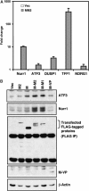
MECT1-MAML2 fusion induces expression of CREB transcriptional _target genes. (A) Expression levels of five _target genes (both known and previously unknown regulated by cAMP signaling) in MECT1-MAML2-transfected cells, relative to the empty vector-transfected cells, were determined by SYBR green real-time PCR. (B) Induction of two known CREB _targets, ATF3 and Nurr1 genes, wasanalyzed by Western blot analyses in human immortalized parotid HSY cells transfected with the indicated expression constructs. Transfected FLAG-tagged proteins except MECT1-VP16 (its band overlaps with IgG light chain, and the expression is shown on the whole-cell lysate blot) were detected after immunoprecipitation (IP) with anti-FLAG antibodies. M2, M, M-M2, M-M1, and M-VP stand for MAML2, MECT1, MECT1-MAML2, MECT1-MAML1, and MECT1-VP16, respectively.
Figure 3
Figure 3
Disruption of CREB activity reduced the abilities of the MECT1-MAML2 fusion to activate the CREB pathway and to induce colony formation in RK3E. (A) Expression of A-CREB in RK3E cells transduced with A-CREB viruses was determined by Western blot analysis. (B) RK3E cells expressing A-CREB and GFP, and the control cells expressing GFP alone, were transfected with either vector or the MECT1-MAML2 expression construct. The expression levels of two CREB _target genes, ATF3 and Nurr1, were analyzed by Western blot analysis. (C) A-CREB-expressing RK3E cells and the control cells were transfected with either vector only or the expression construct encoding MECT1-MAML2 fusion, and the colonies were scored 3 weeks after transfection. (D) 293T cells were cotransfected with the MECT1-MAML2 expression vector and CREB RNAi (or control RNAi), and the levels of ATF3, Nurr1, CREB, and transfected MECT1-MAML2 fusion were analyzed 48 h after transfection by Western blotting. (E) Expression levels of CREB, and five fusion _target genes in 293T cells cotransfectd with MECT1-MAML2 and CREB RNAi, relative to cells cotransfected with MECT1-MAML2 and control RNAi were determined by SYBR green real-time PCR.
Figure 4
Figure 4
Disruption of CREB activity significantly suppressed the growth of two human MECT1-MAML2-positive MEC cell lines. (A) A representative diagram showing the changes in the percentage of GFP-positive cells between cell populations expressing A-CREB and GFP and their control counterparts expressing GFP only. Two MECT1-MAML2-expressing MEC cell lines, H292 and H3118, along with the immortalized normal parotid cell line HSY (MECT1-MAML2 negative), were transduced with A-CREB or control GFP viruses. The percentages of GFP-positive cells were determined by FACS analysis at 3–4 days intervals, for a total of 15 days. The percentage of GFP-positive cells at day 2 postinfection was considered as 100%, and the remaining data were normalized. (B) A representative growth curve of cells expressing A-CREB plus GFP versus controls expressing GFP only. The cell number was determined using crystal blue staining daily for 6 days. (C) Cell cycle distribution of HSY, H3118, and H292 cells transduced with A-CREB viruses or vector control viruses as determined by propidium iodide straining followed by FACS analysis.
Figure 5
Figure 5
MECT1-MAML2 interacts with the CREB pathway regulatory molecule p300. (A) MECT1-MAML2 and p300 colocalize in the nuclei. U20S cells were transfected with the GFP-tagged MECT1-MAML2 and HA-tagged p300, and stained with an anti-HA antibody for p300 expression. The DAPI staining labels the nuclei of the same cells. (B) MECT1-MAML2 post-translationally modifies p300. 293T cells were transfected with HA-tagged p300 and either empty vector, FLAG-tagged MECT1-MAML2, MECT1, or MAML2 TAD (173-1153). p300 expression was detected by IP with anti-HA antibodies followed by Western blot analysis with anti-HA. The expression levels of the FLAG-tagged proteins were determined by probing with anti-FLAG antibodies. (C) p300 modifies MECT1-MAML2 via acetylation. 293T cells were transfected with FLAG-tagged MECT1-MAML2 and either empty vector or HA-tagged p300. Expression levels of the acetylate, total MECT1-MAML2 fusion proteins, and p300 were analyzed by Western blot analyses followed by immunoprecipitation. (D) Expression of MECT1-MAML2 enhances p300 transcriptional activity. U20S cells were transfected with 0.5 μg of a pG5luc firefly luciferase construct containing GAL4-binding sites, 0.5 μg of the plasmid encoding DB fused to p300, increasing amounts of expression constructs expressing FLAG-tagged MECT1-MAML2 (or MAML2-TAD, or MECT1), and 10 ng of a pRL-TK control plasmid expressing Renilla luciferase. pG5luc reporter firefly luciferase activity, corrected for Renilla luciferase activity, is expressed as fold activation relative to cells not expressing MECT1-MAML2. (E) Expression of p300, not the HAT defective mutant, enhances MECT1-MAML2 transcriptional activity. U20S cells were transfected with 0.5 μg of pG5luc reporter and 0.5 μg of plasmid encoding DB fused to MECT1-MAML2 and increasing amounts of expression constructs expressing p300 or p300 with deficient HAT activity. The firefly luciferase activity, normalized to Renilla luciferase expressed from the pBIND plasmid, was expressed as fold activation relative to cells not expressing p300.
Figure 6
Figure 6
p300-binding-deficient mutant exhibits a reduction in the ability to activate the CREB pathway and to induce colony formation in RK3E. (A) The MECT1-MAML2 mutant, Δ48–222, is unable to immunoprecipitate with p300. 293T cells were transfected with constructs expressing HA-tagged p300 and FLAG-tagged full-length or truncation mutants of MECT1-MAML2. The anti-HA immunoprecipitates were immunoblotted with anti-FLAG antibodies. (B) The MECT1-MAML2 (Δ48–222) mutant is not acetylated by p300. 293T cells were transfected with constructs expressing HA-tagged p300 and FLAG-tagged full-length or truncation mutants of MECT1-MAML2. The anti-FLAG immunoprecipitates were immunoblotted with anti-acetylated lysine and anti-FLAG antibodies. (C) The p300-binding-deficient mutant, MECT1-MAML2 (Δ48–222), induced less Nurr1 and ATF3 expression, as compared to the full-length MECT1-MAML2. (D) The p300-binding-deficient MECT1-MAML2 mutant generated a reduced number of colonies in RK3E colony formation assays. The data were based on three independent experiments.
Figure 7
Figure 7
MECT1-MAML2 enhances the recruitment of p300/CBP to the transcriptional complex bound to the CRE site of the Nurr1 promoter. 293T cells were transfected with empty vector or FLAG-tagged MECT1-MAML2 for 24 h. Cells were then fixed with formaldehyde and chromatin lysates were prepared. Chromatin lysates were subjected to immunoprecipitation with antibodies against p300, CBP, CREB, and anti-FLAG (M2) antibodies. Rabbit IgG was used as a negative control. The DNA associated with immunoprecipitates was isolated and used as templates to amplify the Nurr1 promoter region containing CRE. PCR amplification of the GAPDH promoter was used as a negative control. The PCR products were resolved by agarose gel electrophoresis and stained with ethidium bromide.
Figure 8
Figure 8
A proposed model to explain the mechanism by which the MECT1-MAML2 fusion protein constitutively activates the CREB pathway. (A) In a signal-dependent fashion, signal-mediated activation of the cAMP/PKA pathway leads to phosphorylation of CREB at Ser133, which allows the recruitment of p300/CBP and subsequently enhances transcriptional activation. (B) The MECT1-MAML2 fusion contains two functional domains, the CREB-binding domain from MECT1 and the TAD domain from MAML2. The CREB-binding domain enables the MECT1-MAML2 fusion to localize to the CREB complex on the promoters of _target genes, and the TAD domain is responsible for subsequent strong transcription by interacting with p300/CBP coactivators and other factors. Thus, the fusion protein is able to bypass signaling requirements and activate transcription of CREB _target genes that mediate its transforming activities.

Similar articles

Cited by

References

    1. Ahn S, Olive M, Aggarwal S, Krylov D, Ginty DD, Vinson C (1998) A dominant-negative inhibitor of CREB reveals that it is a general mediator of stimulus-dependent transcription of c-fos. Mol Cell Biol 18: 967–977 - PMC - PubMed
    1. Artavanis-Tsakonas S, Rand MD, Lake RJ (1999) Notch signaling: cell fate control and signal integration in development. Science 284: 770–776 - PubMed
    1. Bittinger MA, McWhinnie E, Meltzer J, Iourgenko V, Latario B, Liu X, Chen CH, Song C, Garza D, Labow M (2004) Activation of cAMP response element-mediated gene expression by regulated nuclear transport of TORC proteins. Curr Biol 14: 2156–2161 - PubMed
    1. Borrow J, Stanton VP Jr, Andresen JM, Becher R, Behm FG, Chaganti RS, Civin CI, Disteche C, Dube I, Frischauf AM, Horsman D, Mitelman F, Volinia S, Watmore AE, Housman DE (1996) The translocation t(8;16)(p11;p13) of acute myeloid leukaemia fuses a putative acetyltransferase to the CREB-binding protein. Nat Genet 14: 33–41 - PubMed
    1. Carlesso N, Aster JC, Sklar J, Scadden DT (1999) Notch1-induced delay of human hematopoietic progenitor cell differentiation is associated with altered cell cycle kinetics. Blood 93: 838–848 - PubMed

Publication types

MeSH terms

  NODES
twitter 2