. 2005 Nov;25(22):10040-51.
doi: 10.1128/MCB.25.22.10040-10051.2005.

Intrinsic function of the aryl hydrocarbon (dioxin) receptor as a key factor in female reproduction

Affiliations

Intrinsic function of the aryl hydrocarbon (dioxin) receptor as a key factor in female reproduction

Takashi Baba et al. Mol Cell Biol. 2005 Nov.

Abstract

Dioxins exert a variety of adverse effects on organisms, including teratogenesis, immunosuppression, tumor promotion, and estrogenic action. Studies using aryl hydrocarbon receptor (AhR)-deficient mice suggest that the majority of these toxic effects are mediated by the AhR. In spite of the adverse effects mediated by this receptor, the AhR gene is conserved among a number of animal species, ranging from invertebrates to vertebrates. This high degree of conservation strongly suggests that AhR possesses an important physiologic function, and a critical function is also supported by the reduced fertility observed with AhR-null female mice. We demonstrate that AhR plays a crucial role in female reproduction by regulating the expression of ovarian P450 aromatase (Cyp19), a key enzyme in estrogen synthesis. As revealed by in vitro reporter gene assay and in vivo chromatin immunoprecipitation assay, AhR cooperates with an orphan nuclear receptor, Ad4BP/SF-1, to activate Cyp19 gene transcription in ovarian granulosa cells. Administration to female mice of an AhR ligand, DMBA (9,10-dimethyl-1,2-benzanthracene), induced ovarian Cyp19 gene expression, irrespective of the intrinsic phase of the estrus cycle. In addition to elucidating a physiological function for AhR, our studies also suggest a possible mechanism for the toxic effects of exogenous AhR ligands as endocrine disruptors.


Figures

FIG. 1.
FIG. 1.
Estrus cycle and folliculogenesis affected in AhR−/− ovaries. (A) Ovarian and uterine wet weights of AhR+/+ and AhR−/− females. The ovaries and uteri isolated from 9-week-old mice were weighed. The ratios of ovarian or uterine wet weight to total body weight are also indicated. Three AhR+/+ and AhR−/− mice each were examined in this experiment. (B) Morphologies of the reproductive tracts of 9-week-old AhR+/+ and AhR−/− females. The ovaries are outlined in broken yellow lines. Ov, Od, and Ut indicate the ovary, oviduct, and uterus, respectively. Bar, 1 mm. (C) Histological analysis of the ovaries of AhR+/+ and AhR−/− mice. Five-micrometer paraffin-embedded sections of AhR+/+ and AhR−/− ovaries were stained with hematoxylin-eosin. CL indicates the corpus luteum. Bar, 0.5 mm. (D) Disordered estrus cycles in AhR−/− females. Vaginal smears from AhR+/+, AhR+/−, and AhR−/− female mice were collected for 21 consecutive days and stained with Giemsa solution. +, proestrus or estrus; −, metestrus or diestrus.
FIG. 2.
FIG. 2.
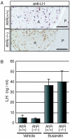
Functionally normal gonadotropes of AhR−/− pituitaries. (A) Presence of gonadotrophs in the pituitary anterior lobes of AhR−/− females. Cryosections of pituitaries isolated from AhR+/+ and AhR−/− mice were treated with an anti-LH antibody, which should specifically stain pituitary gonadotrophs. A and P represent the anterior and posterior lobes, respectively. Bar, 0.1 mm. (B) Secretion of LH from the gonadotrophs of AhR−/− pituitaries. A GnRH agonist, buserelin (2 μg), or vehicle alone was injected subcutaneously into ovariectomized AhR+/+ and AhR−/− females. One hour after injection, serum levels of LH were determined. Values are represented as means ± SD for three to four mice.
FIG. 3.
FIG. 3.
Concentrations of intraovarian steroids in AhR−/− females and the rescue of ovulation by estradiol treatment. (A) Schematic representation of the experimental procedure used to determine intraovarian steroid concentrations during the preovulatory period. (B) Intraovarian estradiol concentrations in AhR+/+ and AhR−/− females. The ovaries of at least three AhR+/+ and three AhR−/− female mice were collected at the times indicated in panel A. Estradiol concentrations were then determined by liquid chromatography-mass spectrometry analysis. *, P < 0.10; **, P < 0.05. (C) Intraovarian testosterone concentrations were determined as described for panel B. (D) Schematic representation of the experimental estradiol administration procedure used to rescue the ovulation of AhR−/− mice. Mice treated with PMSG at day 1 were divided into two groups. One group was treated with various quantities of estradiol, while the other group was given vehicle alone on day 2. The mice from both groups were treated with hCG on day 3, and ovulation was assessed at day 4. (E) Effects of estradiol administration on ovulation in AhR−/− females. After the treatment of AhR+/+ and AhR−/− females with PMSG and hCG, the oocytes released by ovulation were counted (open bars). AhR−/− females were also given an intraperitoneal injection of 5 to 20 ng 17β-estradiol (E2) or vehicle alone (filled bars) as described for panel D prior to counting the ovulated oocytes. *, P < 0.025; **, P < 0.005.
FIG. 4.
FIG. 4.
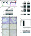
AhR regulates the expression of ovarian Cyp19 during the preovulatory period. (A) Schematic representation of the experimental procedure. The estrus cycle was induced artificially by intraperitoneal injection of PMSG at 1700 h on day 1 and of hCG at 1700 h on day 3. Ovaries were collected 40 and 48 h after PMSG injection or 4, 6, 7, and 12 h after hCG injection (indicated by arrows). (B) Profiles of mRNA expression for AhR, AhRR, and Cyp19 during the preovulatory period. Total RNA samples, prepared from ovaries derived from hormone-treated mice at the indicated times (top), were subjected to RT-PCR with primers sets specific for AhR, AhRR, and Cyp19. β-Actin mRNA was used as a control. (C) Expression of mRNAs encoding steroidogenic enzymes and proteins involved in ovarian folliculogenesis. Total RNA samples, prepared from the ovaries of hormone-treated AhR+/+ and AhR−/− mice at the indicated times (top), were used for RT-PCR with the PCR primers. (D) Quantification of Cyp19 mRNA levels. Total RNA samples, prepared from the ovaries isolated 4 and 7 h after hCG injection, were subjected to quantitative RT-PCR analyses. Three animals were used for this experiment. (E) Expression of Cyp19 protein within AhR+/+ and AhR−/− ovaries during the preovulatory period. Whole-cell extracts (10 μg), prepared from the ovaries of hormone-treated (hCG + 5 h) mice, were subjected to Western blot analysis with an anti-Cyp19 antibody. Three AhR+/+ and three AhR−/− animals were used for these experiments. (F) Immunohistochemical staining of Cyp19 and AhR in the granulosa cells of AhR+/+ and AhR−/− ovaries. Five-micrometer paraffin sections were prepared from the ovaries of hormone-treated (hCG + 5 h) mice. Sections were stained with hematoxylin-eosin (HE) or with anti-AhR or anti-Cyp19 antibody.
FIG. 5.
FIG. 5.
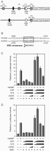
Cooperative activation of AhR and Ad4BP/SF-1 on the Cyp19/CYP19 promoter. (A) Schematic representation of the mouse Cyp19 and human CYP19 gene promoter regions. The square boxes indicate the first exons, exons I.2 (Ex 1e), I.6, I.3 (Ex 1c), and PII (Ex 1d), expressed specifically in the placenta, bone, adipose tissue, and ovary, respectively. The filled and open ovals represent the AhR/Arnt-binding (XRE) and Ad4BP/SF-1-binding (Ad4) sequences, respectively. The ovary-specific transcription start site is numbered as +1, and the positions of the XRE and Ad4 sites were numbered as the negative values of their distances from the transcription start site. (B) Nucleotide sequences containing the XRE site from the mouse Cyp19 and human CYP19 gene upstream regions. The consensus XRE sequence is indicated in bold letters. (C) Cooperative activation of AhR and Ad4BP/SF-1 on the human CYP19 gene promoter. Expression plasmids encoding AhR, Arnt, AhRR, and Ad4BP/SF-1 were cotransfected into 293 cells with a reporter plasmid, in which luciferase expression is driven by the CYP19 promoter (hCYP19-3853Luc), in the presence (+) or absence (−) of 3MC. After a 48-h incubation, cells were recovered and subjected to luciferase assays. All values are the means ± SD for three experiments. (D) Cooperative activation of AhR and Ad4BP/SF-1 on the mouse Cyp19 promoter. mCyp19-5335Luc was used for this assay. All other conditions were as specified for panel C.
FIG. 6.
FIG. 6.
Interaction of AhR with Ad4BP/SF-1 on the Cyp19 promoter. (A) Detection of a physical interaction between AhR and Ad4BP/SF-1 by coimmunoprecipitation. FLAG-tagged proteins from whole-cell extracts of 293 cells transfected with 3×FLAG-AhR, Arnt, and EGFP-Ad4BP were immunoprecipitated (IP) with an anti-FLAG antibody. The immunoprecipitates were then subjected to immunoblotting with an anti-GFP antibody. An EGFP expression vector was transfected as a control. An arrow and an arrowhead indicate the EGFP-Ad4BP and EGFP samples, respectively. (B) Schematic representation of the location of primers used in the ChIP assays. Three sets of primers were used to amplify DNA regions containing the XRE site at −5058 and the Ad4/SF-1 sequence at −92 and a third unrelated region (−2740 to −2441), containing neither of them, as a control. (C) Binding of AhR to the promoter region of the Cyp19 gene, revealed by ChIP assays. Soluble chromatin, prepared from preovulatory granulosa cells (hCG + 2 h), was subjected to ChIP assay with an anti-AhR antibody. β-Actin was used as a negative control. (D) Interaction between AhR and Ad4BP/SF-1 on the Cyp19 gene promoter. Chromatin isolated from preovulatory granulosa cells was incubated with anti-AhR or anti-Ad4BP/SF-1 antibody and then subjected to PCR with two sets of primers amplifying the XRE and Ad4 sites. A primer pair specific for the sequence from −2740 to about −2441 was used as a control. (E) Binding of Ad4BP/SF-1 to the XRE and Ad4 sites in the presence or absence of AhR, revealed by ChIP assays. Chromatin isolated from preovulatory granulosa cells of the AhR+/+ and AhR−/− ovaries was incubated with anti-Ad4BP/SF-1 or control antibody and then subjected to PCR to amplify the XRE and Ad4 sites. IgG, immunoglobulin G.
FIG. 7.
FIG. 7.
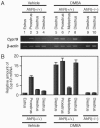
Upregulation of Cyp19 expression by an exogenous AhR ligand, DMBA. (A) Expression of Cyp19 induced by intraperitoneal injection of DMBA. AhR+/+ (lanes 1 to 8) or AhR−/− (lanes 9 and 10) female mice were injected intraperitoneally with DMBA (50 mg/kg of body weight) or vehicle alone. Five hours after injection, we prepared total RNA from the ovaries. The amounts of Cyp19 mRNA were then evaluated by RT-PCR. The estrus cycle phase of each animal was determined by observing vaginal smears collected just before injection of DMBA. β-Actin was used as a control. (B) Quantitative representation of Cyp19 mRNA levels. Quantification of the Cyp19 transcript was performed by using a 7500 real-time PCR system (Applied Biosystems, Japan).

Similar articles

Cited by

References

    1. Abbott, B. D., J. E. Schmid, J. A. Pitt, A. R. Buckalew, C. R. Wood, G. A. Held, and J. J. Diliberto. 1999. Adverse reproductive outcomes in the transgenic Ah receptor-deficient mouse. Toxicol. Appl. Pharmacol. 155:62-70. - PubMed
    1. Baba, T., J. Mimura, K. Gradin, A. Kuroiwa, T. Watanabe, Y. Matsuda, J. Inazawa, K. Sogawa, and Y. Fujii-Kuriyama. 2001. Structure and expression of the Ah receptor repressor gene. J. Biol. Chem. 276:33101-33110. - PubMed
    1. Benedict, J. C., T. M. Lin, I. K. Loeffler, R. E. Peterson, and J. A. Flaws. 2000. Physiological role of the aryl hydrocarbon receptor in mouse ovary development. Toxicol. Sci. 56:382-388. - PubMed
    1. Benedict, J. C., K. P. Miller, T. M. Lin, C. Greenfeld, J. K. Babus, R. E. Peterson, and J. A. Flaws. 2003. Aryl hydrocarbon receptor regulates growth, but not atresia, of mouse preantral and antral follicles. Biol. Reprod. 68:1511-1517. - PubMed
    1. Botrugno, O. A., E. Fayard, J. S. Annicotte, C. Haby, T. Brennan, O. Wendling, T. Tanaka, T. Kodama, W. Thomas, J. Auwerx, and K. Schoonjans. 2004. Synergy between LRH-1 and beta-catenin induces G1 cyclin-mediated cell proliferation. Mol. Cell 15:499-509. - PubMed

Publication types

MeSH terms

  NODES
admin 3
Association 1
twitter 2