. 2005 Dec 21;24(24):4368-80.
doi: 10.1038/sj.emboj.7600903. Epub 2005 Dec 15.

XBP-1 is required for biogenesis of cellular secretory machinery of exocrine glands

Affiliations

XBP-1 is required for biogenesis of cellular secretory machinery of exocrine glands

Ann-Hwee Lee et al. EMBO J. .

Abstract

The secretory function of cells relies on the capacity of the endoplasmic reticulum (ER) to fold and modify nascent polypeptides and to synthesize phospholipids for the subsequent trafficking of secretory proteins through the ER-Golgi network. We have previously demonstrated that the transcription factor XBP-1 activates the expression of certain ER chaperone genes and initiates ER biogenesis. Here, we have rescued the embryonic lethality of XBP-1 deficient fetuses by _targeting an XBP-1 transgene selectively to hepatocytes (XBP-1-/-;LivXBP1). XBP-1-/-;LivXBP1 mice displayed abnormalities exclusively in secretory organs such as exocrine pancreas and salivary gland that led to early postnatal lethality from impaired production of pancreatic digestive enzymes. The ER was poorly developed in pancreatic and salivary gland acinar cells, accompanied by decreased expression of ER chaperone genes. Marked apoptosis of pancreatic acinar cells was observed during embryogenesis. Thus, the absence of XBP-1 results in an imbalance between the cargo load on the ER and its capacity to handle it, leading to the activation of ER stress-mediated proapoptotic pathways. These data lead us to propose that XBP-1 is both necessary and sufficient for the full biogenesis of the secretory machinery in exocrine cells.


Figures
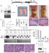
Figure 1
Figure 1
Rescue of embryonic lethality by liver specific expression of an XBP-1 transgene. (A) Transgenic construct to drive liver-specific XBP-1 expression. A mouse XBP-1 cDNA was inserted into the pLiv.7 vector using the promoter and the polyadenylation element of the apolipoprotein E gene (Miyake et al, 2001; Simonet et al, 1993). (B) Total RNA was isolated from the liver of 2-day-old mice of different genotypes for Northern blot analysis. XBP-1 mRNA species produced from the WT and the XBP-1 null allele are indicated. LivXBP1 mice expressed transgenic XBP-1 mRNA at the expected size of ∼1.7 kb as well as another species of ∼2.2 kb, which could be generated by alternative mRNA splicing. (C) A pair of newborn littermates, XBP-1+/− and XBP-1−/−;LivXBP1 mice. The milk spot on their left sides indicates that both have fed since birth. (D) Body weights of XBP-1+/− and XBP-1−/−;LivXBP1 mice were measured at day 2 and 5 after birth (n⩾4). (E) Blood glucose levels of XBP-1+/− (n=6), XBP-1+/−;LivXBP1 (n=11), and XBP-1−/−;LivXBP1 (n=14) mice measured with FreeStyle™ glucometer.(F, G) Abdominal contents of 3-day-old control and XBP-1−/−;LivXBP1 mice. Liver (Lv), stomach (St), small intestine (SI) are indicated. In XBP-1−/−;LivXBP1 mice, the small intestine is markedly distended (resulting in the rostral displacement of the liver) and pale in color, reflecting large amounts of undigested milk. (H, I) Hematoxylin and eosin (H&E) staining of the duodenum of heterozygous XBP-1+/− and XBP-1−/−;LivXBP1 mice. Note the eosinophilic staining of undigested milk in the XBP-1−/−;LivXBP1 mice (I). (J) Total RNAs isolated from various organs of WT and the XBP-1−/−;LivXBP1 mice were subjected to Northern blot analysis to measure the expression level of XBP-1. Bands corresponding to the WT, null and the transgenic XBP-1 mRNA are indicated. Ethidium bromide staining of the gel is shown at the bottom as a loading control. (K) Total RNA was isolated from the submandibular salivary gland and the adrenal gland. XBP-1 mRNA levels were measure by real time PCR analysis. The primers used do not recognize the mutant mRNA produced from the XBP-1 null allele. (L–N) H&E staining of E 18.5 liver of XBP-1+/− (L), XBP-1+/−;LivXBP1 (M) and XBP-1−/−;LivXBP1 rescued mice (N) reveals normal liver histology. (O) Pancreas lysates were tested for the expression of XBP-1s protein by Western blot analysis using anti XBP-1 antibody. Scale (H, I) 150 μm, (L–N) 40 μm.
Figure 2
Figure 2
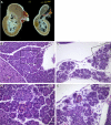
Development of the exocrine pancreas is impaired in the absence of XBP-1. (A) The stomach (St), duodenum (D), spleen (Sp) and pancreas (P) were removed en bloc from P2 mice for photography. Dashed lines outline the superior border of the control pancreas. Asterisks indicate additional areas of pancreatic parenchyma. (B–E) Histologic sections of control heterozygous XBP-1+/− (B, D) and XBP-1−/−;LivXBP1 (C, E) pancreas. Boxed regions in B and C are shown at higher magnifications in D and E, respectively. (L) Liver; (I) Islet. Dashed line outlines a representative acinar unit (A) in control and mutant. Scale (B,C) 100 μm, (D,E) 30 μm.
Figure 3
Figure 3
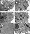
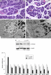
Ultrastructure of pancreatic acini reveals defects in zymogen granules and endoplasmic reticulum. Electron micrographs of sections of acini from WT and XBP-1−/−;LivXBP1 mice. (A–D) While pancreatic cells from WT mice (A, C) have abundant apically located membrane-bound zymogen granules (asterisks), pancreatic acinar cells of the XBP-1−/−;LivXBP1 mice have only a few, small apical granules (B, arrows), as well as immature granule precursors located inside of the endoplasmic reticulum (ER) lumen (D, arrowheads). N, nucleus. L, lumen. (E, F) Electron micrographs of the basolateral portion of acinar cells from WT and mutant mice. The ER contains elaborately organized long, thin, densely packed cisternae. In mutants, the ER is poorly organized and sparse. Scale, 2 μm in A and B; 500 nm in C–F.
Figure 4
Figure 4
Reduced production of pancreatic digestive enzymes in XBP-1−/−;LivXBP1 mice. (A) Western blot analysis of whole-pancreas lysates to detect α-amylase and trypsin. The blot was stained with Ponceau S after transfer. (B) Measurement of zymogen mRNAs. Total RNAs were isolated from WT, XBP-1+/−;LivXBP1, and XBP-1−/−;LivXBP1 pancreas. Expression of the indicated zymogen mRNAs was measured by real time PCR analysis. Values were normalized to β-actin. (C) Expression of select XBP-1 _target genes and pancreas specific chaperone genes, as well as (D) CHOP were measured by real time PCR analysis in WT and XBP-1−/−;LivXBP1 pancreas. The expression levels of each gene are shown as fold induction over WT. Asterisks indicate genes whose expression was significantly impaired in the absence of XBP-1. N=2–4 mice per group.
Figure 5
Figure 5
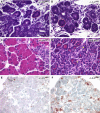
XBP-1-deficient pancreatic acinar cells undergo marked apoptosis during embryonic development. (A, B) Hematoxylin and eosin staining of XBP-1+/−;LivXBP1 control and XBP-1−/−;LivXBP1 E15.5 pancreas. At this stage, acini (red dashed circles) have begun to bud from ducts but granules have only begun to accumulate. (C, D) At E18.5, bright eosinophilic granules show extensive accumulation in the XBP-1+/−; LivXBP1 pancreas but are drastically reduced in mutant acini. Apoptotic cells can be seen in the mutant. (E, F) TUNEL staining of XBP-1+/−; LivXBP1 and XBP-1−/−;LivXBP1 E18.5 pancreas highlights numerous apoptotic acinar cells in the mutant but few in wild type. Scale bar (A–F), 30 μm.
Figure 6
Figure 6
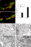
Normal development of the endocrine pancreas in the absence of XBP-1. (A, B) Immunostaining of WT and the XBP-1−/−;LivXBP1 islets with insulin (green) and glucagon (red) antibodies. (C) Expression level of insulin and glucagon mRNA. Total RNA was isolated from whole pancreas of mice with the indicated genotypes and transcript levels quantified by real time PCR analysis. Values were normalized to β-actin. (D, E) α-Cells containing numerous homogeneous electron-dense glucagon granules (red arrowheads) were identified by electron microscopy in WT and XBP-1−/−;LivXBP1 mice. (F, G) β-Cells containing slightly heterogeneous and angular insulin-containing granules (green arrowheads) could be found in both WT and mutant XBP-1−/−;LivXBP1 mice. Scale (D–G, 500 nm).
Figure 7
Figure 7
Impaired development of salivary glands in XBP-1−/−;LivXBP1 mice. (A, B) Hematoxylin and eosin staining of the submandibular salivary glands in the control XBP-1+/−;LivXBP1 and mutant XBP-1−/−;LivXBP1 mice. The lobules comprised of secretory acini (A, red dashed lines) appear hypoplastic in the mutant compared to controls. (D) Secretory ducts. (C, D) Electron micrographs of serous salivary glands of control and mutant XBP-1−/−;LivXBP1 mice. Similar to pancreatic acini, these salivary gland acini have well-developed endoplasmic reticulum and apically located zymogen granules (Z). In the mutant, the ER is less well developed but some zymogen granules are produced. (E) Whole-cell lysates of the submandibular salivary glands were subjected to Western blot analysis to measure amylase expression. Scale A and B, 50 μm; C and D, 2 μm. (F) Expression of select XBP-1 _target genes and CHOP were measured by real time PCR analysis in WT and XBP-1−/−;LivXBP1 salivary glands. The expression levels of each gene are shown as fold induction over WT. Asterisks indicate genes whose expression was significantly impaired in the absence of XBP-1.
Figure 8
Figure 8
Forced expression of XBP-1 in progenitor B cells results in elevated immunoglobulin production in vivo. Lethally irradiated 8-week old Rag2−/− hosts were reconstituted with WT hematopoietic stem cells (HSCs) infected with RVGFP-XBP-1s retrovirus or empty RVGFP-control retrovirus, N=4–6 mice per group. Reconstituted animals were analyzed after 9 weeks by harvesting sera to measure levels of IgM, IgG1 and IgG2a by ELISA. The P-values are shown.

Similar articles

Cited by

References

    1. Aridor M, Balch WE (1999) Integration of endoplasmic reticulum signaling in health and disease. Nat Med 5: 745–751 - PubMed
    1. Beaudoin AR, Grondin G (1991) Secretory pathways in animal cells: with emphasis on pancreatic acinar cells. J Electron Microsc Tech 17: 51–69 - PubMed
    1. Bertolotti A, Zhang Y, Hendershot LM, Harding HP, Ron D (2000) Dynamic interaction of BiP and ER stress transducers in the unfolded-protein response. Nat Cell Biol 2: 326–332 - PubMed
    1. Calfon M, Zeng H, Urano F, Till JH, Hubbard SR, Harding HP, Clark SG, Ron D (2002) IRE1 couples endoplasmic reticulum load to secretory capacity by processing the XBP-1 mRNA. Nature 415: 92–96 - PubMed
    1. Castle JD, Castle AM (1993) Sorting and secretion of salivary proteins. Crit Rev Oral Biol Med 4: 393–398 - PubMed

Publication types

MeSH terms

  NODES
Note 1
twitter 2