CD8+ T cells but not polymorphonuclear leukocytes are required to limit chronic oral carriage of Candida albicans in transgenic mice expressing human immunodeficiency virus type 1
- PMID: 16552068
- PMCID: PMC1418920
- DOI: 10.1128/IAI.74.4.2382-2391.2006
CD8+ T cells but not polymorphonuclear leukocytes are required to limit chronic oral carriage of Candida albicans in transgenic mice expressing human immunodeficiency virus type 1
Abstract
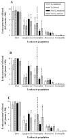
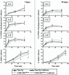
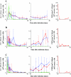
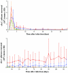
Candida albicans causes oropharyngeal candidiasis (OPC) but rarely disseminates to deep organs in human immunodeficiency virus (HIV) infection. Here, we used a model of OPC in CD4C/HIV(Mut) transgenic (Tg) mice to investigate the role of polymorphonuclear leukocytes (PMNs) and CD8+ T cells in limiting candidiasis to the mucosa. Numbers of circulating PMNs and their oxidative burst were both augmented in CD4C/HIV(MutA) Tg mice expressing rev, env, and nef of HIV type 1 (HIV-1), while phagocytosis and killing of C. albicans were largely unimpaired compared to those in non-Tg mice. Depletion of PMNs in these Tg mice did not alter oral or gastrointestinal burdens of C. albicans or cause systemic dissemination. However, oral burdens of C. albicans were increased in CD4C/HIV(MutG) Tg mice expressing only the nef gene of HIV-1 and bred on a CD8 gene-deficient background (CD8-/-), compared to control or heterozygous CD8+/- CD4C/HIV(MutG) Tg mice. Thus, CD8+ T cells contribute to the host defense against oral candidiasis in vivo, specifically in the context of nef expression in a subset of immune cells.
Figures


Similar articles
-
Altered CD4+ T cell phenotype and function determine the susceptibility to mucosal candidiasis in transgenic mice expressing HIV-1.J Immunol. 2006 Jul 1;177(1):479-91. doi: 10.4049/jimmunol.177.1.479. J Immunol. 2006. PMID: 16785545
-
Macrophage-mediated responses to Candida albicans in mice expressing the human immunodeficiency virus type 1 transgene.Infect Immun. 2009 Sep;77(9):4136-49. doi: 10.1128/IAI.00453-09. Epub 2009 Jun 29. Infect Immun. 2009. PMID: 19564379 Free PMC article.
-
Oral mucosal cell response to Candida albicans in transgenic mice expressing HIV-1.Methods Mol Biol. 2009;470:359-68. doi: 10.1007/978-1-59745-204-5_25. Methods Mol Biol. 2009. PMID: 19089395
-
Candida-host interactions in HIV disease: relationships in oropharyngeal candidiasis.Adv Dent Res. 2006 Apr 1;19(1):80-4. doi: 10.1177/154407370601900116. Adv Dent Res. 2006. PMID: 16672555 Review.
-
Candida and candidiasis in HIV-infected patients: where commensalism, opportunistic behavior and frank pathogenicity lose their borders.AIDS. 2012 Jul 31;26(12):1457-72. doi: 10.1097/QAD.0b013e3283536ba8. AIDS. 2012. PMID: 22472853 Review.
Cited by
-
Altered immune response differentially enhances susceptibility to Cryptococcus neoformans and Cryptococcus gattii infection in mice expressing the HIV-1 transgene.Infect Immun. 2013 Apr;81(4):1100-13. doi: 10.1128/IAI.01339-12. Epub 2013 Jan 22. Infect Immun. 2013. PMID: 23340313 Free PMC article.
-
Total-Body Irradiation Exacerbates Dissemination of Cutaneous Candida Albicans Infection.Radiat Res. 2016 Nov;186(5):436-446. doi: 10.1667/RR14295.1. Epub 2016 Oct 6. Radiat Res. 2016. PMID: 27710703 Free PMC article.
-
Adaptive immunity to fungi.Cold Spring Harb Perspect Med. 2014 Nov 6;5(3):a019612. doi: 10.1101/cshperspect.a019612. Cold Spring Harb Perspect Med. 2014. PMID: 25377140 Free PMC article. Review.
-
Mouse model of oropharyngeal candidiasis.Nat Protoc. 2012 Mar 8;7(4):637-42. doi: 10.1038/nprot.2012.011. Nat Protoc. 2012. PMID: 22402633 Free PMC article.
-
Defective IL-17- and IL-22-dependent mucosal host response to Candida albicans determines susceptibility to oral candidiasis in mice expressing the HIV-1 transgene.BMC Immunol. 2014 Oct 26;15:49. doi: 10.1186/s12865-014-0049-9. BMC Immunol. 2014. PMID: 25344377 Free PMC article.
References
-
- Balish, E., J. Jensen, T. Warner, J. Brekke, and B. Leonard. 1993. Mucosal and disseminated candidiasis in gnotobiotic SCID mice. J. Med. Vet. Mycol. 31:143-154. - PubMed
-
- Bandres, J. C., J. Trial, D. M. Musher, and R. D. Rossen. 1993. Increased phagocytosis and generation of reactive oxygen products by neutrophils and monocytes of men with stage 1 human immunodeficiency virus infection. J. Infect. Dis. 168:75-83. - PubMed
-
- Beno, D. W., A. G. Stover, and H. L. Mathews. 1995. Growth inhibition of Candida albicans hyphae by CD8+ lymphocytes. J. Immunol. 154:5273-5281. - PubMed
Publication types
MeSH terms
Substances
LinkOut - more resources
Full Text Sources
Medical
Molecular Biology Databases
Research Materials
Miscellaneous

