. 2006 May;2(5):e39.
doi: 10.1371/journal.ppat.0020039. Epub 2006 May 12.

HIV-1 Vpu promotes release and prevents endocytosis of nascent retrovirus particles from the plasma membrane

Affiliations

HIV-1 Vpu promotes release and prevents endocytosis of nascent retrovirus particles from the plasma membrane

Stuart J D Neil et al. PLoS Pathog. 2006 May.

Abstract

The human immunodeficiency virus (HIV) type-1 viral protein U (Vpu) protein enhances the release of diverse retroviruses from human, but not monkey, cells and is thought to do so by ablating a dominant restriction to particle release. Here, we determined how Vpu expression affects the subcellular distribution of HIV-1 and murine leukemia virus (MLV) Gag proteins in human cells where Vpu is, or is not, required for efficient particle release. In HeLa cells, where Vpu enhances HIV-1 and MLV release approximately 10-fold, concentrations of HIV-1 Gag and MLV Gag fused to cyan fluorescent protein (CFP) were initially detected at the plasma membrane, but then accumulated over time in early and late endosomes. Endosomal accumulation of Gag-CFP was prevented by Vpu expression and, importantly, inhibition of plasma membrane to early endosome transport by dominant negative mutants of Rab5a, dynamin, and EPS-15. Additionally, accumulation of both HIV and MLV Gag in endosomes required a functional late-budding domain. In human HOS cells, where HIV-1 and MLV release was efficient even in the absence of Vpu, Gag proteins were localized predominantly at the plasma membrane, irrespective of Vpu expression or manipulation of endocytic transport. While these data indicated that Vpu inhibits nascent virion endocytosis, Vpu did not affect transferrin endocytosis. Moreover, inhibition of endocytosis did not restore Vpu-defective HIV-1 release in HeLa cells, but instead resulted in accumulation of mature virions that could be released from the cell surface by protease treatment. Thus, these findings suggest that a specific activity that is present in HeLa cells, but not in HOS cells, and is counteracted by Vpu, traps assembled retrovirus particles at the cell surface. This entrapment leads to subsequent endocytosis by a Rab5a- and clathrin-dependent mechanism and intracellular sequestration of virions in endosomes.


Conflict of interest statement

Competing interests. The authors have declared that no competing interests exist.

Figures

Figure 1
Figure 1. Cell Type–Dependent Effects of Vpu on Retroviral Particle Release
Human HeLa and HOS cells, together with AGM Vero cells, were transfected with HIV-1 NL4.3 and NL4.3delVpu proviral plasmids, or an MLV Gag-Pol–expression vector, as indicated, in the presence of plasmids expressing either no Vpu (none), codon-optimized Vpu, or an inactive Vpu mutant containing a CD8 TM domain (V8). (A and B) Western blot analyses of cell and virion lysates, probed using antibodies to HIV-1 (A) or MLV CA (B). (C and D) Results of chemiluminsecent β-galactosidase assays following inoculation of HeLa-TZM indicator cells with supernatants derived from HeLa, HOS, or Vero cells transfected with NL4.3 (white bars) or NL4.3delVpu (black bars) proviral plasmids (C) or plasmids expressing MLV Gag-Pol, a packageable HIV-1 Tat–expression vector, and VSV-G (D). Vpu and control (GFP)–expression plasmids were included, as indicated, and results plotted as relative light units (RLU) ± standard deviation of the mean.
Figure 2
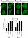
Figure 2. Expression of Vpu Prevents Intracellular Accumulation of Retroviral Gag-CFP Fusion Proteins in HeLa Cells
Cells were transfected with the indicated expression vectors, fixed 18 h after transfection, and observed by deconvolution microscopy. (A and B) Localization of HIV-1 Gag-CFP or MLV Gag-CFP in HeLa cells (A) or HOS cells (B) in the absence of Vpu. Two examples of each cell type and each Gag-CFP protein are shown. White bars in the micrographs indicate a distance of 10 μm. (C) Quantitative analysis of these effects. Ten random fields of HeLa and HOS cells expressing HIV-1 or MLV Gag-CFP proteins in the presence or absence of Vpu were inspected, and the numbers of cells in which Gag-CFP was observed as accumulations at the PM only (black bars), at intracellular sites as well as at the PM (white bars), or as diffuse cytoplasmic fluorescence only (grey bars) were counted. Enumeration of cells with each pattern of Gag-CFP localization is expressed as a percentage the total of the cells counted, which was 75 ± 7 for each condition and is a representative example of three separate experiments. (D) Examples of HeLa cells expressing HIV-1 Gag-CFP (upper panels) or MLV Gag CFP (lower panels) in the presence of co-expressed Vpu. (E and F) The Vpu TM domain is required to prevent intracellular accumulation of HIV-1 Gag-CFP. (E) Examples of HeLa cells expressing HIV-1 Gag-CFP and V8. White bars in the micrographs indicate a distance of 10 μm. (F) Cells expressing HIV-1 Gag-CFP only, Gag-CFP and Vpu, or Gag-CFP and V8 were inspected and enumerated as described in (C), except that cells containing only diffuse Gag-CFP and no visible accumulations were excluded from the analysis.
Figure 3
Figure 3. Intracellular HIV-1 Gag-CFP Partly Co-localizes with both Early and Late Endosomal Markers in HeLa Cells
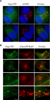
(A) HeLa cells expressing HIV-1 Gag-CFP (green) were fixed 18 h post-transfection and stained with a monoclonal antibody specific for human CD63, and a secondary anti-mouse Alexa-Fluor-588 conjugate (red). Nuclei were counter-stained with DAPI (blue). (B) HeLa cells expressing HIV-1 Gag-CFP (green) were co-transfected with CherryFP-Rab5a (red), a marker for early endosomal structures. Cells were fixed and images acquired at 18 h post-transfection. The lower set of images presented in (B) are shown at approximately 5-fold higher magnification to give a clearer indication of the juxtaposition of the HIV-1 Gag-CFP and CherryFP-Rab5a signals. White bars in the micrographs indicate a distance of 10 μm, except in the lower set of images in (B), where the bar indicates 2 μm
Figure 4
Figure 4. HIV-1 Gag-CFP Accumulation at the PM Precedes Accumulation at Intracellular Sites in HeLa Cells in the Absence of Vpu Expression
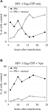
(A and B) HeLa cells were transfected with HIV-1 Gag-CFP and either an irrelevant plasmid control (A) or a Vpu-expression plasmid (B). The cells were washed 5 h post-transfection and then fixed at 2-h intervals thereafter. The number of cells in ten fields that exhibited HIV-1 Gag-CFP accumulations at the PM only or at the PM and intracellular sites, or which expressed only diffuse Gag-CFP, were enumerated as in Figure 1C and plotted as the percentage of the total. Cells containing only diffuse Gag-CFP have been omitted from the charts for simplicity.
Figure 5
Figure 5. DN Mutants of Cellular Proteins that Mediate PM-to-Early-Endosome Transport Affect Gag-CFP Localization in HeLa, but Not HOS Cells
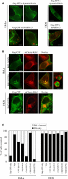
(A) HeLa cells (left panels) or HOS cells (right panels) were transfected with plasmids expressing HIV-1 Gag-CFP (green) and either dynamin K44A (no color) or dnEPS-15-YFP (red). Cells were fixed at 18 h post-transfection and analyzed using deconvolution microscopy. (B) HeLa cells (upper and center panels) expressing HIV-1 Gag-CFP (green), and either DN (S34N) or constitutively active (Q79L) mutants of CherryFP-Rab5a (red), were fixed and images acquired 18 h post-transfection. The far-right-center panel shows an example of how localization of HIV-1 Gag-CFP in swollen CherryRab5aQ79L early endosomes could be resolved as apparently particulate fluorescence within a vesicle lumen. (B, lower panels) shows a similar analysis HIV-1 Gag-CFP localization (green) in the presence of CherryFP-Rab5a(Q79L) in HOS cells. White bars in the micrographs indicate a distance of 10 μm, except in the far-right-center panel of (B) where a distance of 1 μm is indicated. (C) The numbers of HIV-1 Gag-CFP–expressing cells in ten fields exhibiting PM Gag-CFP accumulation only (black bars) or additional intracellular accumulations (white bars) in the presence of the indicated co-expressed cellular proteins was counted, as in Figure 2C, and is plotted as a percentage of the total number of cells with Gag-CFP accumulations.
Figure 6
Figure 6. Immunofluorescent Localization of HIV-1 Gag Expressed from a Proviral Construct and Effects of Vpu and Endocytosis Inhibitors
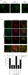
(A) HeLa cells (left and center panels) or HOS cells (right panel) were transfected with NL4.3 or NL4.3delVpu proviral plasmids, as indicated. Gag localization was determined by immunofluorescence using an α-CA antibody as described in Materials and Methods. (B) HeLa cells expressing Gag from the NL4.3delVpu proviral plasmid, and either DN (S34N, upper panels) or constitutively active (Q79L center and lower panels) mutants of CherryFP-Rab5a (red), were fixed and subjected to immunofluorescence to detect HIV-1 Gag (green) at 20 h post-transfection. White bars in the micrographs indicate a distance of 10 μm. The lower three images in (B) show expanded views of a portion of the overlay panels (indicated by the dashed squares) to exemplify how HIV-1 Gag and the CherryRab5a proteins are juxtaposed, Note that Gag sometimes appears to be within the lumen of the CherryFP-Rab5a(Q79L)–marked endosomes. (C) The numbers of HIV-1 Gag–expressing cells in ten fields exhibiting PM Gag immunofluorescence only (black bars) or additional intracellular accumulations (white bars) after transfection with NL4.3 or NL4.3delVpu proviral plasmids in the presence of the indicated co-expressed cellular proteins was counted, as in Figure 2C. Data are plotted as a percentage of the total number of cells with visible Gag accumulations.
Figure 7
Figure 7. Electron Microscopy Examination of HIV-1 Assembly and Effects of Vpu and Endocytosis Inhibition
Hela cells expressing Gag-Pol in the presence or absence of Vpu or Rab5a(S34N) were examined. (A–C) Examples of cells expressing Gag-Pol only, revealing mature particles at both the PM (A and C) and within internal, apparently membrane-bound compartments (arrows in A and B). Immature budding structure are also observed at the PM but only rarely within endosomes. (D) Cells co-expressing Gag-Pol and Vpu with reduced numbers of cell-associated particles, and absence of particles from endosomes. (E and F) Cells co-expressing Gag-Pol and Rab5a(S34N), with exclusively surface-accumulated virions.
Figure 8
Figure 8. Vpu Expression Has No Effect on Transferrin Endocytosis
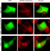
HeLa cells were transfected with a control plasmid, or with plasmids expressing dynamin K44A, or Vpu together with a GFP-expression plasmid to mark transfected cells. Five hours after transfection, cells were transferred to serum-free medium. After overnight incubation, cells were then washed and incubated in fresh medium containing 5 μg/ml of transferrin conjugated to Alexa-Fluor-568. After 15 min of incubation, cells were fixed and images acquired.
Figure 9
Figure 9. Inhibition of Endocytosis or Early Endosome Function Does Not Restore Vpu-Deficient HIV-1 Release
HeLa cells were transfected with NL4.3 (A) or NL4.3delVpu (B) proviral plasmids along with plasmids expressing GFP or Vpu (as controls), dynamin K44A, DN EPS-15, or wild-type and mutant Rab5a proteins. Cell and virus lysates were harvested 48 h post-transfection, and analyzed by Western blotting with an HIV-1 CA-specific monoclonal antibody
Figure 10
Figure 10. Endosomal Accumulation of HIV-1 and MLV Gag-CFP Requires a Functional L-Domain
HeLa cells were transfected with plasmids expressing HIV-1 (A) or MLV Gag-CFP (B), or mutant variants thereof lacking functional L-domains (HIV-1 Gag dLD-CFP and MLV Gag dPY-CFP, respectively) in the presence or absence of co-expressed Vpu, as indicated. The cells were fixed at 18 h post-transfection and images acquired. Representative examples are shown in panels (A) and (B) and a quantitative assessment of Gag-CFP localization, enumerated as in Figure 1, is shown (C). The numbers of HIV-1 Gag-CFP–expressing cells in ten fields exhibiting Gag-CFP accumulation only at the PM (black bars) or at intracellular sites (white bars) were counted, as in Figure 2C, and are plotted as a percentage of the total number of cells with Gag-CFP accumulations.
Figure 11
Figure 11. Absence of Vpu Results in Cell-Surface Entrapment of Mature Virions
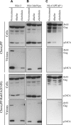
Subtilisin-induced release of mature, Vpu-defective HIV-1 particles from HeLa cells. Hela cells were transfected with NL4.3 (A), NL4.3delVpu (B), or NL4.3 (PTAP) (C) proviral plasmids as indicated, in the presence of CherryFP-Rab5a(S34N) or CherryFP-expression vectors. Cells and supernatants were harvested 48 h post-transfection and analyzed directly (no treatment), while cells in replicate wells were either treated with 1 mg/ml subtilisin (+ subtilisin) or a buffer control (+ buffer) for 15 min at 37 °C. Virions released by buffer or subtilisin and corresponding cell lysates were harvested and analyzed by Western blotting with an anti p24CA antibody as before.
Figure 12
Figure 12. Model for the Role of Vpu in Retroviral Particle Release
Retroviral Gag molecules _targeted to the PM (by unknown mechanisms) require the recruitment of class E VPS factors to enable budding of mature virions. At this point, nascent viral particles are subject to an unknown, subtilisin-sensitive, host-cell type-specific restriction (depicted as a question mark) that results in their retention at the cell surface. Subsequently, endocytosis by a dynamin-, EPS-15-, and Rab5a-dependent process result in intracellular accumulation in early/late endosomes, where they may be simply sequestered, or perhaps destroyed, in lysosomes. Vpu overcomes this restriction, leading to more efficient release of virions from the cell surface. In contrast, inhibition of endocytosis by DN mutants of dynamin, EPS-15, or Rab5a prevents the accumulation of virions in endosomes, but does not enhance virion release, suggesting Vpu inhibits a specific host-restriction activity, rather than imposing a generalized inhibition on endocytosis.

Similar articles

Cited by

References

    1. Cohen EA, Terwilliger EF, Sodroski JG, Haseltine WA. Identification of a protein encoded by the Vpu gene of HIV-1. Nature. 1988;334:532–534. - PubMed
    1. Strebel K, Klimkait T, Martin MA. A novel gene of HIV-1, Vpu, and its 16-kilodalton product. Science. 1988;241:1221–1223. - PubMed
    1. Huet T, Cheynier R, Meyerhans A, Roelants G, Wain-Hobson S. Genetic organization of a chimpanzee lentivirus related to HIV-1. Nature. 1990;345:356–359. - PubMed
    1. Courgnaud V, Salemi M, Pourrut X, Mpoudi-Ngole E, Abela B, et al. Characterization of a novel simian immunodeficiency virus with a Vpu gene from greater spot-nosed monkeys (Cercopithecus nictitans) provides new insights into simian/human immunodeficiency virus phylogeny. J Virol. 2002;76:8298–8309. - PMC - PubMed
    1. Courgnaud V, Abela B, Pourrut X, Mpoudi-Ngole E, Loul S, et al. Identification of a new simian immunodeficiency virus lineage with a Vpu gene present among different Cercopithecus monkeys (C. mona, C. cephus, and C. nictitans) from Cameroon. J Virol. 2003;77:12523–12534. - PMC - PubMed

Publication types

MeSH terms

Substances

  NODES
INTERN 2
Note 1
twitter 2