. 2007 Jan;27(1):54-64.
doi: 10.1128/MCB.01365-06. Epub 2006 Oct 30.

TBP is differentially regulated by c-Jun N-terminal kinase 1 (JNK1) and JNK2 through Elk-1, controlling c-Jun expression and cell proliferation

Affiliations

TBP is differentially regulated by c-Jun N-terminal kinase 1 (JNK1) and JNK2 through Elk-1, controlling c-Jun expression and cell proliferation

Shuping Zhong et al. Mol Cell Biol. 2007 Jan.

Abstract

Emerging evidence supports the idea that the c-Jun N-terminal kinases (JNKs) possess overlapping but distinct functions. The potential roles of the ubiquitously expressed JNK1 and JNK2 in regulating expression of the central transcription initiation factor, TATA-binding protein (TBP), were examined. Relative to wild-type fibroblasts, TBP was decreased in Jnk1(-/-) cells and increased in Jnk2(-/-) cells. Similarly, reduction of JNK1 in human hepatoma cells decreased TBP expression, whereas reduction of JNK2 enhanced it. JNK-mediated regulation of TBP expression occurs at the transcriptional level through their ability to _target Elk-1, which directly regulates the TBP promoter in response to epidermal growth factor stimulation. JNK1 increases, whereas JNK2 decreases, the phosphorylation state of Elk-1, which differentially affects Elk-1 occupancy at a defined site within the TBP promoter. These JNK-mediated alterations in TBP expression, alone, serve to regulate c-Jun expression and fibroblast proliferation rates. These studies uncovered several new molecular events that distinguish the functions of JNK1 and JNK2 that are critical for their regulation of cellular proliferation.


Figures

FIG. 1.
FIG. 1.
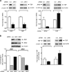
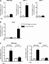
TBP expression is differentially regulated in mouse embryo fibroblasts deficient for JNK1 or JNK2 (A) JNK1 and JNK2 expression in MEFs. RT-PCR was performed using RNA isolated from each of the MEFs and specific primers for JNK1 and JNK2. (B) TBP mRNA expression in MEFs. Serum-starved cells were treated with or without EGF, and RT-PCR was performed using total RNA and specific primers for TBP or β-actin as indicated. (C) TBP levels in MEFs. Cells were treated with or without anisomycin. Protein lysates from each cell line were isolated, and immunoblot analysis was performed to detect the two phosphorylated isoforms each of JNK1 (p-JNK1) and JNK2 (p-JNK2), total JNK1 and JNK2, or β-actin antibodies as designated. A representative result of at least three independent determinations is shown. (D) Induction of TBP promoter activity requires JNK1. Wild-type, Jnk1−/−, or Jnk2−/− MEFs were transfected with a human TBP promoter-luciferase construct (p−4500/+66hTBP-Luc) alone or together with either JNK1 or JNK2 expression plasmids as indicated. Cells were treated with or without EGF (top panel) or anisomycin (bottom panel). Protein lysates were prepared, and luciferase activity was measured. The change in TBP promoter activity was calculated relative to that in nontreated cells. (E) Expression of HA-JNK1, HA-JNK2, TBP, and β-actin in cells deficient for JNK1 or JNK2. Jnk1−/− or Jnk2−/− MEFs were transfected with expression plasmids for HA-JNK1 or HA-JNK2. Protein lysates were isolated, and immunoblot analysis was used to detect HA-JNKs using antibodies against HA. The membranes were stripped and reprobed with antibodies against TBP or β-actin.
FIG. 2.
FIG. 2.
TBP expression is differentially regulated by reducing JNK1 or JNK2 expression in Huh-7 cells (A) Analysis of changes in TBP mRNA expression upon reduction of JNK1 or JNK2. Huh-7 cells were transfected with an siRNA selective for JNK1, JNK2, or mismatch siRNAs. RT-PCR was performed using RNA isolated from the Huh-7 cells and specific primers for JNK1, JNK2, TBP, or β-actin. (B) Reduction of JNK1 or JNK2 has opposing effects on TBP levels. Protein lysates derived from the siRNA-transfected cells were subjected to immunoblot analysis using antibodies against JNKs and β-actin (left) or TBP and β-actin (right) as indicated.
FIG. 3.
FIG. 3.
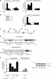
Elk-1 directly modulates EGF-induced TBP promoter activity and is differentially regulated by JNK1 and JNK2. (A) JNK1 and JNK2 regulate TBP promoter activity through a common Ets transcription factor binding site. Huh-7 cells were transfected with either a p−736/+66hTBP-Luc or p−736/+66hEtsmutTBP-Luc construct. In addition, cells were transfected with mismatch siRNA (−) or JNK1 siRNA (+) (left panel) or mismatch siRNA (−) or JNK2 siRNA (+) (right panel). Where designated, cells were additionally treated with EGF. Protein lysates were prepared, and luciferase activity was measured. Changes (fold) were calculated based on the control (TBP promoter plus mm siRNA) and normalized to total protein. (B) Elk-1 regulates TBP expression. Huh7 cells were transfected with either mismatch siRNA or Elk-1 siRNA. Immunoblot analysis was performed using lysates prepared from transfected cells using antibodies against Elk-1, TBP, and β-actin (left panel). Cells were additionally transfected with either p−736/+66hTBP-Luc or p−736/+66hEtsmutTBP-Luc (right panel), and TBP promoter activity was measured as described for panel A. (C) Schematic diagram of a genomic fragment containing the human TBP promoter. The boxed Ets motif designates the Ets binding site that is mutated in the p−736/+66hEtsmutTBP-Luc construct used. The arrows depict the relative location of the PCR primers used for the ChIP analysis. (D) Elk-1 binds to the TBP promoter upon EGF stimulation. ChIP assays were performed as described in Materials and Methods. The resultant chromatin was immunoprecipitated with Elk-1 antibody or a control antibody. Specific DNA fragments were quantified by PCR using primers _targeting the regions within the TBP promoter specified in panel C. “Input” represents primer-specific amplification of 10% of total chromatin isolated for each sample. “NTC” designates reactions performed without added template. IgG, immunoglobulin G. (E) JNK1 increases, whereas JNK2 decreases, Elk-1 occupancy on the TBP promoter. Huh-7 cells were transfected with mismatch, JNK1, or JNK2 siRNAs. Where designated, cells were additionally treated with EGF. Immunoblot analysis was performed using lysates derived from the transfected cells (right panel). ChIP assays were performed using the transfected cells, and real-time PCR was used to quantify the amplified DNA. The change (fold) in TBP occupancy was calculated based on the control (TBP promoter plus mm siRNA). The results shown were derived from three independent chromatin preparations.
FIG. 4.
FIG. 4.
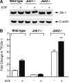
JNK1 and JNK2 differentially regulate Elk-1 phosphorylation (A) JNK1 and JNK2 do not change Elk-1 protein levels. Wild-type, Jnk1−/−, and Jnk2−/− MEFs were serum starved overnight and then incubated with or without EGF. Protein lysates were isolated, and immunoblot analysis was performed using Elk-1 or β-actin antibodies. (B) JNK1 increases, whereas JNK2 decreases, the total phosphorylation state of Elk-1. MEFs were cultured as described for panel A. In addition, cells were incubated with 32Pi (0.5 mCi/ml) for 3 h prior to EGF treatment. The resultant cell lysates were immunoprecipitated with Elk-1 antibody and subjected to sodium dodecyl sulfate-polyacrylamide gel electrophoresis. Autoradiography was used to examine the amount of 32P-incorporated Elk-1. The values shown are the means ± SEM of three independent experiments where the values were normalized to the total amount of Elk-1 protein.
FIG. 5.
FIG. 5.
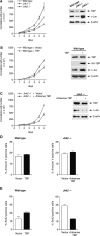
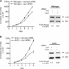
JNK-mediated changes in TBP levels alter the accumulation rates of MEFs (A) The accumulation rates of wild-type, Jnk1−/−, and Jnk−/− MEFs correlate with c-Jun and TBP levels. Wild-type, Jnk1−/−, and Jnk2−/− MEFs were plated in triplicate, and cell viability and total cell numbers were measured daily for 6 days (left panel). The results represent three independent experiments. Protein lysates derived from each of the cell lines were subjected to immunoblot analysis using antibodies against TBP, c-Jun, or β-actin as indicated (right panel). (B) Increased TBP expression enhances c-Jun expression and the accumulation of wild-type MEFs. Wild-type MEFs were stably transfected with an HA-tagged human TBP expression vector (Wild type + TBP) or empty vector (Wild type + vector). The accumulation rates of these cells were measured as described for panel A (left panel). The results represent two independent experiments resulting from two nonclonally selected populations of each stable cell line. Protein lysates derived from these cells were subjected to immunoblot analysis using antibodies against TBP, c-Jun, hemagglutinin, or β-actin as designated (right panel). (C) Reduced expression of TBP decreases the c-Jun expression and accumulation rate of Jnk2−/− MEFs. Stable cell lines were established by transfecting Jnk2−/− MEFs with an antisense TBP expression construct (Jnk2−/− + Antisense TBP) or vector alone (Jnk2−/− + Vector). The accumulation rates of these cells were measured as described for panel A (left panel). TBP, c-Jun, and β-actin protein levels were determined by immunoblot analysis (right panel). (D) Alterations in TBP levels do not affect apoptotic rates of MEFs. Stable cells of the wild-type-plus-TBP (left panel) and Jnk2−/−-plus-antisense TBP cell lines (right panel) were stained with annexin V, and rates of apoptosis were assessed as described in Materials and Methods. (E) Alterations in TBP levels change cellular proliferation rates of MEFs. Stable cells of the wild-type-plus-TBP (left panel) and Jnk2−/−-plus-antisense TBP cell lines (right panel) were labeled with BrdU, and S-phase-labeled cells were quantified by flow cytometry. The values shown are the means ± SEM of three independent experiments.
FIG. 6.
FIG. 6.
Cellular TBP amounts regulate c-Jun and AP-1 promoter activities in Huh-7 cells and MEFs (A) c-Jun promoter activity is differentially responsive to increases in TBP in wild-type and Jnk1- and Jnk2-deficient cells. MEFs were transiently cotransfected with human c-jun promoter-luciferase and human TBP expression plasmids, and luciferase activity was measured. The values shown are the means ± SEM of at least four independent experiments. (B) Increasing TBP expression enhances c-Jun promoter activity in Huh-7 cells. Human hepatoma Huh-7 cells were transiently cotransfected with the c-jun promoter-luciferase construct or a β3-integrin-luciferase construct together with the human TBP expression plasmid or empty vector. The change in c-jun promoter activity was calculated relative to that with vector alone. (C) Increasing TBP expression enhances AP-1-dependent promoter activity in wild-type MEFs and Huh-7 cells. Wild-type MEFs or Huh-7 cells were transiently cotransfected with a human AP-1-dependent promoter-luciferase construct, or the same promoter containing a mutated AP-1 site and the human TBP expression plasmid. The change in AP-1-dependent promoter activity was calculated relative to that with vector alone. The values shown are the means ± SEM of three independent experiments.
FIG. 7.
FIG. 7.
Decreased expression of c-Jun in MEFs reduces accumulation rates but does not affect TBP expression. Wild-type (A) or Jnk2−/− (B) MEFs were transfected with either c-Jun siRNA or an mm siRNA. MEFs were plated in triplicate, and cell viability and total cell numbers were measured daily for 6 days (left panel). The results represent three independent experiments. Protein lysates derived from each of the cell lines 72 h after transfection with the siRNAs were subjected to immunoblot analysis using antibodies against TBP and c-Jun. The changes in c-Jun levels were normalized to the total amount of TBP.
FIG. 8.
FIG. 8.
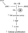
Model for opposing functions of JNK1 and JNK2. JNK1 promotes Elk-1 phosphorylation in response to EGF stimulation, whereas JNK2 negatively regulates the phosphorylation state of Elk-1 in the absence of stimuli. Phosphorylation of Elk-1 enhances it ability to be recruited to the TBP promoter, thereby inducing TBP promoter activity and its expression. Increased TBP levels then modulate c-Jun promoter activity and expression, ultimately dictating cellular proliferation rates.

Similar articles

Cited by

References

    1. Cavigelli, M., F. Dolfi, F. X. Claret, and M. Karin. 1995. Induction of c-fos expression through JNK-mediated TCF/Elk-1 phosphorylation. EMBO J. 14:5957-5964. - PMC - PubMed
    1. Chang, L., Y. Jones, M. H. Ellisman, L. S. Goldstein, and M. Karin. 2003. JNK1 is required for maintenance of neuronal microtubules and controls phosphorylation of microtubule-associated proteins. Dev. Cell 4:521-533. - PubMed
    1. Chang, L., and M. Karin. 2001. Mammalian MAP kinase signalling cascades. Nature 410:37-40. - PubMed
    1. Choi, B. Y., H. S. Choi, K. Ko, Y. Cho, F. Zhu, B. S. Kang, S. P. Ermakova, W. Y. Ma, A. M. Bode, and Z. Dong. 2005. The tumor suppressor p16(INK4a) prevents cell transformation through inhibition of c-Jun phosphorylation and AP-1 activity. Nat. Struct. Mol. Biol. 12:699-707. - PubMed
    1. Colgan, J., and J. L. Manley. 1992. TFIID can be rate limiting in vivo for TATA-containing but not TATA-lacking, RNA pol II promoters. Genes Dev. 6:304-315. - PubMed

Publication types

MeSH terms

Substances

  NODES
design 6
eth 5
see 3
Story 1
twitter 2