. 2007 Jan;81(2):860-71.
doi: 10.1128/JVI.01167-06. Epub 2006 Nov 1.

Coordinated regulation and widespread cellular expression of interferon-stimulated genes (ISG) ISG-49, ISG-54, and ISG-56 in the central nervous system after infection with distinct viruses

Affiliations

Coordinated regulation and widespread cellular expression of interferon-stimulated genes (ISG) ISG-49, ISG-54, and ISG-56 in the central nervous system after infection with distinct viruses

Christie Wacher et al. J Virol. 2007 Jan.

Abstract

The interferon (IFN)-stimulated genes (ISGs) ISG-49, ISG-54, and ISG-56 are highly responsive to viral infection, yet the regulation and function of these genes in vivo are unknown. We examined the simultaneous regulation of these ISGs in the brains of mice during infection with either lymphocytic choriomeningitis virus (LCMV) or West Nile virus (WNV). Expression of the ISG-49 and ISG-56 genes increased significantly during LCMV infection, being widespread and localized predominantly to common as well as distinct neuronal populations. Expression of the ISG-54 gene also increased but to lower levels and with a more restricted distribution. Although expression of the ISG-49, ISG-54, and ISG-56 genes was increased in the brains of LCMV-infected STAT1 and STAT2 knockout (KO) mice, this was blunted, delayed, and restricted to the choroid plexus, meninges, and endothelium. ISG-56 protein was regulated in parallel with the corresponding RNA transcript in the brain during LCMV infection in wild-type and STAT KO mice. Similar changes in ISG-49, ISG-54, and ISG-56 RNA levels and ISG-56 protein levels were observed in the brains of wild-type mice following infection with WNV. Thus, the ISG-49, ISG-54, and ISG-56 genes are coordinately upregulated in the brain during LCMV and WNV infection; this upregulation, in the case of LCMV, was totally (neurons) or partially (non-neurons) dependent on the IFN-signaling molecules STAT1 and STAT2. These findings suggest a dominant role for the ISG-49, ISG-54, and ISG-56 genes in the host response to different viruses in the central nervous system, where, particularly in neurons, these genes may have nonredundant functions.


Figures
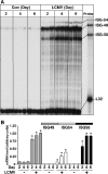
FIG. 1.
FIG. 1.
ISG-49, ISG-54, and ISG-56 mRNA levels in brains of sham- or LCMV-infected WT mice. Mice (C57BL/6) were injected i.c. with vehicle or vehicle plus 250 PFU of LCMV (ARM). At the times shown, mice were euthanized, poly(A)+ RNA was isolated from the brains, and 2 μg was analyzed by RPA as described in Materials and Methods. (A) Low constitutive levels of ISG-49 mRNA transcripts were present in normal, uninfected brain, while ISG-54 and ISG-56 mRNA transcripts were undetectable. The levels of ISG-49, ISG-54, and ISG-56 mRNA transcripts increased significantly (P < 0.05) by day 2 postinfection and continued to increase to day 4 postinfection. At day 6 postinfection, the levels of ISG-49 and ISG-54 mRNA continued to increase significantly from those seen at day 4, while levels of ISG-56 mRNA transcripts remained unchanged between days 4 and 6 postinfection. (B) Quantification of ISG mRNA levels in LCMV-infected WT mice. Densitometric analysis of each lane was performed on scanned autoradiographs with NIH Image software (version 1.63), with each individual mRNA density normalized to that of the corresponding L32 loading control. Statistical analysis was performed with Student's t test. *, significant increase compared to uninfected mice (P < 0.05).
FIG. 2.
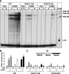
FIG. 2.
ISG-49, ISG-54, and ISG-56 mRNA levels in brains of sham- or LCMV-infected WT, STAT1 KO, and STAT2 KO mice. Mice (129/Sv) were manipulated as described in the legend to Fig. 1. (A) In WT mice, ISG-49, ISG-54, and ISG-56 mRNA levels were significantly (P < 0.05) increased by day 4 post-LCMV infection and continued to increase to day 6. There was a significant increase in the level of ISG-49, ISG-54, and ISG-56 mRNAs at day 4 postinfection in the STAT1 KO mice, which decreased but remained significantly above control levels by day 6. A minor but significant increase occurred in the level of ISG-49 mRNA only at day 4 postinfection in the STAT2 KO brain. At day 6 postinfection in STAT2 KO mice, ISG-49, ISG-54, and ISG-56 mRNA transcripts all increased slightly. (B, C, and D) Quantification of ISG expression in WT, STAT1 KO, and STAT2 KO mice, respectively. Densitometric analysis of each lane was performed on scanned autoradiographs with NIH Image software (version 1.63), with each individual mRNA density normalized to that of the corresponding L32 loading control. Statistical analysis was performed with Student's t test. *, significant increase compared to uninfected mice (P < 0.05).
FIG. 3.
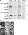
FIG. 3.
Anatomic localization of ISG-49, ISG-54, ISG-56, and LCMV NP RNA in the brain. WT, STAT1 KO, and STAT2 KO mice were injected i.c. as described in the legend to Fig. 1, and brains were removed at days 4 and 6 for in situ hybridization. Paraffin-embedded sagittal sections (8 μm) were hybridized with 33P-labeled cRNA probes transcribed from linearized ISG-49, ISG-54, ISG-56, or LCMV NP RNA plasmids and exposed to Kodak MR film for 3 days as outlined in Materials and Methods. (A) No hybridization above background of the ISG-49, ISG-54, ISG-56, or LCMV NP probes was evident in uninfected (control) brains. Following LCMV infection, in WT mice high levels of ISG-49 and ISG-56 hybridization were evident throughout the brain at day 4 and increased further by day 6. In contrast, there was a lower and more restricted level of ISG-54 hybridization. Increased ISG-49 and ISG-56 hybridization was also evident in the brain of STAT1 KO mice at day 4 but not day 6 postinfection, particularly in the meninges and ventricles. A small increase in ISG-49 hybridization was evident in the meninges and choroid plexus in the brain of STAT2 KO mice at day 6 postinfection. (B) Differential patterns of ISG-49, ISG-54, and ISG-56 hybridization at day 6 postinfection. In the Purkinje cell layer (arrowhead) of the cerebellum (cb), high levels of ISG-49 but low levels of ISG-54 and ISG-56 hybridization were seen. In the CA1 pyramidal neurons (arrow) and dentate gyrus (arrowhead) of the hippocampus (hc), high levels of ISG-56 but low levels of ISG-49 and ISG-54 hybridization were seen.
FIG. 4.
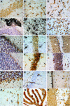
FIG. 4.
Cellular localization of ISG-49, ISG-54, and ISG-56 RNA in the brain. Mice were injected i.c. with saline or 250 PFU of LCMV (ARM), and the brain was removed at day 4 and day 6 postinfection for in situ hybridization and immunohistochemistry. Eight-micrometer-thick paraffin-embedded sections were hybridized with 33P-labeled cRNA probes transcribed from linearized ISG-49 or ISG-56 RPA plasmids and then processed for immunohistochemistry for cell-specific markers to identify astrocytes, microglia, and neurons as outlined in Materials and Methods. Slides were coated with photographic emulsion, developed after 2 weeks, and visualized with bright-field microscopy. Photomicrographs are of uninfected (A) and day 6 LCMV-infected (B to L) brain sections from WT mice hybridized for ISG-49 (A to I) or ISG-56 (J to L). ISG-49 hybridization was localized to astrocytes (B, arrows, GFAP stained) and microglia (C, arrows, lectin stained), gigantocellular neurons (E, arrows, NeuN stained), cortical neurons (F, arrows, NeuN stained), Purkinje cells (G, arrows NeuN stained), and hippocampal hilus (H, arrows, NeuN stained). Lower magnification revealed high-level expression of ISG-49 (D, NeuN stained) in the choroid plexus (asterisk) and ependymal layer (arrowhead). In the hippocampus, there was low ISG-49 hybridization in neurons of the CA1 region (H, asterisk, NeuN stained) and little hybridization to dentate gyrus (I, asterisk, NeuN stained). By contrast, ISG-56 hybridization was very high in both CA1 (K, asterisk, NeuN stained) and dentate gyrus (L, asterisk, NeuN stained) neurons of the hippocampus but low in Purkinje cells (J, NeuN stained) and neurons of the hippocampal hilus (K, arrows, NeuN stained). In LCMV infection of STAT1 KO mice at day 4 postinfection, ISG-49 hybridization was localized to vascular endothelium (M and N, arrows) and meninges (O, arrows, NeuN stained). A similar cellular localization was found for ISG-56 hybridization, which was seen predominantly in vascular endothelium and microglia (P, arrows, lectin stained) and choroid plexus (Q, lectin stained). Note the lack of hybridization for both ISG-49 (M to O) and ISG-56 (R) to neurons. Bars, 50 μm (A-C, E-G, J, M, O, and P), 100 μm (H, I, K, L, N, and R), 150 μm (Q), and 300 μm (D).
FIG. 5.
FIG. 5.
Localization and levels of ISG-56 protein in the brains of sham- or LCMV-infected mice. Mice were injected i.c. with saline or 250 PFU of LCMV (ARM), and the brains were removed at day 4 and day 6 postinfection and processed for immunohistochemistry for ISG-56 protein as outlined in Materials and Methods. (A) Sham-infected WT control specimen. ISG-56 protein immunostaining was not detectable and was comparable to adjacent sections treated with a preimmune rabbit sera. (B to E) LCMV-infected WT specimens (day 6). Prominent neuronal staining was evident in the cortex (B, arrows) and CA2 region of the hippocampus (C, arrows). Strong immunostaining for ISG-56 was also seen in the choroid plexus (D, arrow), meninges (E, arrow), and vascular endothelium (E, arrowhead). (F) LCMV-infected STAT1 KO specimen (day 6). Compared with LCMV-infected WT mice, ISG-56 immunostaining was reduced considerably. In this representative section, positive immunostaining can be seen in the vascular endothelium (arrow) and some scattered cells (arrowhead) likely to be microglia. Total magnification, ×100 (A and D) and ×200 (B, C, E, and F).
FIG. 6.
FIG. 6.
Regulation of ISG-49, ISG-54, and ISG-56 mRNA and ISG-56 protein in the brains of WNV-infected WT mice. Mice were inoculated intranasally with vehicle or vehicle plus 6 × 10⁴ PFU of WNV (Sarafend strain). At day 7 postinfection, mice were euthanized and the brains were removed and analyzed as described in Materials and Methods. (A) RNase protection analysis revealed that compared with sham-inoculated controls the levels of ISG-49, ISG-54, and ISG-56 mRNA transcripts increased significantly (*, P < 0.05) in WNV infection. (B) Anatomic localization of ISG RNA transcripts in the brain by in situ hybridization. Compared with sham-inoculated controls, during WNV infection the levels of all three ISG RNAs increased markedly and were distributed widely throughout the brain. (C) Immunohistochemical localization of the WNV nonstructural protein NS1 compared with the ISG-56 protein. Infection of many neurons by WNV is evident in most regions of the cerebrum but was largely absent from the choroid plexus (left middle panel, arrows) and cerebellar neurons. ISG-56 protein was found to be more widespread in the majority of neurons in both cerebrum and cerebellum, as well as in the choroid plexus (right middle panel, arrowhead). Total magnification, ×100.

Similar articles

Cited by

References

    1. Asensio, V. C., C. Kincaid, and I. L. Campbell. 1999. Chemokines and the inflammatory response to viral infection in the central nervous system with a focus on lymphocytic choriomeningitis virus. J. Neurovirol. 5:65-75. - PubMed
    1. Badley, J. E., G. A. Bishop, T. St. John, and J. A. Frelinger. 1988. A simple, rapid method for the purification of poly A+ RNA. BioTechniques 6:114-116. - PubMed
    1. Bluyssen, H. A., R. J. Vlietstra, P. W. Faber, E. M. Smit, A. Hagemeijer, and J. Trapman. 1994. Structure, chromosome localization, and regulation of expression of the interferon-regulated mouse Ifi54/Ifi56 gene family. Genomics 24:137-148. - PubMed
    1. Buchmeier, M. J., R. M. Welsh, F. J. Dutko, and M. B. Oldstone. 1980. The virology and immunobiology of lymphocytic choriomeningitis virus infection. Adv. Immunol. 30:275-331. - PubMed
    1. Campbell, I. L., M. V. Hobbs, P. Kemper, and M. B. A. Oldstone. 1994. Cerebral expression of multiple cytokine genes in mice with lymphocytic choriomeningitis. J. Immunol. 152:716-723. - PubMed

Publication types

MeSH terms

  NODES
Note 1
twitter 2