. 2006 Dec 1;127(5):1015-26.
doi: 10.1016/j.cell.2006.09.044.

Force sensing by mechanical extension of the Src family kinase substrate p130Cas

Affiliations

Force sensing by mechanical extension of the Src family kinase substrate p130Cas

Yasuhiro Sawada et al. Cell. .

Abstract

How physical force is sensed by cells and transduced into cellular signaling pathways is poorly understood. Previously, we showed that tyrosine phosphorylation of p130Cas (Cas) in a cytoskeletal complex is involved in force-dependent activation of the small GTPase Rap1. Here, we mechanically extended bacterially expressed Cas substrate domain protein (CasSD) in vitro and found a remarkable enhancement of phosphorylation by Src family kinases with no apparent change in kinase activity. Using an antibody that recognized extended CasSD in vitro, we observed Cas extension in intact cells in the peripheral regions of spreading cells, where higher traction forces are expected and where phosphorylated Cas was detected, suggesting that the in vitro extension and phosphorylation of CasSD are relevant to physiological force transduction. Thus, we propose that Cas acts as a primary force sensor, transducing force into mechanical extension and thereby priming phosphorylation and activation of downstream signaling.


Figures
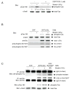
Figure 1
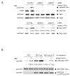
Figure 1. SFK- and Stretch-dependent Tyrosine Phosphorylation of Cas In Vivo
(A) Stretch-dependent tyrosine phosphorylation of Cas in intact cells. 2 x 10⁵ HEK293 cells on the collagen (Type I)-coated stretchable silicone dish were treated with either CGP77675 (1 μM) or its vehicle (0.01% DMSO) and were either stretched (biaxially, 10% in each dimension) or left unstretched. 1 min after stretching or without stretching, the cells were solubilized with 1 x SDS sample buffer containing 20 mM DTT and analyzed for Cas phosphorylation by anti-phospho-Cas (pCas-165) and anti-Cas (αCas3) immunoblotting. Quantification of phosphorylation (phospho-Cas / total Cas) was scaled with unstretched control set at 1 and noted below the pCas-165 blot with s.d. (n = 4). (B) Cell stretching increases Src-dependent phosphorylation of Cas without apparent change in phosphorylation levels of activating and inhibiting tyrosines of c-Src. 4 x 10⁵ SYF cells (triple knock-out cells of c-src, c-yes, and fyn) or SYF cells stably expressing c-Src were either stretched or left unstretched. 1 min after stretching or without stretching, cells were solubilized with SDS sample buffer and equivalent portions of each sample were subjected to SDS-PAGE followed by pCas-165, αCas3, anti-Src, anti-phospho-Src Y416, and anti-phospho-Src Y527 immunoblotting. (C) Cell stretching increases Src-dependent phosphorylation of Cas without apparent change in Src kinase activity. 4 x 10⁵ SYF cells or SYF cells stably expressing c-Src were either stretched or left unstretched. 1 min after stretching or without stretching, cells were lysed and subjected to immunoprecipitation followed by an in vitro kinase assay using acid-treated enolase as a substrate. Src kinase activity was analyzed by measuring the phosphorylation of enolase with anti-phospho-tyrosine immunoblotting (top panel). Immunoprecipitated Src, i.e. Src protein in the kinase reaction, was quantified by anti-Src immunoblotting (second panel). Equivalent small portions of each lysate was mixed with SDS sample buffer and subjected directly to SDS-PAGE followed by pCas-165 and αCas3 immunoblotting to analyze for Cas phosphorylation (third and fourth panels). Kinase reactions for the lane 3 and 4 samples appeared not to be saturated because the sample prepared from SYF/SrcY527F cells (SYF cells expressing SrcY527F, the highly active mutant form of c-Src) cultured on a plastic plate following the same protocol gave more phosphorylation of enolase (lane 5). The intense bands above enolase (top panel) and below Src (second panel) represent IgG (heavy chain) from the anti-Src antibody.
Figure 2
Figure 2. Significant Role of Cas Phosphorylation in Physiological Force Transduction
(A) Cas is involved in stretch-dependent Rap1 activation in intact cells. RNAi experiments were performed as described in Experimental Procedures. siRNA used were Stealth RNAi Negative Control Med GC (N-CTRL: lanes 1 and 2), BCAR1-HSS114272 (HSS272: lanes 3 and 4), and BCAR1-HSS114273 (HSS273: lanes 5 and 6) (Invitrogen). 24 h after transfection, HEK293 cells were either stretched or left unstretched. To determine the level of Cas expression and phosphorylation, cells were solubilized with SDS sample buffer 1 min after stretching or without stretching and equivalent portions of each sample were subjected to SDS-PAGE followed by anti-phospho-Cas (pCas-165), anti-Cas (αCas3), anti-Rap1, and anti-actin immunoblotting (upper panel). To measure stretch-dependent Rap1 activity, cells were solubilized with lysis buffer for GST pull-down assay (see Experimental Procedures) 5 min after stretching or without stretching. Rap1 was quantified by anti-Rap1 immunoblotting. Rap1 activity (Rap1•GTP / Rap1 input) was scaled with the unstretched control set at 1 and noted below the Rap1•GTP blot with s.d. (n = 4) (lower panel). The data shown in Figure 2A (upper and lower panels) was obtained with siRNA transfection performed at the same time. (B) Significant role of tyrosine phosphorylation of Cas in stretch-dependent Rap1 activation. RFP, RFP-Cas or RFP-Cas15YF was co-transfected with GFP-Rap1 into HEK293 cells (1 x 10⁵ / dish). 24 h after transfection, cells were either stretched or left unstretched. 5 min after stretching or without stretching, cells were solubilized and subjected to the GST pull-down assay. GFP-Rap1 was quantified by anti-GFP immunoblotting. GFP-Rap1 activity (GFP-Rap1•GTP / GFP-Rap1 input) was scaled with the unstretched RFP-transfected cells set at 1 and noted below the GFP-Rap1•GTP blot with s.d. (n = 4).
Figure 3
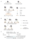
Figure 3. In Vitro Protein Extension (IPE) System
(A) Scheme of NC-biotinylated CasSD, C-biotinylated CasSD, and the process of mechanical extension of CasSD in the IPE system. (B) Schematic description of YFP amino-terminal swapping. (C) His6-YFP-N binds to extended NY/CY-NC-biotinylated CasSD, but not to NY/CY-C-biotinylated CasSD or NC-biotinylated CasSD. Biotinylated CasSD proteins, either extended or unextended on latex membrane, were incubated with His6-YFP-N in the buffer containing 1% Triton X-100 and 1% BSA. After washing, bound complex was solubilized and subjected to SDS-PAGE followed by anti-polyHistidine immunoblotting or avidin affinity blotting.
Figure 4
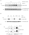
Figure 4. Extension-dependent Phosphorylation of CasSD by Tyrosine Kinases In Vitro
(A) CasSD is tyrosine phosphorylated by recombinant c-Src in an extension-dependent manner. NC-biotinylated or C-biotinylated CasSD was either extended or left unextended on latex membrane, incubated with recombinant c-Src for 2 min, washed, solubilized, and analyzed for tyrosine phosphorylation by anti-phospho-Cas (αP-Cas460Y) immunoblotting and avidin affinity blotting. The magnitude of the latex membrane stretching is described as the % change of length in each dimension. Quantification of phosphorylation of CasSD was scaled with unextended NC-biotinylated CasSD set at 1 and noted below the anti-phospho-Cas blot with s.d. (n = 4). (B) Kinase specificity of extension-dependent tyrosine phosphorylation of CasSD. NC-biotinylated CasSD was either extended (100%) or left unextended and then incubated with recombinant c-Src, Csk, Abl1, ZAP-70 or FynT for 2 min at room temperature. Tyrosine phosphorylation of CasSD was analyzed as in A. (C) Extension-dependent phosphorylation of CasSD by c-Src measured by two different anti-phospho-Cas antibodies. Samples were prepared as in A except for the extent of the latex membrane stretching (40% in the left panel and 100% in the right panel). Equivalent portions of each sample were subjected to SDS-PAGE followed by pCas-165 and pCas-410 immunoblotting and avidin affinity blotting. Quantification of phosphorylation of CasSD was scaled with unextended NC-biotinylated CasSD set at 1 and noted below the anti-phospho-Cas blots with s.d. (n = 4).
Figure 5
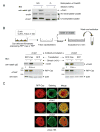
Figure 5. Extension of Cas In Situ and In Vivo
(A) αCas1 recognizes extended CasSD in vitro. NC-biotinylated or C-biotinylated CasSD was either extended (100%) or left unextended in the IPE system. After blocking, CasSD proteins were incubated with αCas1, washed, and solubilized with SDS sample buffer containing 0.12 M DTT. Equivalent portions of each sample were analyzed for quantification of bound αCas1 by anti-rabbit IgG immunoblotting. The amount of NC-biotinylated and C-biotinylated CasSD in each sample was quantified by avidin affinity blotting and αCas1 immunoblotting. Note that the difference in the relative signal intensity between avidin and αCas1 blots is consistent with the molar ratio of biotinylation (NC-biotinylated CasSD : C-biotinylated CasSD = 2 : 1). (B) Stretch-dependence of αCas1 and αCas3 binding to Cas in Triton cytoskeletons. Triton cytoskeletons were prepared from Cas-deficient fibroblasts transfected with RFP-Cas or RFP alone, either stretched or left unstretched, and incubated with either αCas1 or αCas3 as shown in the diagram. Quantification of bound antibody by anti-rabbit IgG immunoblotting was scaled with unstretched control set at 1 and noted with s.d. (n = 4). (C) αCas1 preferentially binds to Cas where higher traction forces are expected in vivo. Cas-deficient fibroblasts expressing RFP-Cas were plated, fixed after 20 min, and then stained with αCas1, αCas3 or pCas-165. Confocal images are shown for RFP-Cas (left, red channel), immunostaining (center, green channel) and merged (right). Scale bars represent 10 μm.
Figure 6
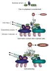
Figure 6. Model of Extension of Cas and Signaling at Cell-matrix Contact Sites
The top and middle panels represent a Cas molecule with unextended configuration of substrate domain in the cytoplasm and with moderate extension of substrate domain at the cell-matrix contact site of spread cells, respectively. The bottom panel represents the extension-dependent phosphorylation of Cas substrate domain by SFK and enhancement of its downstream signaling. SH3 and SB represent the SH3 and the Src-binding domains of Cas, respectively.

Comment in

Similar articles

Cited by

References

    1. Balaban NQ, Schwarz US, Riveline D, Goichberg P, Tzur G, Sabanay I, Mahalu D, Safran S, Bershadsky A, Addadi L, Geiger B. Force and focal adhesion assembly: a close relationship studied using elastic micropatterned substrates. Nat Cell Biol. 2001;3:466–472. - PubMed
    1. Ballestrem C, Erez N, Kirchner J, Kam Z, Bershadsky A, Geiger B. Molecular mapping of tyrosine-phosphorylated proteins in focal adhesions using fluorescence resonance energy transfer. J Cell Sci. 2006;119:866–875. - PubMed
    1. Bougeret C, Rothhut B, Jullien P, Fischer S, Benarous R. Recombinant Csk expressed in Escherichia coli is autophosphorylated on tyrosine residue(s) Oncogene. 1993;8:1241–1247. - PubMed
    1. Briknarova K, Nasertorabi F, Havert ML, Eggleston E, Hoyt DW, Li C, Olson AJ, Vuori K, Ely KR. The serine-rich domain from Crk-associated substrate (p130Cas) is a four-helix bundle. J Biol Chem. 2005;280:21908–21914. - PubMed
    1. Carrion-Vazquez M, Oberhauser AF, Fowler SB, Marszalek PE, Broedel SE, Clarke J, Fernandez JM. Mechanical and chemical unfolding of a single protein: a comparison. Proc Natl Acad Sci U S A. 1999;96:3694–3699. - PMC - PubMed

Publication types

MeSH terms

LinkOut - more resources

  NODES
Note 7
twitter 2