Abnormal sperm in mice lacking the Taf7l gene
- PMID: 17242199
- PMCID: PMC1899882
- DOI: 10.1128/MCB.01722-06
Abnormal sperm in mice lacking the Taf7l gene
Abstract
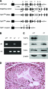
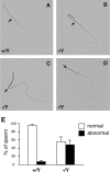
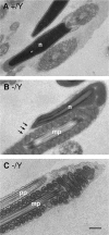
TFIID is a general transcription factor required for transcription of most protein-coding genes by RNA polymerase II. TAF7L is an X-linked germ cell-specific paralogue of TAF7, which is a generally expressed component of TFIID. Here, we report the generation of Taf7l mutant mice by homologous recombination in embryonic stem cells by using the Cre-loxP strategy. While spermatogenesis was completed in Taf7l(-/Y) mice, the weight of Taf7l(-/Y) testis decreased and the amount of sperm in the epididymides was sharply reduced. Mutant epididymal sperm exhibited abnormal morphology, including folded tails. Sperm motility was significantly reduced, and Taf7l(-/Y) males were fertile with reduced litter size. Microarray profiling revealed that the abundance of six gene transcripts (including Fscn1) in Taf7l(-/Y) testes decreased more than twofold. In particular, FSCN1 is an F-action-bundling protein and thus may be critical for normal sperm morphology and sperm motility. Although deficiency of Taf7l may be compensated in part by Taf7, Taf7l has apparently evolved new specialized functions in the gene-selective transcription in male germ cell differentiation. Our mouse studies suggest that mutations in the human TAF7L gene might be implicated in X-linked oligozoospermia in men.
Figures


Similar articles
-
The intracellular localisation of TAF7L, a paralogue of transcription factor TFIID subunit TAF7, is developmentally regulated during male germ-cell differentiation.J Cell Sci. 2003 May 1;116(Pt 9):1847-58. doi: 10.1242/jcs.00391. J Cell Sci. 2003. PMID: 12665565
-
Taf7l cooperates with Trf2 to regulate spermiogenesis.Proc Natl Acad Sci U S A. 2013 Oct 15;110(42):16886-91. doi: 10.1073/pnas.1317034110. Epub 2013 Sep 30. Proc Natl Acad Sci U S A. 2013. PMID: 24082143 Free PMC article.
-
Basonuclin 1 deficiency causes testicular premature aging: BNC1 cooperates with TAF7L to regulate spermatogenesis.J Mol Cell Biol. 2020 Jan 22;12(1):71-83. doi: 10.1093/jmcb/mjz035. J Mol Cell Biol. 2020. PMID: 31065688 Free PMC article.
-
Testis-specific transcription mechanisms promoting male germ-cell differentiation.Reproduction. 2004 Jul;128(1):5-12. doi: 10.1530/rep.1.00170. Reproduction. 2004. PMID: 15232059 Review.
-
Regulation of male fertility by X-linked genes.J Androl. 2010 Jan-Feb;31(1):79-85. doi: 10.2164/jandrol.109.008193. Epub 2009 Oct 29. J Androl. 2010. PMID: 19875494 Free PMC article. Review.
Cited by
-
Differential protein repertoires related to sperm function identified in extracellular vesicles (EVs) in seminal plasma of distinct fertility buffalo (Bubalus bubalis) bulls.Front Cell Dev Biol. 2024 Jul 29;12:1400323. doi: 10.3389/fcell.2024.1400323. eCollection 2024. Front Cell Dev Biol. 2024. PMID: 39135778 Free PMC article.
-
TAF7L regulates early stages of male germ cell development in the rat.FASEB J. 2024 Jan;38(1):e23376. doi: 10.1096/fj.202301716RR. FASEB J. 2024. PMID: 38112167 Free PMC article.
-
Loss of the importin Kpna2 causes infertility in male mice by disrupting the translocation of testis-specific transcription factors.iScience. 2023 Jun 16;26(7):107134. doi: 10.1016/j.isci.2023.107134. eCollection 2023 Jul 21. iScience. 2023. PMID: 37456838 Free PMC article.
-
Fascin-1: Updated biological functions and therapeutic implications in cancer biology.BBA Adv. 2022 May 17;2:100052. doi: 10.1016/j.bbadva.2022.100052. eCollection 2022. BBA Adv. 2022. PMID: 37082587 Free PMC article. Review.
-
MicroRNA profiling reveals the role of miR-133b-3p in promoting apoptosis and inhibiting cell proliferation and testosterone synthesis in mouse TM3 cells.In Vitro Cell Dev Biol Anim. 2023 Jan;59(1):63-75. doi: 10.1007/s11626-022-00745-z. Epub 2023 Jan 30. In Vitro Cell Dev Biol Anim. 2023. PMID: 36715892
References
-
- Adams, J. C. 2004. Roles of fascin in cell adhesion and motility. Curr. Opin. Cell Biol. 16:590-596. - PubMed
-
- Albright, S. R., and R. Tjian. 2000. TAFs revisited: more data reveal new twists and confirm old ideas. Gene 242:1-13. - PubMed
-
- Aumüller, G., and J. Seitz. 1988. Immunocytochemical localization of actin and tubulin in rat testis and spermatozoa. Histochemistry 89:261-267. - PubMed
-
- Bell, B., E. Scheer, and L. Tora. 2001. Identification of hTAF(II)80 delta links apoptotic signaling pathways to transcription factor TFIID function. Mol. Cell 8:591-600. - PubMed
-
- Breitbart, H., G. Cohen, and S. Rubinstein. 2005. Role of actin cytoskeleton in mammalian sperm capacitation and the acrosome reaction. Reproduction 129:263-268. - PubMed
Publication types
MeSH terms
Substances
Associated data
- Actions
Grants and funding
LinkOut - more resources
Full Text Sources
Molecular Biology Databases
Miscellaneous
