. 2007 Apr;27(7):2661-75.
doi: 10.1128/MCB.01098-06. Epub 2007 Feb 5.

An acetylation/deacetylation-SUMOylation switch through a phylogenetically conserved psiKXEP motif in the tumor suppressor HIC1 regulates transcriptional repression activity

Affiliations

An acetylation/deacetylation-SUMOylation switch through a phylogenetically conserved psiKXEP motif in the tumor suppressor HIC1 regulates transcriptional repression activity

Nicolas Stankovic-Valentin et al. Mol Cell Biol. 2007 Apr.

Abstract

Tumor suppressor HIC1 (hypermethylated in cancer 1) is a gene that is essential for mammalian development, epigenetically silenced in many human tumors, and involved in a complex pathway regulating P53 tumor suppression activity. HIC1 encodes a sequence-specific transcriptional repressor containing five Krüppel-like C(2)H(2) zinc fingers and an N-terminal BTB/POZ repression domain. Here, we show that endogenous HIC1 is SUMOylated in vivo on a phylogenetically conserved lysine, K314, located in the central region which is a second repression domain. K314R mutation does not influence HIC1 subnuclear localization but significantly reduces its transcriptional repression potential, as does the mutation of the other conserved residue in the psiKXE consensus, E316A, or the overexpression of the deSUMOylase SSP3/SENP2. Furthermore, HIC1 is acetylated in vitro by P300/CBP. Strikingly, the K314R mutant is less acetylated than wild-type HIC1, suggesting that this lysine is a _target for both SUMOylation and acetylation. We further show that HIC1 transcriptional repression activity is positively controlled by two types of deacetylases, SIRT1 and HDAC4, which increase the deacetylation and SUMOylation, respectively, of K314. Knockdown of endogenous SIRT1 by the transfection of short interfering RNA causes a significant loss of HIC1 SUMOylation. Thus, this dual-deacetylase complex induces either a phosphorylation-dependent acetylation-SUMOylation switch through a psiKXEXXSP motif, as previously shown for MEF2, or a phosphorylation-independent switch through a psiKXEP motif, as shown here for HIC1, since P317A mutation severely impairs HIC1 acetylation. Finally, our results demonstrate that HIC1 is a _target of the class III deacetylase SIRT1 and identify a new posttranslational modification step in the P53-HIC1-SIRT1 regulatory loop.


Figures

FIG. 1.
FIG. 1.
Identification of a new conserved motif in the central repression domain of HIC1: a SUMOylation consensus. Schematic structure of the human HIC1 protein. The BTB/POZ domain and the five C2H2 zinc fingers are represented as dotted and gray boxes, respectively. The evolutionarily conserved CtBP-interaction domain (CID) (16) and the SUMOylation motif identified in this study (ψKXE) are represented as dotted and solid lines, respectively. Sequences from the various HIC1 proteins (starting at the conserved BTB/POZ domain) were aligned with CLUSTAL/Jalview (EMBL) and default parameters. Only the regions surrounding the CtBP interaction domain and the newly defined conserved region are shown. In the consensus lane (Cons), identical residues are shown as asterisks under the aligned sequences. The residues conforming to the CID consensus (GLDLS) and the SUMOylation consensus (ψKXE) are bold and underlined. Notably, the adjacent proline-directed phosphorylation site (SP) found in the recently defined PDSM or SAS ψKXEXXSP is not present in HIC1 (26). Hu, human; Mu, murine; Ck, chicken; Zf, zebrafish (Danio rerio); Fu, Fugu rubipres.
FIG. 2.
FIG. 2.
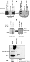
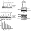
HIC1 is SUMOylated in vivo. (A) In vivo SUMOylation of HIC1 in a stable inducible cell line. Cos-7 cells were transfected with expression vectors for FLAG-HIC1 and SUMO-1 (lane 1) as a positive control, whereas the parental EcR-CHO cell line (lane 2) and the stable inducible EcR-CHO-pIND-FLAG-HIC1 clone 6 cell line were treated with 10 μM ponasterone A for 48 h (16). Cell lysates were prepared in the presence of NEM and then subjected to immunoprecipitation (IP) with an anti-HIC1 polyclonal serum directed against a C-terminal peptide of HIC1 (325) (16). The immunoprecipitates were resolved by SDS-PAGE and immunoblotted with anti-FLAG M2 monoclonal antibody (left panel). The blot was stripped and probed with an anti-SUMO-1 monoclonal antibody (right panel, lanes 1′, 2′, and 3′). A remnant of the wild-type HIC1 proteins resistant to the stripping procedure yielded a convenient size control (*). (B) In vivo SUMOylation of endogenous HIC1 in the DAOY medulloblastoma cell line. Lysates were immunoprecipitated with antibodies raised against the C-terminal end of HIC1 (amino acids 590 to 714 fused to glutathione S-transferase [GST]) (2563) (16) (lane 2) and by the same immune serum adsorbed with an excess of the purified GST-HIC1 fusion protein used to immunize the rabbit (Ads-GST-HIC1, lane 1). The immunoprecipitates were resolved by SDS-PAGE and analyzed by Western blotting (WB) with the anti-HIC1 325 antibody. After stripping, the blot was reprobed with an anti-SUMO-1 monoclonal antibody (right panel, lanes 1′ and 2′). (C) Detection of endogenous SUMOylated HIC1 proteins in the DAOY medulloblastoma cell line. Cells were directly lysed in Laemmli loading buffer. The lysates were immediately boiled for 10 min, and equal amounts were resolved by SDS-PAGE and analyzed by Western blotting using the polyclonal anti-HIC1 antibodies (2563) or the same antibodies but adsorbed with an excess of the purified GST-HIC1 polypeptide used to immunize the rabbit (2563 Ads GST-HIC1).
FIG. 3.
FIG. 3.
The evolutionarily conserved lysine 314 is the major SUMO-1 modification site in HIC1 in vitro and in vivo. (A) In vitro-translated, [35S]-labeled, full-length FLAG-HIC1 (lanes 1 to 3) or FLAG-HIC1 K314R (lanes 4 to 6) was analyzed by SDS-PAGE directly (I, Input; lanes 1 and 4) or was subjected to an in vitro modification reaction by incubation with a mix containing a fraction of HeLa cells (as a source of E1 activity), recombinant Ubc9, and ATP in either the presence (+; lanes 2 and 5) or the absence (−; lanes 3 and 6) of recombinant SUMO-1 before SDS-PAGE analysis and autoradiography. (B) Cos-7 cells were cotransfected with the indicated expression vectors and an empty vector (−) or a vector expressing His-tagged SUMO-1 (+). Forty-eight hours after transfection, cell lysates were subjected to nickel-agarose precipitation. Twenty percent of the lysates (lanes 1 to 6) and the proteins retained on nickel-agarose beads (Ni-ppt; lanes 7 to 12) were separated by SDS-PAGE and immunoblotted with the rabbit anti-HIC1 (αHIC1) (325) polyclonal antibody. (C) The SUMOylation of the wt HIC1 protein or the E316A mutant (a point mutation of the other strictly conserved residue in SUMOylation motifs) was analyzed as described for panel B, except that the FLAG monoclonal antibody was used to reveal the HIC1 proteins. (D) Cos-7 cells were cotransfected with expression vectors for wt HIC1 and His-SUMO1 and with an empty vector (−) or a vector expressing the deSUMOylase SSP3 (+). Forty-eight hours after transfection, cell lysates were subjected to nickel-agarose precipitation (lanes 1 and 2), separated by SDS-PAGE, and immunoblotted with the FLAG monoclonal antibody. WB, Western blot.
FIG. 4.
FIG. 4.
PIAS family members (but not Pc2) are E3 ligases for HIC1 in vivo. (A) Cos-7 cells were transfected with 1.25 μg of FLAG-HIC1 and 1.25 μg His-SUMO-1 expression vectors (lane 1) or 125 ng of His-SUMO-1 expression vector alone (lane 2) or together with the expression vectors for PIAS1, PIASγ, PIASXβ, and PIASXα (lanes 3 to 6). Forty-eight hours after transfection, cell lysates were subjected to nickel-agarose precipitation, separated by SDS-PAGE, and immunoblotted with the anti-FLAG monoclonal antibody. The lack of HIC1 SUMOylation in the presence of PIASγ and PIASXβ (lanes 4 and 5) is due to the fact that these overexpressed E3 ligases conjugate the limiting amount of SUMO-1 onto their own endogenous substrates, thus precluding the SUMOylation of HIC1. (B) Cos-7 cells were transfected with 1.25 μg of FLAG-HIC1 expression vector alone (lane 1) with 1.25 μg His-SUMO-1 expression vector (lane 2) or 125 ng of His-SUMO-1 expression vector together with the expression vectors for PIAS1Xα or Pc2 (lanes 3 and 4). Forty-eight hours after transfection, cells were lysed in buffer (RIPA) containing 20 mM NEM. Lysates were subjected to immunoprecipitation (IP) with the polyclonal anti-HIC1 antibody, separated by SDS-PAGE, and immunoblotted with the anti-FLAG monoclonal antibody. WB, Western blot.
FIG. 5.
FIG. 5.
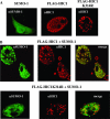
wt FLAG-HIC1 but not FLAG-HIC1 K314R colocalizes with SUMO-1 on nuclear dots in transfected Cos-7 cells. (A) The mutation of the SUMOylation consensus does not impinge on the subnuclear localization of ectopically expressed HIC1 proteins. Cos-7 cells were transfected with expression vectors for the above-indicated proteins and fixed 24 h after transfection. Cells were labeled with the SUMO-1 monoclonal antibody (αSUMO-1), followed by fluorescein isothiocyanate-conjugated anti-mouse immunoglobulin G (IgG) antibody, or with the 325 polyclonal antibody (FLAG-HIC1 and FLAG-HIC1 K314R), followed by Texas Red-conjugated anti-rabbit IgG antibody. Confocal images are shown. (B) HIC1 but not HIC1 K314R colocalizes at nuclear dots with SUMO-1. After the coexpression of FLAG-HIC1 (top) or FLAG-HIC1 K314R (bottom) with SUMO-1 in Cos-7 cells, SUMO-1 and FLAG-HIC1 proteins were visualized as described for panel A. Each horizontal lane represents the same cells immunostained with the monoclonal anti-SUMO-1 antibody and then the polyclonal anti-HIC1 325 antibody, and finally, all signals are merged in the last picture.
FIG. 6.
FIG. 6.
SUMO-1 modification modulates HIC1-mediated transcriptional repression. (A) Schematic structure of the two HIC1 BTB-CR-Gal4 chimeras. Numbers refer to human HIC1 residues. The BTB/POZ domain, the CR, and the Gal4 DBD domain are represented as dotted, white, and gray boxes, respectively. The SUMOylation consensus site is shown as a black line, and the mutated SUMOylation consensus site is shown as a hatched line. A nuclear localization signal (NLS) and an HA epitope tagged at the C-terminal part of the chimeras are shown as black triangles. (B) The BTB-CR-G4 chimera, but not the mutated BTB-CR-G4 K314R chimera, is SUMOylated in vivo. Whole-cell extracts prepared from Cos-7 cells transfected with vectors expressing His-tagged SUMO-1, and the different Gal4 chimeras were either immunoblotted with anti-HA antibody directly (left panel, lanes 1 to 3) or subjected to Ni affinity chromatography prior to Western blot (WB) analysis (right panel, lanes 4 to 6). The arrowhead indicates the position of the SUMO-1-modified BTB-CR-G4 protein. (C) The mutation of K314 in the SUMOylation site reduces the repression capacity of the BTB-CR-Gal4 chimeras. RK13 cells were transiently transfected in triplicate with 200 ng of the indicated Gal4 chimeras and 250 ng of the pG5-luc reporter (schematically drawn in panel A). The luciferase activity was normalized to the β-galactosidase activity of a cotransfected β-OS-lacZ construct (50 ng). After normalization, the data were expressed as Luc activity relative to the activity of pG5-luc with Gal4-NLS-HA expression vector (G4), which was given an arbitrary value of 1. The results are the mean values and standard deviations (error bars) from one independent transfection performed in triplicate that is representative of three independent experiments. (D) The mutation of the consensus E316 residue in the SUMOylation site also reduces the repression capacity of the BTB-CR-Gal4 chimera. A similar experiment was conducted as described above but with expression vectors for the wt BTB-CR-Gal4 chimera or the E316A point mutant. Error bars indicate standard deviations. (E) Expression of the deSUMOylase SENP2/SSP3 impairs the repression potential of the wt and non-SUMOylable K314R BTB-CR-Gal4 chimeras. RK13 cells were transiently transfected in triplicate with 150 ng of the indicated Gal4 chimeras and 200 ng of the pG5-luc reporter alone (−) or in the presence (+) of 200 ng of the SSP3 expression vector. The luciferase activity was normalized to the β-galactosidase activity of a cotransfected β-OS-lacZ construct (50 ng). After normalization, the data were expressed as Luc activity relative to the activity of pG5-luc with the wt chimera in the absence of the SSP3 expression vector, which was given an arbitrary value of 100%. The results are the mean values and standard deviations (error bars) from two independent transfections performed in triplicate.
FIG. 7.
FIG. 7.
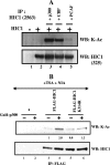
HIC1 is acetylated on several lysine residues, including K314. (A) HIC1 was expressed alone (lane 2) or with the indicated acetyltransferase (lanes 3 to 5) by transient transfection in Cos-7 cells. Cells were treated with 300 nM TSA for 24 h before lysis and immunoprecipitation (IP). Lane 1 represents untransfected cells used as a control. HIC1 acetylation was detected by immunoprecipitation with the polyclonal anti-HIC1 antibody (2563) and Western blot (WB) analysis with the monoclonal pan acetyl-lysine antibody (K-Ac) from Cell Signaling (top panel). Western blotting with another anti-HIC1 polyclonal antibody (325) was used to ascertain the presence of HIC1 proteins (bottom panel). (B) The acetylation levels of wt FLAG-HIC1 and FLAG-HIC1 K314R were determined (as described above) in the presence of HDAC inhibitors (300 nM TSA and 5 mM NIA) added 24 h before lysis without (−; lanes 3 and 5) or with (+; lanes 4 and 6) expression vectors for P300. Lanes 1 and 2 correspond to controls in the absence of HIC1. Western blots were quantified using Syngene Tools. The ratio between wild-type Ac-HIC1 and total wild-type HIC1 with p300 in the absence of inhibitors was arbitrarily set to 1, and the values obtained are indicated between the two panels.
FIG. 8.
FIG. 8.
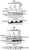
wt HIC1 and the K314R mutant interact with SIRT1 and HDAC4. (A) Cos-7 cells were mock transfected (lane 1) or transfected with expression vectors for FLAG-tagged HIC1 proteins (1.25 μg; wild type or K314R mutant) and SIRT1 (1.25 μg) (lanes 2 to 6). HIC1 proteins were immunoprecipitated (IP) from cell lysates with anti-HIC1 2563 polyclonal antibodies. The resulting immunoprecipitates were then Western blotted and analyzed with the anti-SIRT1 monoclonal antibody (upper panel). The blot was stripped and probed with the rabbit anti-HIC1 polyclonal antibody (325) to ascertain the presence of HIC1 (middle panel). Two percent of each total cell extract (Input) was resolved by SDS-PAGE and immunoblotted with the anti-SIRT1 antibody (lower panel). The asterisk refers to a nonspecific band as specified by the supplier (Upstate). (B) A similar experiment was conducted as described above but with expression vectors for FLAG-HIC1 proteins (wild type or K314R mutant) and HA-tagged HDAC4. HDAC4 proteins were detected by using an anti-HA monoclonal antibody (Babco). −, absence of; +, presence of.
FIG. 9.
FIG. 9.
Class III deacetylase SIRT1 has HIC1 deacetylase activity but not class II deacetylase HDAC4. (A) Cos-7 cells were transfected with expression vectors for FLAG-tagged HIC1 proteins (1.25 μg) in the absence (lane 1) or presence (+; lanes 2 to 4) of expression vectors for the acetyltransferase p300 (1 μg). The class III HDAC SIRT1 or its catalytic dead mutant (H363Y) was cotransfected (1.25 μg; lane 3 or 4, respectively). HIC1 proteins were immunoprecipitated from cell lysates with the anti-HIC1 2563 polyclonal antibodies. The resulting immunoprecipitates (IP) were then Western blotted (WB) and analyzed with the anti-monoacetylated lysine monoclonal antibody (upper panel). The blot was stripped and probed with the FLAG monoclonal antibody to ascertain the presence of HIC1 (middle panel). The ratio between Ac-HIC1 and total HIC1 with p300 (lane 2) was arbitrarily set to 1, and the values obtained are indicated below each lane. One percent of each total cell extract (Input) was resolved by SDS-PAGE and immunoblotted with the anti-SIRT1 antibody (lower panel). The asterisk refers to a nonspecific band as specified by the supplier (Upstate). Note that only SIRT1, not its enzymatic-dead mutant, changes HIC1 acetylation. (B) The acetylation levels of HIC1 in the presence of SIRT1 or HA-HDAC4 were examined as described above. Note that HDAC4 (lane 3) does not change HIC1 acetylation in contrast to SIRT1 (lane 4). (C) A docking model of the MKHEP peptide in the catalytic domain of SIRT1. The K acetylated side chain (C = O group) is strongly hydrogen bonded to the ribose moiety (3′ hydroxyl group) attached to the nicotinamide part of NAD. The catalytic histidine is green.
FIG. 10.
FIG. 10.
SUMOylation of HIC1 K314 is enhanced by HDAC4 and SIRT1 but through different mechanisms. (A) HDAC4 stimulates SUMO-1 conjugation to HIC1 lysine 314. Cos-7 cells were transfected with 1.25 μg of FLAG-HIC1 and 1.25 μg His-SUMO-1 expression vectors (lane 1) or 125 ng of His-SUMO-1 expression vector alone (lane 2) or together with the expression vectors for PIAS1 or PIASγ as a positive or negative control, respectively, of canonical SUMO E3 ligases and HDAC4 as indicated (lanes 3 to 5). Forty-eight hours after transfection, cell lysates were subjected to nickel-agarose precipitation, separated by SDS-PAGE, and immunoblotted with the anti-HIC1 (325) polyclonal antibody. (B) SUMOylation of HIC1 on lysine 314 is regulated positively by the deacetylase SIRT1 and negatively by HDAC inhibitors. Cos-7 cells were transfected with 1.25 μg of FLAG-HIC1 and 125 ng of His-SUMO-1 expression vector alone (lane 1) or together with the expression vectors for wt SIRT1 (lane 2) or the catalytically inactive SIRT1 mutant (mut; H363Y) (lane 3), with 300 nM TSA (lane 4), 5 mM nicotinamide (lane 5), or both HDAC inhibitors (lane 6). Forty-eight hours after transfection, cell lysates were treated as described above. The ratio between the SUMOylated and total HIC1 proteins in the absence of SIRT1 or HDAC inhibitors (lane 1) was arbitrarily set to 100%, and the values obtained in the other cases are represented below as a graph. (C) SIRT1 knockdown in HEK293T cells results in decreased SUMOylation of HIC1 on lysine 314. A SUMOylation test has been performed with HEK293T cells transfected 24 h before with negative control siRNA which did not match any sequence in the human genome (Eurogentec) (left) or with SIRT1 siRNA (Ambion) (right). Whole-cell extracts were subjected to Ni affinity (Ni-ppt) (top panel) chromatography prior to Western blot (WB) analysis with the FLAG antibody. As controls, equal amounts of cell extracts were resolved by SDS-PAGE and immunoblotted with the anti-SIRT1 antibody and with the Hsp60 antibody (loading control). In addition, the detection of the SUMOylated form of RanGAP-1, the major SUMO-1 substrate, with an anti-GST-SUMO-1 antibody (J. Seeler, unpublished results), demontrates that SUMOylation is not globally affected. WB, Western blot.
FIG. 11.
FIG. 11.
KXEP: a core motif for coordinated acetylation and SUMOylation? Consensus motifs for which modifications have been fully validated by functional assays (44, 51, 63) are indicated. We noticed that in all known examples of coordinated SUMOylation and acetylation, the glutamic acid residue in the SUMOylation consensus is immediately followed by a proline, except for Sp3, which contains a stretch of three glutamic acid residues.
FIG. 12.
FIG. 12.
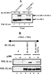
Mutation of proline 317 in the KXEP motif affects the acetylation but not the SUMOylation of HIC1. (A) The SUMOylation of the wt HIC1 protein or of the P317A mutant (a point mutation of the conserved proline residue in the HIC1 ψKXEP SUMOylation motif) was analyzed exactly as described in the legend for Fig. 3. Lanes 1 and 3 correspond to controls with no His-SUMO-1 transfected. HIC1 acetylation was detected by immunoprecipitation with the monoclonal anti-FLAG antibody and Western blot (WB) analysis with the monoclonal pan acetyl-lysine antibody (K-Ac) from Cell Signaling (top panel). Western blotting with the FLAG monoclonal antibody was used to ascertain the presence of HIC1 proteins (bottom panel). (B) The acetylation levels of wt FLAG-HIC1 and FLAG-HIC1 P317A were determined as described in the legend for Fig. 7 in the presence of HDAC inhibitors (300 nM TSA and 5 mM NIA) added 24 h before lysis without (lanes 3 and 5) or with (lanes 4 and 6) p300. Lanes 1 and 2 correspond to controls in the absence of HIC1.

Similar articles

Cited by

References

    1. Bereshchenko, O. R., W. Gu, and R. Dalla-Favera. 2002. Acetylation inactivates the transcriptional repressor BCL6. Nat. Genet. 32:606-613. - PubMed
    1. Bertrand, S., S. Pinte, N. Stankovic-Valentin, S. Deltour-Balerdi, C. Guerardel, A. Begue, V. Laudet, and D. Leprince. 2004. Identification and developmental expression of the zebrafish orthologue of the tumor suppressor gene HIC1. Biochim. Biophys. Acta 1678:57-66. - PubMed
    1. Blander, G., J. Olejnik, E. Krzymanska-Olejnik, T. McDonagh, M. Haigis, M. B. Yaffe, and L. Guarente. 2005. SIRT1 shows no substrate specificity in vitro. J. Biol. Chem. 280:9780-9785. - PubMed
    1. Bossis, G., and F. Melchior. 2006. Regulation of SUMOylation by reversible oxidation of SUMO conjugating enzymes. Mol. Cell 21:349-357. - PubMed
    1. Bourachot, B., M. Yaniv, and C. Muchardt. 2003. Growth inhibition by the mammalian SWI-SNF subunit Brm is regulated by acetylation. EMBO J. 22:6505-6515. - PMC - PubMed

Publication types

MeSH terms

  NODES
INTERN 1
Note 2
twitter 2