. 2007 Jun 29;129(7):1351-63.
doi: 10.1016/j.cell.2007.04.035.

HEF1-dependent Aurora A activation induces disassembly of the primary cilium

Affiliations

HEF1-dependent Aurora A activation induces disassembly of the primary cilium

Elena N Pugacheva et al. Cell. .

Abstract

The mammalian cilium protrudes from the apical/lumenal surface of polarized cells and acts as a sensor of environmental cues. Numerous developmental disorders and pathological conditions have been shown to arise from defects in cilia-associated signaling proteins. Despite mounting evidence that cilia are essential sites for coordination of cell signaling, little is known about the cellular mechanisms controlling their formation and disassembly. Here, we show that interactions between the prometastatic scaffolding protein HEF1/Cas-L/NEDD9 and the oncogenic Aurora A (AurA) kinase at the basal body of cilia causes phosphorylation and activation of HDAC6, a tubulin deacetylase, promoting ciliary disassembly. We show that this pathway is both necessary and sufficient for ciliary resorption and that it constitutes an unexpected nonmitotic activity of AurA in vertebrates. Moreover, we demonstrate that small molecule inhibitors of AurA and HDAC6 selectively stabilize cilia from regulated resorption cues, suggesting a novel mode of action for these clinical agents.


Figures
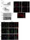
Figure 1
Figure 1. Activation of AurA at the basal body occurs during ciliary disassembly
A. Assembly of cilia. An average of 200 cells were counted in two independent experiments B. Disassembly of cilia induced by serum stimulation. An average of 150 cells were counted in each of 4 experiments. C. Immunofluorescence of quiescent cells with antibody to AurA (green), acetylated α-tubulin (blue), and DNA (red). Scale bar 10 μm. In this and subsequent panels, boxes in main image indicate structures shown at high magnification to right. D. Immunofluorescence of quiescent cells with polyclonal rabbit antibody to HEF1 (green), also visualizing acetylated α-tubulin (blue), and DNA (red); compare also to E. Scale bar 10 μm. E. Immunofluorescence of quiescent cells with monoclonal antibody to HEF1 (green), also visualizing γ-tubulin (blue) and DNA (red). Scale bar 5 μm. See also Supplemental Figure S3A. F. Immunofluorescence of quiescent cells with antibody to phospho-Aura (green), acetylated α-tubulin (blue), and DNA (red). Scale bar 12.5 μm. G. Immunofluorescence of serum-stimulated cells with antibody to phospho-Aura (green), acetylated α-tubulin (blue), and DNA (red). Scale bar 5 μm. H. Western analysis of AurA and HEF1 in hTERT-RPE1 cells after serum stimulation. Western blots shown represent strips and reprobes of a single gel. Higher molecular weight HEF1 band reflects hyperphosphorylation, and coincides with AurA activation and ciliary disassembly at 2 and 24 hours after serum addition (at time point 0). Light gray arrow indicates cross-reactivity of phospho-AurA directed antibody with total AurA; black arrow indicates phospho-AurA. See also Supplemental Figure 1H. I. Immunofluorescence depicting AurA activation in serum-stimulated cells during disassembly of cilia. All images are merged panels of acetylated α-tubulin (red), phospho-AurA or total AurA (green) and DNA (blue).
Figure 2
Figure 2. Activation of AurA is necessary for ciliary resorption
A. Disassembly of cilia in cells treated with siRNA to AurA or HEF1, or with Scrambled (Scr) control siRNA, for 0 to 24 hours after serum addition. Assay performed 3 times, with an average of 100 cells counted/experiment by acetylated tubulin staining. Results were confirmed using a second antibody (anti-glutamylated tubulin) to independently score cilia following depletion (Supplemental Figures S4D, S4E). B. Ciliary disassembly was induced in ciliated cells pre-treated with control (Scr), AurA-_targeted (siA), or HEF1-_targeted (siH) siRNA by supplementing growth media with serum. At 2, 12, and 24 hours after addition of serum, AurA was immunoprecipitated and used for an in vitro kinase assay as in (Pugacheva and Golemis, 2005). Shown, 32P-labelled phosphorylated histone H3 (top) and total histone H3 in the reaction (stained with Coomassie Blue, bottom). C. Length of cilia in untreated hTERT-RPE1 cells (--), or the hTERT-RPE1 cells treated with control (Scr) or HEF1 _targeting siRNA, at the indicated time points. D. Ciliated hTERT-RPE1 cells were treated with AurA inhibitor (PHA-680632) or DMSO, then disassembly of cilia tracked for 24 hours post serum addition. The in vitro IC50 of PHA-680632 is 27 nM for AurA; this compound also less potently inhibits AurC, AurB, and FGFR1 (IC50 120, 185, and 390 nM, respectively, (Soncini et al., 2006)). Results were confirmed using anti-glutamylated tubulin, as shown in Supplemental Figure S4D. E. Analysis performed in parallel with experiments described in D demonstrates PHA-680632 blocks appearance of T288-phospho-AurA (visualized with antibody from BioLegend), and HEF1 phosphorylation (115 kDa form), in reference to DMSO (-) at the 2 and 24 hour time points. Black arrows marks phosphorylated AurA, and hyperphosphorylated (p115) HEF1; gray arrow indicates p105 HEF1. See also Figure S1I. F. Cells were treated with indicated concentrations of the AurA inhibitor PHA-680632, and then AurA immunoprecipitated, and used for in vitro kinase reactions (left) or whole cell lysates used for Western analysis with antibody to total or phosphorylated AurA (right). G. Immunofluorescence analysis of appearance of phospho-AurA at times indicated after serum stimulation in DMSO- or PHA-680632-treated cells. DNA (blue), acetylated α-tubulin (red), and T288-phospho-AurA (green). In 18 hr DMSO/ph-AurA, an asterisk (*) marks a rare observation of phospho-AurA at the base of a shortened cilium. H. FACS analysis of cells treated with DMSO vehicle or PHA-680632 at the times indicated after serum stimulation.
Figure 3
Figure 3. Microinjection of active AurA causes rapid loss of cilia
A. Microinjection of wild type AurA, T288A or D274N mutant AurA, or GST, or PBS buffer, into hTERT-RPE1 cells with pre-formed cilia. (-), uninjected controls. Time reflects minutes from injection to initiating fixation of slides. Experiments repeated 3 times, with >100 injected cells scored in each experiment. B. Cilia 45 minutes post-injection of AurA or D274N. Red, acetylated α-tubulin; blue, glutamylated α-tubulin (a second independent marker of cilia); blue, DNA; green, Dextran488 indicates injected cells. High magnification images to right are from boxed cells; * marks magnification of uninjected cells. C. AurA and mutants (D274N, T288A) were incubated with histone H3 (17 kD) and MBP (22 kD) substrates in an in vitro kinase assay, confirming the activity of kinase. +Lysate indicates that mutants were incubated for 3 hours at 4°C with hTERT-RPE1 cell lysate, then pulled down and used for the kinase assay.
Figure 4
Figure 4. HDAC6 activity is necessary for resorption of cilia
A. Treatment of hTERT-RPE1 cells with histone deacetylase inhibitors prevents ciliary resorption. Cells were incubated with indicated compounds or vehicle (DMSO) at concentrations described in Methods for 2 hours prior to induction of ciliary disassembly. The assay was performed 3 times, with an average of 100 cells counted/time point. B. TSA and tubacin increase intracellular levels of acetylated tubulin. Shown, Western blot with indicated antibodies showing levels of acetylated tubulin in cells treated with TSA, tubacin, niltubacin, or vehicle (DMSO) C. GSK3β inhibitor and farnesyltransferase inhibitor (FTI) do not inhibit ciliary disassembly. D. Depletion of HDAC6 restricts serum-induced disassembly of cilia in hTERT-RPE1 cells transfected for 48 hrs with siRNAs to HDAC6, HDAC2, or a scrambled control. E. Western analysis of hTERT-RPE1 cells treated with siRNA to HDAC6, HDAC2, or scrambled control. F. Active AurA or PBS were microinjected into hTERT-RPE1 cells pretreated for 2 hours with tubacin or DMSO.
Figure 5
Figure 5. Direct phosphorylation by AurA activates HDAC6 tubulin deacetylase activity
A. hTERT-RPE1 whole cell lysate (WCL) of cells treated with AurA inhibitor PHA 680632 (+) or with vehicle (-) was analyzed by Western blot directly, or following immunoprecipitation (IP) with antibody to AurA, using antibodies as indicated. The immunoprecipitation of the slow-migrating form of HDAC6 is not impacted by treatment of cells with PHA-680632, indicating that it most likely represents HDAC6 modified by an additional (unknown) cellular kinase/s. B. AurA phosphorylates HDAC6. In vitro translated and immunoprecipitated HDAC2 or HDAC6 (HD2, HD6), or recombinant GST (-), were mixed with recombinant AurA and used in an in vitro kinase assay. Reaction was split and used for autoradiography (32P) or Western Blot (WB) C. In vitro translated HDAC2 or HDAC6 (HD2, HD6) were immunoprecipitated (IPed). IPs were mixed with AurA (+) or buffer (-), then used for either an in vitro tubulin deacetylation assay, or in an in vitro kinase assay using γ-32P-ATP (see Methods). Reaction mix was visualized by Western blot and by autoradiography, as indicated. D. HDAC6 localizes to disassembling cilia 2 hours after serum treatment. Scale bar, 15 μM.
Figure 6
Figure 6. A role for IFT proteins in AurA-induced ciliary resoprtion
A. Western blot demonstrating siRNA depletion of IFT88 (siIFT88) in ciliated hTERT-RPE1 cells at times following serum treatment, relative to scramble-depleted control. B. Immunofluorescence matching Figure 6A at time 0, indicating relative degree of depletion of IFT88 at the basal body. C. Ciliary disassembly in IFT88- depleted (s88) versus Scr-depleted cells, at 0, 12, or 18 hours after serum treatment, based on the total cell population (gray bars). Black bars (right) indicate % ciliated cells at time 0 calculated specifically from cells confirmed by immunofluorescence to have significant IFT88 staining (88+), or to be well-depleted for IFT88 (88-). D. Cells treated with scrambled (Scr) or AurA-_targeting (siAurA) siRNAs, or with PHA-680632 were fixed 2 hours after serum-initiated disassembly. Shown, immunofluorescence indicating cilia (anti-acetylated α-tubulin, red) and IFT88 (green). Insets are enlargements of boxed ciliary structures; arrows indicate direction of ciliary projection relative to basal body. Scale bars, 10 μm.
Figure 7
Figure 7. Working Model
A. Aurora A (AurA) and low levels of HEF1 are localized to the basal body of quiescent, ciliated cells. B. Our data are consistent with a model in which growth factors induce HEF1 expression, promoting HEF1-dependent activation of Aurora A. This results in phosphorylation of ciliary HDAC6 6 (H6) by Aurora A, thereby inducing ciliary resorption.

Comment in

Similar articles

Cited by

References

    1. Anand S, Penrhyn-Lowe S, Venkitaraman AR. AURORA-A amplification overrides the mitotic spindle assembly checkpoint, inducing resistance to Taxol. Cancer Cell. 2003;3:51–62. - PubMed
    1. Andrews PD. Aurora kinases: shining lights on the therapeutic horizon? Oncogene. 2005;24:5005–5015. - PubMed
    1. Benzing T, Gerke P, Hopker K, Hildebrandt F, Kim E, Walz G. Nephrocystin interacts with Pyk2, p130(Cas), and tensin and triggers phosphorylation of Pyk2. Proc Natl Acad Sci U S A. 2001;98:9784–9789. - PMC - PubMed
    1. Benzing T, Walz G. Cilium-generated signaling: a cellular GPS? Curr Opin Nephrol Hypertens. 2006;15:245–249. - PubMed
    1. Bischoff JR, Anderson L, Zhu Y, Mossie K, Ng L, Souza B, Schryver B, Flanagan P, Clairvoyant F, Ginther C, et al. A homologue of Drosophila aurora kinase is oncogenic and amplified in human colorectal cancers. Embo J. 1998;17:3052–3065. - PMC - PubMed

Publication types

MeSH terms

  NODES
Project 1
twitter 2