Improved glucose homeostasis in mice with muscle-specific deletion of protein-tyrosine phosphatase 1B
- PMID: 17724080
- PMCID: PMC2169063
- DOI: 10.1128/MCB.00959-07
Improved glucose homeostasis in mice with muscle-specific deletion of protein-tyrosine phosphatase 1B
Abstract
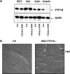
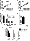
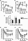
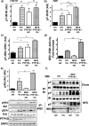
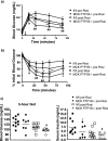
Obesity and type 2 diabetes are characterized by insulin resistance. Mice lacking the protein-tyrosine phosphatase PTP1B in all tissues are hypersensitive to insulin but also have diminished fat stores. Because adiposity affects insulin sensitivity, the extent to which PTP1B directly regulates glucose homeostasis has been unclear. We report that mice lacking PTP1B only in muscle have body weight and adiposity comparable to those of controls on either chow or a high-fat diet (HFD). Muscle triglycerides and serum adipokines are also affected similarly by HFD in both groups. Nevertheless, muscle-specific PTP1B(-/-) mice exhibit increased muscle glucose uptake, improved systemic insulin sensitivity, and enhanced glucose tolerance. These findings correlate with and are most likely caused by increased phosphorylation of the insulin receptor and its downstream signaling components. Thus, muscle PTP1B plays a major role in regulating insulin action and glucose homeostasis, independent of adiposity. In addition, rosiglitazone treatment of HFD-fed control and muscle-specific PTP1B(-/-) mice revealed that rosiglitazone acts additively with PTP1B deletion. Therefore, combining PTP1B inhibition with thiazolidinediones should be more effective than either alone for treating insulin-resistant states.
Figures


Similar articles
-
Liver-specific deletion of protein-tyrosine phosphatase 1B (PTP1B) improves metabolic syndrome and attenuates diet-induced endoplasmic reticulum stress.Diabetes. 2009 Mar;58(3):590-9. doi: 10.2337/db08-0913. Epub 2008 Dec 15. Diabetes. 2009. PMID: 19074988 Free PMC article.
-
Effects of hepatic protein tyrosine phosphatase 1B and methionine restriction on hepatic and whole-body glucose and lipid metabolism in mice.Metabolism. 2015 Feb;64(2):305-14. doi: 10.1016/j.metabol.2014.10.038. Epub 2014 Nov 5. Metabolism. 2015. PMID: 25468142 Free PMC article.
-
Early growth response-1 negative feedback regulates skeletal muscle postprandial insulin sensitivity via activating Ptp1b transcription.FASEB J. 2018 Aug;32(8):4370-4379. doi: 10.1096/fj.201701340R. Epub 2018 Mar 15. FASEB J. 2018. PMID: 29543533
-
PTP1B and TCPTP--nonredundant phosphatases in insulin signaling and glucose homeostasis.FEBS J. 2013 Jan;280(2):445-58. doi: 10.1111/j.1742-4658.2012.08563.x. Epub 2012 Apr 18. FEBS J. 2013. PMID: 22404968 Review.
-
Protein tyrosine phosphatase 1B (PTP1B) and obesity.Vitam Horm. 2013;91:405-24. doi: 10.1016/B978-0-12-407766-9.00017-1. Vitam Horm. 2013. PMID: 23374726 Review.
Cited by
-
T cell protein tyrosine phosphatase (TCPTP) deficiency in muscle does not alter insulin signalling and glucose homeostasis in mice.Diabetologia. 2012 Feb;55(2):468-78. doi: 10.1007/s00125-011-2386-z. Epub 2011 Nov 29. Diabetologia. 2012. PMID: 22124607 Free PMC article.
-
Deficiency of PTP1B in leptin receptor-expressing neurons leads to decreased body weight and adiposity in mice.Endocrinology. 2012 Sep;153(9):4227-37. doi: 10.1210/en.2012-1548. Epub 2012 Jul 16. Endocrinology. 2012. PMID: 22802463 Free PMC article.
-
Protein-tyrosine phosphatase 1B expression is induced by inflammation in vivo.J Biol Chem. 2008 May 23;283(21):14230-41. doi: 10.1074/jbc.M800061200. Epub 2008 Feb 14. J Biol Chem. 2008. PMID: 18281274 Free PMC article.
-
Ventromedial hypothalamus-specific Ptpn1 deletion exacerbates diet-induced obesity in female mice.J Clin Invest. 2014 Sep;124(9):3781-92. doi: 10.1172/JCI68585. Epub 2014 Aug 1. J Clin Invest. 2014. PMID: 25083988 Free PMC article.
-
A novel protein tyrosine phosphatase 1B inhibitor with therapeutic potential for insulin resistance.Br J Pharmacol. 2016 Jun;173(12):1939-49. doi: 10.1111/bph.13483. Epub 2016 May 4. Br J Pharmacol. 2016. PMID: 26990621 Free PMC article.
References
-
- Ahmad, F., and B. J. Goldstein. 1995. Increased abundance of specific skeletal muscle protein-tyrosine phosphatases in a genetic model of insulin-resistant obesity and diabetes mellitus. Metabolism 44: 1175-1184. - PubMed
-
- Bence, K. K., M. Delibegovic, B. Xue, C. Z. Gorgun, G. S. Hotamisligil, B. G. Neel, and B. B. Kahn. 2006. Neuronal PTP1B regulates body weight, adiposity and leptin action. Nat. Med. 12: 917-924. - PubMed
-
- Biddinger, S. B., and C. R. Kahn. 2006. From mice to men: insights into the insulin resistance syndromes. Annu. Rev. Physiol. 68: 123-158. - PubMed
-
- Bruning, J. C., M. D. Michael, J. N. Winnay, T. Hayashi, D. Horsch, D. Accili, L. J. Goodyear, and C. R. Kahn. 1998. A muscle-specific insulin receptor knockout exhibits features of the metabolic syndrome of NIDDM without altering glucose tolerance. Mol. Cell 2: 559-569. - PubMed
-
- Byon, J. C., K. A. Kenner, A. B. Kusari, and J. Kusari. 1997. Regulation of growth factor-induced signaling by protein-tyrosine-phosphatases. Proc. Soc. Exp. Biol. Med. 216: 1-20. - PubMed
Publication types
MeSH terms
Substances
Grants and funding
LinkOut - more resources
Full Text Sources
Other Literature Sources
Molecular Biology Databases
Miscellaneous
