. 2007;35(17):5898-912.
doi: 10.1093/nar/gkm607. Epub 2007 Aug 28.

The checkpoint Saccharomyces cerevisiae Rad9 protein contains a tandem tudor domain that recognizes DNA

Affiliations

The checkpoint Saccharomyces cerevisiae Rad9 protein contains a tandem tudor domain that recognizes DNA

Nathalie Lancelot et al. Nucleic Acids Res. 2007.

Abstract

DNA damage checkpoints are signal transduction pathways that are activated after genotoxic insults to protect genomic integrity. At the site of DNA damage, 'mediator' proteins are in charge of recruiting 'signal transducers' to molecules 'sensing' the damage. Budding yeast Rad9, fission yeast Crb2 and metazoan 53BP1 are presented as mediators involved in the activation of checkpoint kinases. Here we show that, despite low sequence conservation, Rad9 exhibits a tandem tudor domain structurally close to those found in human/mouse 53BP1 and fission yeast Crb2. Moreover, this region is important for the resistance of Saccharomyces cerevisiae to different genotoxic stresses. It does not mediate direct binding to a histone H3 peptide dimethylated on K79, nor to a histone H4 peptide dimethylated on lysine 20, as was demonstrated for 53BP1. However, the tandem tudor region of Rad9 directly interacts with single-stranded DNA and double-stranded DNAs of various lengths and sequences through a positively charged region absent from 53BP1 and Crb2 but present in several yeast Rad9 homologs. Our results argue that the tandem tudor domains of Rad9, Crb2 and 53BP1 mediate chromatin binding next to double-strand breaks. However, their modes of chromatin recognition are different, suggesting that the corresponding interactions are differently regulated.


Figures

Figure 1.
Figure 1.
(A) Ribbon representation of the ScRad9[754–947] 3D structure. Only fragment 762–896 is displayed. Assigned regions are in green, unassigned regions are in purple. The β-sheets are colored in magenta, except the strand β0′ which is in cyan. (B) Superimposition of the 3D structures of ScRad9[754–947] (magenta) and Crb2[358–507] (cyan). (C) Superimposition of the 3D structures of ScRad9[754–947] (magenta) and Mm53BP1[1463–1617] (yellow). (D) Ribbon representation of the 3D structure of the Rad9 fragment 762–896, calculated with three additional hydrogen bond restraints deduced from the structural comparison with Crb2 (see text). Colors are the same as in (A). (E) Alignment of the Rad9 sequence 778–896 with sequences of analogous proteins from 17 yeast species and 9 metazoans. This alignment was deduced from the structural alignment of ScRad9[754–947] with human 53BP1 tandem tudor domain [PDB reference 1XNI, (16); PDB reference 2G3R, (14)], mouse 53BP1 tandem tudor domain [PDB reference 1SSF, (15,31)] and fission yeast Crb2 tandem tudor domain [PDB reference 2FHD, (14)]. Red/blue stars indicate Rad9 solvent-exposed/buried residues whose backbone 15N or 1Hn NMR signals are affected by addition of a 10 mer oligonucleotide. Brown stars indicate Rad9 residues whose side chain 15N or 1Hn NMR signals are affected by the oligonucleotide addition.
Figure 1.
Figure 1.
(A) Ribbon representation of the ScRad9[754–947] 3D structure. Only fragment 762–896 is displayed. Assigned regions are in green, unassigned regions are in purple. The β-sheets are colored in magenta, except the strand β0′ which is in cyan. (B) Superimposition of the 3D structures of ScRad9[754–947] (magenta) and Crb2[358–507] (cyan). (C) Superimposition of the 3D structures of ScRad9[754–947] (magenta) and Mm53BP1[1463–1617] (yellow). (D) Ribbon representation of the 3D structure of the Rad9 fragment 762–896, calculated with three additional hydrogen bond restraints deduced from the structural comparison with Crb2 (see text). Colors are the same as in (A). (E) Alignment of the Rad9 sequence 778–896 with sequences of analogous proteins from 17 yeast species and 9 metazoans. This alignment was deduced from the structural alignment of ScRad9[754–947] with human 53BP1 tandem tudor domain [PDB reference 1XNI, (16); PDB reference 2G3R, (14)], mouse 53BP1 tandem tudor domain [PDB reference 1SSF, (15,31)] and fission yeast Crb2 tandem tudor domain [PDB reference 2FHD, (14)]. Red/blue stars indicate Rad9 solvent-exposed/buried residues whose backbone 15N or 1Hn NMR signals are affected by addition of a 10 mer oligonucleotide. Brown stars indicate Rad9 residues whose side chain 15N or 1Hn NMR signals are affected by the oligonucleotide addition.
Figure 2.
Figure 2.
ScRad9[754–947] is required for cell resistance to DNA damage and for Rad53 phosphorylation in G1 phase. (A) rad9Δ (rad9Δ -L157) transformants containing either an empty vector, the YCp50-Rad9 or the YCp50-Rad9Δ Tudor plasmids were grown to exponential phase in YPD medium and 10-fold serial dilutions were spotted on YPD plates supplemented or not with camptothecin (CPT) or 4-nitroquinoline 1-oxide (4-NQO). Sets of YPD plates were also UV- or X-irradiated. (B) rad9Δ (rad9Δ -L157) transformants containing either an empty vector, the YCp50-Rad9 or the YCp50-Rad9Δ Tudor plasmids were arrested in G1 with α-factor (αF), UV irradiated (120 J/m2) and maintained in G1 phase in the continuous presence of α-factor. Aliquots were taken at the indicated times after UV irradiation and analyzed by western blotting using anti-Rad53 antibodies.
Figure 3.
Figure 3.
NMR titrations with dimethylated histone peptides. In (A) is displayed the superimposition of the 1H-15N HSQC spectra of H3K79me2 bound (red) versus free (blue) ScRad9[754–947]. In (B), the same superimposition is shown for Mm53BP1[1463–1617]. Similarly, in (C) is displayed the superimposition of the 1H-15N HSQC spectra of yeast H4K20me2 bound (green) versus free (blue) ScRad9[754–947]. In (D), the same superimposition is shown for metazoan H4K20me2 bound (green) versus free (blue) Mm53BP1[1463–1617]. Peptides were added to the protein fragments up to peptide:protein ratios of (A) 4:1 (B) 7:1 (C) 8:1 (D) 4:1. Peaks shifting or disappearing after histone peptide binding are labeled.
Figure 3.
Figure 3.
NMR titrations with dimethylated histone peptides. In (A) is displayed the superimposition of the 1H-15N HSQC spectra of H3K79me2 bound (red) versus free (blue) ScRad9[754–947]. In (B), the same superimposition is shown for Mm53BP1[1463–1617]. Similarly, in (C) is displayed the superimposition of the 1H-15N HSQC spectra of yeast H4K20me2 bound (green) versus free (blue) ScRad9[754–947]. In (D), the same superimposition is shown for metazoan H4K20me2 bound (green) versus free (blue) Mm53BP1[1463–1617]. Peptides were added to the protein fragments up to peptide:protein ratios of (A) 4:1 (B) 7:1 (C) 8:1 (D) 4:1. Peaks shifting or disappearing after histone peptide binding are labeled.
Figure 4.
Figure 4.
EMSA experiments between a 146 bp double-stranded oligonucleotide and (A) Mm53BP1[1463–1617], (B) ScRad9[754–947]. Increasing concentrations of proteins were incubated with a 146 bp DNA fragment at a concentration of 26 nM: 2-fold (lanes 2 and 7), 8-fold (lanes 3 and 8), 32-fold (lanes 4 and 9) and 128-fold (lanes 5 and 10) molar excess of proteins were used. Lanes 1 and 6 indicate the mobility of naked DNA.
Figure 5.
Figure 5.
DNA-binding properties of Rad9 tudor region. (A) EMSA experiment between a 35 bp single-stranded oligonucleotide and ScRad9[754–947]. (B–D) EMSA experiments between a 35 bp double-stranded oligonucleotide and (B) ScRad9[754–947], (C) ScRad9[754–931], (D) ScRad9[754–931,C788A,C811S,C852S]. (E) EMSA experiment between a 357 bp double-stranded oligonucleotide and ScRad9[754–947]. (F) EMSA experiment between a 35 bp double-stranded oligonucleotide with a 3′ terminal biotin and ScRad9[754–947]. For this experiment, DNA is preincubated with 0.5 mg/ml streptavidin 10 min prior to addition of ScRad9[754–947] in order to allow conjugation of biotin. Increasing concentrations of proteins were incubated with a 357 or 35 bp DNA fragment at a concentration of 26 nM: 9-fold (lane 22), 16-fold (lanes 2, 7, 12, 17 and 27), 18-fold (lane 23), 32-fold (lanes 3, 8, 13, 18 and 28), 36-fold (lane 24), 64-fold (lanes 4, 9, 14, 19 and 29), 72-fold (lane 25) and 128-fold (lanes 5, 10, 15, 20 and 30) molar excess of proteins were used. Lanes 1, 6, 11, 16, 21 and 26 indicate the mobility of naked DNA.
Figure 6.
Figure 6.
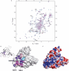
Interaction of ScRad9[754–947] with a 10 bp oligonucleotide. (A)Superimposition of the 1H-15N HSQC spectra of DNA bound (magenta) versus free (cyan) ScRad9[754–947]. DNA/peptide ratio yielded 2.2 in the DNA bound ScRad9[754–947] spectrum. Peak shifting after DNA addition are labeled. (B) Surface representation of ScRad9[754–947] showing the DNA-binding region. When the backbone NH-group chemical shift perturbation (|Δ δ (1H)| + 0.1 × |Δ δ (15N)|) of a residue is higher than the average of all combined shifts plus one SD (0.050 p.p.m.), the residue is colored in purple. When the backbone chemical shift perturbation is comprised between the average value (0.023 p.p.m.) and the average plus 1 SD, the residue is colored in magenta. When the side-chain chemical shift perturbation is higher than 0.023 p.p.m., the residue is colored in pink. The side chain of R862 whose amide is not assigned is shown in cyan. (C) Surface representation of the electrostatic potential of ScRad9[754–947]. The binding and electrostatic surfaces are shown in the same orientation.

Similar articles

Cited by

References

    1. Javaheri A, Wysocki R, Jobin-Robitaille O, Altaf M, Cote J, Kron SJ. Yeast G1 DNA damage checkpoint regulation by H2A phosphorylation is independent of chromatin remodeling. Proc. Natl Acad. Sci. USA. 2006;103:13771–13776. - PMC - PubMed
    1. Vialard JE, Gilbert CS, Green CM, Lowndes NF. The budding yeast Rad9 checkpoint protein is subjected to Mec1/Tel1-dependent hyperphosphorylation and interacts with Rad53 after DNA damage. EMBO J. 1998;17:5679–5688. - PMC - PubMed
    1. Emili A. MEC1-dependent phosphorylation of Rad9p in response to DNA damage. Mol. Cell. 1998;2:183–189. - PubMed
    1. Gilbert CS, Green CM, Lowndes NF. Budding yeast Rad9 is an ATP-dependent Rad53 activating machine. Mol. Cell. 2001;8:129–136. - PubMed
    1. Sun Z, Hsiao J, Fay DS, Stern DF. Rad53 FHA domain associated with phosphorylated Rad9 in the DNA damage checkpoint. Science. 1998;281:272–274. - PubMed

Publication types

MeSH terms

  NODES
twitter 2