Dual roles of autophagy in the survival of Caenorhabditis elegans during starvation
- PMID: 17785524
- PMCID: PMC1950855
- DOI: 10.1101/gad.1573107
Dual roles of autophagy in the survival of Caenorhabditis elegans during starvation
Abstract
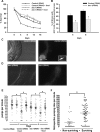
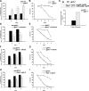
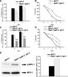
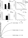
Autophagy is a major pathway used to degrade long-lived proteins and organelles. Autophagy is thought to promote both cell and organism survival by providing fundamental building blocks to maintain energy homeostasis during starvation. Under different conditions, however, autophagy may instead act to promote cell death through an autophagic cell death pathway distinct from apoptosis. Although several recent papers suggest that autophagy plays a role in cell death, it is not known whether autophagy can cause the death of an organism. Furthermore, why autophagy acts in some instances to promote survival but in others to promote death is poorly understood. Here we show that physiological levels of autophagy act to promote survival in Caenorhabditis elegans during starvation, whereas insufficient or excessive levels of autophagy contribute to death. We found that inhibition of autophagy decreases survival of wild-type worms during starvation, and that muscarinic signaling regulates starvation-induced autophagy, at least in part, through the death-associated protein kinase signaling pathway. Furthermore, we found that in gpb-2 mutants, in which muscarinic signaling cannot be down-regulated, starvation induces excessive autophagy in pharyngeal muscles, which in turn, causes damage that may contribute to death. Taken together, our results demonstrate that autophagy can have either prosurvival or prodeath functions in an organism, depending on its level of activation.
Figures


Similar articles
-
Starvation activates MAP kinase through the muscarinic acetylcholine pathway in Caenorhabditis elegans pharynx.Cell Metab. 2006 Apr;3(4):237-45. doi: 10.1016/j.cmet.2006.02.012. Cell Metab. 2006. PMID: 16581001 Free PMC article.
-
Mapping out starvation responses.Cell Metab. 2006 Apr;3(4):235-6. doi: 10.1016/j.cmet.2006.03.002. Cell Metab. 2006. PMID: 16581000
-
To be or not to be, the level of autophagy is the question: dual roles of autophagy in the survival response to starvation.Autophagy. 2008 Jan;4(1):82-4. doi: 10.4161/auto.5154. Epub 2007 Oct 12. Autophagy. 2008. PMID: 17952023 Free PMC article.
-
Death-associated protein kinase (DAPK) and signal transduction: fine-tuning of autophagy in Caenorhabditis elegans homeostasis.FEBS J. 2010 Jan;277(1):66-73. doi: 10.1111/j.1742-4658.2009.07413.x. Epub 2009 Oct 30. FEBS J. 2010. PMID: 19878311 Free PMC article. Review.
-
Role of autophagy in Caenorhabditis elegans.FEBS Lett. 2010 Apr 2;584(7):1335-41. doi: 10.1016/j.febslet.2010.02.002. Epub 2010 Feb 5. FEBS Lett. 2010. PMID: 20138173 Review.
Cited by
-
Autophagy-dependent ribosomal RNA degradation is essential for maintaining nucleotide homeostasis during C. elegans development.Elife. 2018 Aug 13;7:e36588. doi: 10.7554/eLife.36588. Elife. 2018. PMID: 30102152 Free PMC article.
-
ISG20L1 is a p53 family _target gene that modulates genotoxic stress-induced autophagy.Mol Cancer. 2010 Apr 29;9:95. doi: 10.1186/1476-4598-9-95. Mol Cancer. 2010. PMID: 20429933 Free PMC article.
-
Autophagy of germ-granule components, PGL-1 and PGL-3, contributes to DNA damage-induced germ cell apoptosis in C. elegans.PLoS Genet. 2019 May 24;15(5):e1008150. doi: 10.1371/journal.pgen.1008150. eCollection 2019 May. PLoS Genet. 2019. PMID: 31125345 Free PMC article.
-
Autophagy compensates for defects in mitochondrial dynamics.PLoS Genet. 2020 Mar 19;16(3):e1008638. doi: 10.1371/journal.pgen.1008638. eCollection 2020 Mar. PLoS Genet. 2020. PMID: 32191694 Free PMC article.
-
Ubiquitin-mediated response to microsporidia and virus infection in C. elegans.PLoS Pathog. 2014 Jun 19;10(6):e1004200. doi: 10.1371/journal.ppat.1004200. eCollection 2014 Jun. PLoS Pathog. 2014. PMID: 24945527 Free PMC article.
References
-
- Chen C.H., Wang W.J., Kuo J.C., Tsai H.C., Lin J.R., Chang Z.F., Chen R.H., Wang W.J., Kuo J.C., Tsai H.C., Lin J.R., Chang Z.F., Chen R.H., Kuo J.C., Tsai H.C., Lin J.R., Chang Z.F., Chen R.H., Tsai H.C., Lin J.R., Chang Z.F., Chen R.H., Lin J.R., Chang Z.F., Chen R.H., Chang Z.F., Chen R.H., Chen R.H. Bidirectional signals transduced by DAPK–ERK interaction promote the apoptotic effect of DAPK. EMBO J. 2005;24:294–304. - PMC - PubMed
-
- Codogno P., Meijer A.J., Meijer A.J. Autophagy and signaling: Their role in cell survival and cell death. Cell Death Differ. 2005;12 (Suppl. 2):1509–1518. - PubMed
-
- Cuervo A.M. Autophagy: In sickness and in health. Trends Cell Biol. 2004;14:70–77. - PubMed
-
- Inbal B., Bialik S., Sabanay I., Shani G., Kimchi A., Bialik S., Sabanay I., Shani G., Kimchi A., Sabanay I., Shani G., Kimchi A., Shani G., Kimchi A., Kimchi A. DAP kinase and DRP-1 mediate membrane blebbing and the formation of autophagic vesicles during programmed cell death. J. Cell Biol. 2002;157:455–468. - PMC - PubMed
Publication types
MeSH terms
Substances
Grants and funding
LinkOut - more resources
Full Text Sources
Other Literature Sources
Molecular Biology Databases
