. 2008 Jan 1;22(1):37-49.
doi: 10.1101/gad.1609708.

Deubiquitylation of histone H2A activates transcriptional initiation via trans-histone cross-talk with H3K4 di- and trimethylation

Affiliations

Deubiquitylation of histone H2A activates transcriptional initiation via trans-histone cross-talk with H3K4 di- and trimethylation

Takeya Nakagawa et al. Genes Dev. .

Abstract

Transcriptional initiation is a key step in the control of mRNA synthesis and is intimately related to chromatin structure and histone modification. Here, we show that the ubiquitylation of H2A (ubH2A) correlates with silent chromatin and regulates transcriptional initiation. The levels of ubH2A vary during hepatocyte regeneration, and based on microarray expression data from regenerating liver, we identified USP21, a ubiquitin-specific protease that catalyzes the hydrolysis of ubH2A. When chromatin is assembled in vitro, ubH2A, but not H2A, specifically represses the di- and trimethylation of H3K4. USP21 relieves this ubH2A-specific repression. In addition, in vitro transcription analysis revealed that ubH2A represses transcriptional initiation, but not transcriptional elongation, by inhibiting H3K4 methylation. Notably, ubH2A-mediated repression was not observed when H3 Lys 4 was changed to arginine. Furthermore, overexpression of USP21 in the liver up-regulates a gene that is normally down-regulated during hepatocyte regeneration. Our studies revealed a novel mode of trans-histone cross-talk, in which H2A ubiquitylation controls the di- and trimethylation of H3K4, resulting in regulation of transcriptional initiation.


Figures
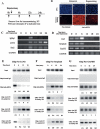
Figure 1.
Figure 1.
Histone H2A ubiquitylation plays a role in gene silencing during hepatocyte regeneration. (A) Experimental scheme. Hepatectomized liver was used for quiescent (0 h) specimens, whereas residual mouse liver was removed and analyzed as regenerating liver at the indicated times after hepatectomy. (B) ubH2A was detected by anti-ubH2A antibodies with quiescent and regenerating hepatocytes, which were obtained by hepatectomy and residual liver resected 12 h after hepatectomy, respectively. Although most of the nuclear ubH2A disappeared 12 h after hepatectomy, a significant amount of ubH2A still persisted in the regenerating nuclei. (C,D) RT–PCR analysis of six representative genes, with altered expression during hepatocyte regeneration, based on expression microarray data together with GAPDH that exists in multiple loci in the mammalian genome. cDNAs for RT–PCR were obtained from mouse livers at the indicated times after hepatectomy, and the primers for RT–PCR are described in Supplemental Table 1. (EG) ChIP analysis was carried out using quiescent and regenerating hepatocytes at the indicated time points. Samples were homogenized and immunoprecipitated using 25 μg of rabbit anti-ubH2A (Supplemental Fig. 1), 25 μg of anti-H3 dimethyl K4 (Supplemental Fig. 2), 5 μL of anti-H3 trimethyl K4 serum (Upstate Biotechnology #07-473), and 25 μg of anti-H3 trimethyl K9 (Supplemental Fig. 3) antibodies. The promoter regions of LCN2 and Serpina6 were analyzed by ChIP assays together with GAPDH that exists in multiple loci as a control. Nonspecific IgG was used as a control for immunoprecipitation. Primers for PCR are described in Supplemental Table 1.
Figure 2.
Figure 2.
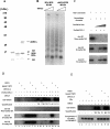
Recombinant USP21 specifically hydrolyzes an isopeptide bond of ubiquitylated H2A in nucleosome form. (A) Expression of USP21 and USP4 in E. coli. (B) Recombinant USP21 (0.013–0.1 pmol) and the same amount of USP4 were incubated with native liver chromatin, prepared by glycerol gradient centrifugation after micrococcal nuclease digestion. After a 2-h incubation, the components were analyzed by Western blotting with a 1:250 dilution of mouse anti-ubH2A (Upstate Biotechnology #06-678) as primary antibody, 2 ng/μL of rabbit anti-mouse (Zymed #61-6400) as secondary antibody, and 125I protein A for detection. As negative control, buffer was used. Amidoblack staining of the immunoblot filter is shown to rule out the degradation of the core histones by the USPs. (C) Recombinant USP21 (0.16 pmol) was incubated with native liver chromatin and an equal amount of free core histones for the indicated times in minutes.
Figure 3.
Figure 3.
Chromatin assembled with ubH2A directly inhibits H3K4 di- and trimethylation by MLL. (A) Coomassie staining of purified H3–H4 tetramer, H2A–H2B, and ubiquitylated H2A–H2B. Separation of ubH2A–H2B from H2A–H2B was performed with rabbit anti-ubH2A antibody purification and elution with bifurcated peptides. Both H2A–H2B and ubH2A–H2B were then further purified by SP Sepharose chromatography. (B) Chromatin assembly with native purified histones by NAP-1/ACF. The samples were partially digested with various concentrations of micrococcal nuclease and then deproteinized. For each set of reaction conditions, the different lanes represent increasing concentrations, from left to right, of micrococcal nuclease used to digest the chromatin. The resulting DNA fragments were subjected to agarose gel electrophoresis and visualized by staining with ethidium bromide. (C) Purified MLL3 was tested for its activity. Antibodies used to detect H3K4 mono- and trimethylation by Western blotting were Upstate Biotechnology #07-436 and Upstate Biotechnology #07-473, respectively. Antibodies used to detect H3K4 dimethylation by Western blotting are characterized in Supplemental Figure 1. As a positive control, native chromatin was used. For H3K4 methylation, a chromatin template containing 300 ng of all four core histones was incubated with purified mouse MLL in TKM buffer (50 mM Tris-Cl at pH 8.0, 20 mM KCl, 1 mM MgCl2) in the presence of 200 μM of SAM. (D,E) A chromatin template was made with the pGIE4 plasmid and purified H3–H4 in addition to H2A–H2B or ubiquitylated H2A–H2B, as indicated. To determine whether ubH2A is hydrolyzed by 0.16 pmol of USP21 or heat-inactivated USP21, Western blotting was performed with anti-ubH2A antibodies. (D) For H3K4 methylation, a chromatin template containing 300 ng of all four core histones was incubated with purified mouse MLL, USP21, heated USP21, and USP21CA, as indicated. H3K4 methylation was detected by Western blotting with anti-H3 mono-, di-, and trimethyl K4 antibodies. (E) For the methylation of H3K9&K27, a chromatin template containing 300 ng of all four core histones was incubated with purified mouse MLL, USP21, heated USP21, and USP21CA, as indicated. Then the template was subsequently incubated with 50 ng of G9a in TKM buffer in the presence of 10 kBq of 3H SAM. To detect the incorporation of 3H methyl groups, fluorography was performed.
Figure 4.
Figure 4.
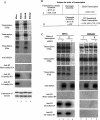
ubH2A represses transcriptional initiation but not elongation, and the H3K4R mutation cancels this repression in vitro. (A) H3wt, cloned in pET11a (gift from James T. Kadonaga), was mutated into alanine(H3K4A), arginine(H3K4R), and glutamine(H3K4Q). The H3–H4 tetramer was expressed and purified, as shown in the lowest panel, basically as described previously (Aihara et al. 2004). Chromatin was assembled using recombinant H3–H4 and native H2A–H2B, and was subjected to GAL4-VP16-mediated transcription. Simultaneously, the chromatin was examined by Western blotting using anti-H3 mono-, di-, and trimethyl K4 antibodies. At the bottom, purified recombinant H3–H4 is shown. For primer extension to test transcription including elongation, oligonucleotide primers were designed to detect different length of transcripts (Supplemental Table 1). (B) Scheme of the order of transcription. For order I, the transcription complex was formed in the presence of GAL4-VP16, nuclear extract, and with limited nucleotides before chromatin assembly and subsequent transcription. For order II, the chromatin was assembled and then GAL4-VP16-mediated transcription was performed. (C) In vitro transcription was performed with chromatin made with H3wt or H3K4R by a different order of transcription, as illustrated in the scheme. Transcription was detected by primer extension as shown in A, and the template was examined by Western blotting with anti-ubH2A and anti-H3 mono-, di-, and trimethyl K4 antibodies after transcription.
Figure 5.
Figure 5.
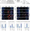
USP21 regulates transcription in vivo. (A) Scheme for DsRed-USP fusion or control constructs that were introduced into liver by in vivo electroporation. (B) The fluorescent protein construct was introduced into liver 48 h before hepatectomy. Samples removed by hepatectomy were designated as quiescent hepatocytes, and the residual liver removed 12 h after hepatectomy was designated as regenerating hepatocytes. (C) Quiescent and regenerating hepatocytes that expressed fluorescent protein are shown, together with DNA staining and immunostaining by anti-ubH2A antibodies, and these three panels are merged, as shown in the rows designated I–V. The arrow indicates a cell with ubH2A decreased by the introduction of USP21wt in rows II and III, and without a significant change in ubH2A by the introduction of USP21CA in rows IV and V. “Q” and “R” indicate quiescent and regenerating hepatocytes, respectively. (D) Quiescent and regenerating hepatocytes that expressed fluorescent protein are shown, together with DNA staining and immunostaining by anti-Serpina6 antibodies, and three panels are merged, as shown in the rows designated I–V. The arrowhead indicates a cell with increased Serpina6 expression by the introduction of USP21wt in row III and with decreased Serpina6 expression by the introduction of USP21CA in row IV. (E,G) USP21 decreased H2A ubiquitylation in both quiescent and regenerating hepatocytes. (F) USP21CA down-regulates Serpina6 in quiescent hepatocytes. (H) USP21wt up-regulates Serpina6 in regenerating liver. (EH) Fifty positive nuclei, together with at least two associated controls, were counted for each experiment, and an error bar is indicated in each graph.
Figure 6.
Figure 6.
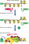
Model for transcriptional initiation from a chromatin template. Deubiquitylation of histone H2A by USP21 activates transcriptional initiation via trans-histone cross-talk with H3K4 di- and trimethylation. The designations “ub” and “me” indicate histone H2A ubiquitylation and H3K4 di- and trimethylation, respectively.

Similar articles

Cited by

References

    1. Aihara H., Nakagawa T., Yasui K., Ohta T., Hirose S., Dhomae N., Takio K., Kaneko M., Takeshima Y., Muramatsu M., et al. Nucleosomal histone kinase-1 phosphorylates H2A Thr 119 during mitosis in the early Drosophila embryo. Genes & Dev. 2004;18:877–888. - PMC - PubMed
    1. Bannister A.J., Kouzarides T. Reversing histone methylation. Nature. 2005;436:1103–1106. - PubMed
    1. Berger S.L. Histone modifications in transcriptional regulation. Curr. Opin. Genet. Dev. 2002;12:142–148. - PubMed
    1. Bernstein B.E., Kamal M., Lindblad-Toh K., Bekiranov S., Bailey D.K., Huebert D.J., McMahon S., Karlsson E.K., Kulbokas E.J., Gingeras T.R., et al. Genomic maps and comparative analysis of histone modifications in human and mouse. Cell. 2005;120:169–181. - PubMed
    1. Briggs S.D., Xiao T., Sun Z.W., Caldwell J.A., Shabanowitz J., Hunt D.F., Allis C.D., Strahl B.D. Gene silencing: Trans-histone regulatory pathway in chromatin. Nature. 2002;418:498. doi: 10.1038/nature00970. - DOI - PubMed
  NODES
twitter 2