Munc13-2-/- baseline secretion defect reveals source of oligomeric mucins in mouse airways
- PMID: 18258655
- PMCID: PMC2375724
- DOI: 10.1113/jphysiol.2007.149310
Munc13-2-/- baseline secretion defect reveals source of oligomeric mucins in mouse airways
Abstract
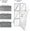
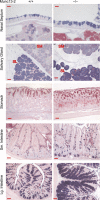
Since the airways of control mouse lungs contain few alcian blue/periodic acid-Schiff's (AB/PAS)+ staining 'goblet' cells in the absence of an inflammatory stimulus such as allergen sensitization, it was surprising to find that the lungs of mice deficient for the exocytic priming protein Munc13-2 stain prominently with AB/PAS under control conditions. Purinergic agonists (ATP/UTP) stimulated release of accumulated mucins in the Munc13-2-deficient airways, suggesting that the other airway isoform, Munc13-4, supports agonist-regulated secretion. Notably, however, not all of the mucins in Munc13-2-deficient airways were secreted, suggesting a strict Munc13-2 priming requirement for a population of secretory granules. AB/PAS+ staining of Munc13-2-deficient airways was not caused by an inflammatory, metaplastic-like response: bronchial-alveolar lavage leucocyte numbers, Muc5ac and Muc5b mRNA levels, and Clara cell ultrastructure (except for increased secretory granule numbers) were all normal. A Muc5b-specific antibody indicated the presence of this mucin in Clara cells of wildtype (WT) control mice, and increased amounts in Munc13-2-deficient mice. Munc13-2 therefore appears to prime a regulated, baseline secretory pathway, such that Clara cell Muc5b, normally secreted soon after synthesis, accumulates in the gene-deficient animals, making them stain AB/PAS+. The defective priming phenotype is widespread, as goblet cells of several mucosal tissues appear engorged and Clara cells accumulated Clara cell secretory protein (CCSP) in Munc13-2-deficient mice. Additionally, because in the human airways, MUC5AC localizes to the surface epithelium and MUC5B to submucosal glands, the finding that Muc5b is secreted by Clara cells under control conditions may indicate that it is also secreted tonically from human bronchiolar Clara cells.
Figures


Similar articles
-
Mucin is produced by clara cells in the proximal airways of antigen-challenged mice.Am J Respir Cell Mol Biol. 2004 Oct;31(4):382-94. doi: 10.1165/rcmb.2004-0060OC. Epub 2004 Jun 10. Am J Respir Cell Mol Biol. 2004. PMID: 15191915 Free PMC article.
-
Mucins MUC5AC and MUC5B Are Variably Packaged in the Same and in Separate Secretory Granules.Am J Respir Crit Care Med. 2022 Nov 1;206(9):1081-1095. doi: 10.1164/rccm.202202-0309OC. Am J Respir Crit Care Med. 2022. PMID: 35776514 Free PMC article.
-
Localization of Secretory Mucins MUC5AC and MUC5B in Normal/Healthy Human Airways.Am J Respir Crit Care Med. 2019 Mar 15;199(6):715-727. doi: 10.1164/rccm.201804-0734OC. Am J Respir Crit Care Med. 2019. PMID: 30352166 Free PMC article.
-
Regulated airway goblet cell mucin secretion.Annu Rev Physiol. 2008;70:487-512. doi: 10.1146/annurev.physiol.70.113006.100638. Annu Rev Physiol. 2008. PMID: 17988208 Review.
-
Mucus hypersecretion in asthma: causes and effects.Curr Opin Pulm Med. 2009 Jan;15(1):4-11. doi: 10.1097/MCP.0b013e32831da8d3. Curr Opin Pulm Med. 2009. PMID: 19077699 Free PMC article. Review.
Cited by
-
Recent progress in histochemistry and cell biology.Histochem Cell Biol. 2012 Apr;137(4):403-57. doi: 10.1007/s00418-012-0933-4. Epub 2012 Feb 25. Histochem Cell Biol. 2012. PMID: 22366957 Review.
-
Epithelial tethering of MUC5AC-rich mucus impairs mucociliary transport in asthma.J Clin Invest. 2016 Jun 1;126(6):2367-71. doi: 10.1172/JCI84910. Epub 2016 May 16. J Clin Invest. 2016. PMID: 27183390 Free PMC article.
-
Mucin production during prenatal and postnatal murine lung development.Am J Respir Cell Mol Biol. 2011 Jun;44(6):755-60. doi: 10.1165/rcmb.2010-0020oc. Am J Respir Cell Mol Biol. 2011. PMID: 21653907 Free PMC article.
-
Autophagy proteins control goblet cell function by potentiating reactive oxygen species production.EMBO J. 2013 Dec 11;32(24):3130-44. doi: 10.1038/emboj.2013.233. Epub 2013 Nov 1. EMBO J. 2013. PMID: 24185898 Free PMC article.
-
Club cell CREB regulates the goblet cell transcriptional network and pro-mucin effects of IL-1B.Front Physiol. 2023 Dec 20;14:1323865. doi: 10.3389/fphys.2023.1323865. eCollection 2023. Front Physiol. 2023. PMID: 38173934 Free PMC article.
References
-
- Abdullah LH, Bundy JT, Ehre C, Davis CW. Mucin secretion and PKC isoforms in SPOC1 goblet cells: differential activation by purinergic agonist and PMA. Am J Physiol Lung Cell Mol Physiol. 2003;285:L149–L160. - PubMed
-
- Abdullah LH, Conway JD, Cohn JA, Davis CW. Protein kinase C and Ca2+ activation of mucin secretion in airway goblet cells. Am J Physiol Lung Cell Mol Physiol. 1997;273:L201–L210. - PubMed
-
- Andrews-Zwilling YS, Kawabe H, Reim K, Varoqueaux F, Brose N. Binding to RAB3A-interacting molecule RIM regulates the presynaptic recruitment of Munc13–1 and ubMunc13–2. J Biol Chem. 2006;281:19720–19731. - PubMed
Publication types
MeSH terms
Substances
Grants and funding
LinkOut - more resources
Full Text Sources
Molecular Biology Databases
Miscellaneous

