. 2008 Jun 2:8:158.
doi: 10.1186/1471-2407-8-158.

Vav3 oncogene activates estrogen receptor and its overexpression may be involved in human breast cancer

Affiliations

Vav3 oncogene activates estrogen receptor and its overexpression may be involved in human breast cancer

Kiwon Lee et al. BMC Cancer. .

Abstract

Background: Our previous study revealed that Vav3 oncogene is overexpressed in human prostate cancer, activates androgen receptor, and stimulates growth in prostate cancer cells. The current study is to determine a potential role of Vav3 oncogene in human breast cancer and impact on estrogen receptor a (ERalpha)-mediated signaling axis.

Methods: Immunohistochemistry analysis was performed in 43 breast cancer specimens and western blot analysis was used for human breast cancer cell lines to determine the expression level of Vav3 protein. The impact of Vav3 on breast cancer cell growth was determined by siRNA knockdown of Vav3 expression. The role of Vav3 in ERalpha activation was examined in luciferase reporter assays. Deletion mutation analysis of Vav3 protein was performed to localize the functional domain involved in ERalpha activation. Finally, the interaction of Vav3 and ERalpha was assessed by GST pull-down analysis.

Results: We found that Vav3 was overexpressed in 81% of human breast cancer specimens, particularly in poorly differentiated lesions. Vav3 activated ERalpha partially via PI3K-Akt signaling and stimulated growth of breast cancer cells. Vav3 also potentiated EGF activity for cell growth and ERalpha activation in breast cancer cells. More interestingly, we found that Vav3 complexed with ERalpha. Consistent with its function for AR, the DH domain of Vav3 was essential for ERalpha activation.

Conclusion: Vav3 oncogene is overexpressed in human breast cancer. Vav3 complexes with ERalpha and enhances ERalpha activity. These findings suggest that Vav3 overexpression may aberrantly enhance ERalpha-mediated signaling axis and play a role in breast cancer development and/or progression.


Figures

Figure 1
Figure 1
Overexpression of Vav3 in human breast cancer. (A) The normal breast epithelial cells reveal negative immunoreactivity for Vav3 (200× magnification). Breast adenocarcinomas with well-differentiation (B), moderately differentiation(C), and poorly differentiation (D) show positive immunoreactivity (brown staining) for Vav3 in both nucleus and cytoplasm. Arrows indicate the breast epithelial and cancer cells. The microphotographs indicate nucleus feature of the breast epithelial and breast cancer cells (400× magnification). For moderately and poorly differentiation breast cancer cells, the nuclei are significantly enlarged and hyperchromatic with coarse clumping of chromatin, prominent nucleoli, and irregular nuclear membrane.
Figure 2
Figure 2
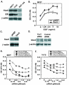
Vav3 is involved in growth of breast cancer cells. (A) Expression analysis of Vav3 and ERα in breast cancer MCF7 and T47D cells and nontumoral breast epithelial MCF-10A cells. The cell extracts were prepared from T47D, MCF7, and MCF-10A cells and subjected to western blot analysis for Vav3 and ERα. β-actin was served as loading control. (B) MCF7 cells were transiently transfected with Vav3 expression vector or control empty vector and then cultured in stripped medium in the absence or presence of EGF for 5 days, followed by MTT assay. The data was presented as absorbance at OD 570 nM. (C) MCF7 cells were transiently transfected with Vav3 expression vector or empty pHEF vector for 3 days, followed by cell extracts preparation and western blot analysis for Vav3. β-actin was served as loading control. (D) Knock down expression of Vav3 upon transfection of Vav3 siRNA. T47D cells were transfected with 5 pM/well of siVav3-247 or control-247 in 6-well plate for 3 days. The cell extracts were prepared and subjected to western blot analysis for Vav3. β-actin was served as loading control. (E and F) MCF7 cells and T47D cells (2500 cells/well in 96-well plate) were transiently transfected with siVav3-247 or control-247 at the concentrations of 0, 0.15, 0.3, 0.6, 1.2, 2.5, 5.0 pM/well using Lipofectamine 2000 for overnight. Then, the cells were cultured in stripped medium without or with E2 (10-9 M) for 5 days, followed by MTT assay. The data was presented as absorbance at OD 570 nM.
Figure 3
Figure 3
Vav3 enhances ERα activity. (A) Hela cells (105 cells/well in 12-well plate) were cotransfected with ERE-Luc (0.5 ug), expression vectors for Vav3, Vav3*, or empty vector pHEF (200 ng) and ERα (50 ng), respectively. Then, the cells were treated without or with E2 (10-9 M) and without or with Tamoxifen. (B and C) MCF7 cells (105 cells/well in 12-well plate) were cotransfected with ERE-Luc (0.5 ug) (B) or pS2-Luc (0.5 ug) (C), and expression vector (0.25 ug) for Vav3*, or empty vector pHEF, respectively. Then, the cells were treated with E2. All transfection and drug treatment are in stripped medium for 24 hours, followed by luciferase assay. Renilla luciferase as an internal control was used to normalize the data. Data are presented as the mean (± SD) of duplicate values of a representative experiment that was independently repeated for five times.
Figure 4
Figure 4
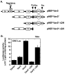
Determination of the functional domain of Vav3 involved in ERα activation. (A) Deletion constructs of Vav3. (B) Hela cells (105 cells/well in 12-well plate) were cotransfected with ERE-Luc (0.5 ug) and expression vectors (200 ng) for Vav3, Vav3*, Vav3*-ΔDH, Vav3*-ΔSH, or empty vector pHEF, as well as expression vector (50 ng) for ERα, respectively. All transfection and drug treatment are in stripped medium for 24 hours, followed by luciferase assay. Renilla luciferase as an internal control was used to normalize the data. Data are presented as the mean (± SD) of duplicate values of a representative experiment that was independently repeated for five times.
Figure 5
Figure 5
Vav3 activates ERα partially via PI3K-Akt signaling and potentiates EGF for ERα activation. (A) Hela cells were cotransfected with ERE-Luc (0.25 ug), expression vectors ERα (25 ng), Vav3* or empty vector pHEF (50 ng), p85+p110 or empty vector pCR3.1 (50 ng). (B) Hela cells were cotransfected with ERE-Luc reporter (0.25 ug/well in 12-well plate), expression vectors ERα (25 ng), dominant-negative Akt expression vector or empty vector pCR3.1 (0.1 ug), and Vav3* expression vector or empty vector pHEF (0.1 ug), respectively. (C) T47D cells were cotransfected with ERE-Luc (0.5 ug) and expression vector for Vav3 or empty vector pHEF (0.25 ug). Then, the cells were treated with Wortmannin (0.5 um). (D) T47D cells were cotransfected with pS2-Luc (0.5 ug) and expression vector for Vav3 or empty vector pHEF (0.25 ug). Then, the cells were treated with EGF (20 ng/ml). All transfection and drug treatment are in stripped medium for 24 hours, followed by luciferase assay. Renilla luciferase as an internal control was used to normalize the data. Data are presented as the mean (± SD) of duplicate values of a representative experiment that was independently repeated for five times.
Figure 6
Figure 6
Sequence analysis of Vav3 and Vav1 genes. (A) The consensus sequences of LXXLL motifs I, II, and III were identified in the DH domain of Vav3 as indicated. Mutation of LLLQELV sequence overlapping with LXXLL motifs II and III has been shown to abolish the GEF activity of Vav3. (B) A consensus sequence of the nucleus localization signal (NLS) in Vav3 was localized in the PH domain.
Figure 7
Figure 7
Vav3 complexes with ERα by GST pull down analysis. (A) GST-Vav3-DH+PH fusion protein. (B) GST-Vav3-DH+PH fusion protein (lane 3 and 4) and control GST protein (lane 1 and 2) were subjected to pull down reaction in the absence (lane 1 and 3) and presence (land 2 and 4) of cell extract derived from MCF7 cells. The samples were fractionated in SDS-PAGE and stained with Coomassie Blue. (C) The same samples were subjected to western blot analysis for ERα.

Similar articles

Cited by

References

    1. Bustelo XR. Regulatory and signaling properties of the Vav family. Mol Cell Biol. 2000;20:1461–1477. doi: 10.1128/MCB.20.5.1461-1477.2000. - DOI - PMC - PubMed
    1. Bustelo XR. Vav proteins, adaptors and cell signaling. Oncogene. 2001;20:6372–6381. doi: 10.1038/sj.onc.1204780. - DOI - PubMed
    1. Katzav S, Martin-Zanca D, Barbacid M. vav, a novel human oncogene derived from a locus ubiquitously expressed in hematopoietic cells. Embo J. 1989;8:2283–2290. - PMC - PubMed
    1. Zugaza JL, Lopez-Lago MA, Caloca MJ, Dosil M, Movilla N, Bustelo XR. Structural determinants for the biological activity of Vav proteins. J Biol Chem. 2002;277:45377–45392. doi: 10.1074/jbc.M208039200. - DOI - PubMed
    1. Moores SL, Selfors LM, Fredericks J, Breit T, Fujikawa K, Alt FW, Brugge JS, Swat W. Vav family proteins couple to diverse cell surface receptors. Mol Cell Biol. 2000;20:6364–6373. doi: 10.1128/MCB.20.17.6364-6373.2000. - DOI - PMC - PubMed

Publication types

MeSH terms

Substances

  NODES
Association 1
INTERN 3
twitter 2