Correlation of enhanced thrombospondin-1 expression, TGF-beta signalling and proteinuria in human type-2 diabetic nephropathy
- PMID: 18676351
- PMCID: PMC2639063
- DOI: 10.1093/ndt/gfn399
Correlation of enhanced thrombospondin-1 expression, TGF-beta signalling and proteinuria in human type-2 diabetic nephropathy
Abstract
Background: Activation of the thrombospondin-1 (TSP-1)-TGF-beta pathway by glucose and the relevance of TSP-1-dependent activation of TGF-beta for renal matrix expansion, renal fibrosis and sclerosis have previously been demonstrated by our group in in vivo and in vitro studies. Design and methods. We investigated renal biopsies (n = 40) and clinical data (n = 30) of patients with diabetic nephropathy. Ten kidneys without evidence of renal disease served as controls. Glomerular and cortical expression of TSP-1, p-smad2/3, fibrosis and glomerular sclerosis (PAS) were assessed by immunhistochemical staining and related with clinical data.
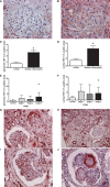
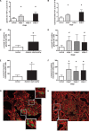
Results: Glomerular (g) and cortical (c) TSP-1 were increased during diabetic nephropathy (g: 2.62 +/- 2.65; c: 4.5 +/- 4.2) compared to controls (g: 0.67 +/- 0.7; c: 1.5 +/- 1.2). P-smad2/3 was significantly increased (g: 16.7 +/- 12.9; c: 148.7 +/- 92.8) compared to controls (g: 7.1 +/- 3.6; c: 55 +/- 25; P < 0.05). TSP-1 was coexpressed with p-smad2/3 as an indicator of TGF-beta activation. TSP-1 correlated with enhanced tubulointerstitial p-smad2/3 positivity (r = 0.39 and r = 0.4, P < 0.05) and glomerular p-smad2/3 correlated with proteinuria (r = 0.35, P < 0.05).
Conclusions: In summary, the present study suggests a functional activity of the TSP-1/TGF-beta axis, especially in the tubulointerstitium of patients with diabetic nephropathy. The positive correlation of glomerular p-smad2/3 positivity with proteinuria further supports the importance of the TSP-1/TGF-beta system as a relevant mechanism for progression of human type-2 diabetic nephropathy.
Figures


Similar articles
-
Thrombospondin-1 is an endogenous activator of TGF-beta in experimental diabetic nephropathy in vivo.Diabetes. 2007 Dec;56(12):2982-9. doi: 10.2337/db07-0551. Epub 2007 Sep 18. Diabetes. 2007. PMID: 17878288
-
Blockade of TSP1-dependent TGF-β activity reduces renal injury and proteinuria in a murine model of diabetic nephropathy.Am J Pathol. 2011 Jun;178(6):2573-86. doi: 10.1016/j.ajpath.2011.02.039. Am J Pathol. 2011. PMID: 21641382 Free PMC article.
-
Activation of the TGF-beta/Smad signaling pathway in focal segmental glomerulosclerosis.Kidney Int. 2003 Nov;64(5):1715-21. doi: 10.1046/j.1523-1755.2003.00288.x. Kidney Int. 2003. PMID: 14531804
-
Diabetic nephropathy and transforming growth factor-beta: transforming our view of glomerulosclerosis and fibrosis build-up.Semin Nephrol. 2003 Nov;23(6):532-43. doi: 10.1053/s0270-9295(03)00132-3. Semin Nephrol. 2003. PMID: 14631561 Review.
-
Transforming growth factor-β/Smad signalling in diabetic nephropathy.Clin Exp Pharmacol Physiol. 2012 Aug;39(8):731-8. doi: 10.1111/j.1440-1681.2011.05663.x. Clin Exp Pharmacol Physiol. 2012. PMID: 22211842 Review.
Cited by
-
Stem cell-derived and circulating exosomal microRNAs as new potential tools for diabetic nephropathy management.Stem Cell Res Ther. 2022 Jan 24;13(1):25. doi: 10.1186/s13287-021-02696-w. Stem Cell Res Ther. 2022. PMID: 35073973 Free PMC article. Review.
-
Prevention of diabetic nephropathy in Ins2(+/)⁻(AkitaJ) mice by the mitochondria-_targeted therapy MitoQ.Biochem J. 2010 Nov 15;432(1):9-19. doi: 10.1042/BJ20100308. Biochem J. 2010. PMID: 20825366 Free PMC article.
-
Molecular profiling of urinary extracellular vesicles in chronic kidney disease and renal fibrosis.Front Pharmacol. 2023 Jan 12;13:1041327. doi: 10.3389/fphar.2022.1041327. eCollection 2022. Front Pharmacol. 2023. PMID: 36712680 Free PMC article. Review.
-
Thrombospondin 1 mediates renal dysfunction in a mouse model of high-fat diet-induced obesity.Am J Physiol Renal Physiol. 2013 Sep 15;305(6):F871-80. doi: 10.1152/ajprenal.00209.2013. Epub 2013 Jul 17. Am J Physiol Renal Physiol. 2013. PMID: 23863467 Free PMC article.
-
Activated CD47 regulates multiple vascular and stress responses: implications for acute kidney injury and its management.Am J Physiol Renal Physiol. 2012 Oct 15;303(8):F1117-25. doi: 10.1152/ajprenal.00359.2012. Epub 2012 Aug 8. Am J Physiol Renal Physiol. 2012. PMID: 22874763 Free PMC article. Review.
References
-
- Ritz E, Rychlik I, Locatelli F, et al. End-stage renal failure in type 2 diabetes: a medical catastrophe of worldwide dimensions. Am J Kidney Dis. 1999;34:795–808. - PubMed
-
- Ritz E, Stefanski A. Diabetic nephropathy in type II diabetes. Am J Kidney Dis. 1996;27:167–194. - PubMed
-
- Wolf G, Ziyadeh FN. Molecular mechanisms of diabetic renal hypertrophy. Kidney Int. 1999;56:393–405. - PubMed
-
- Wolf G, Ziyadeh FN. Cellular and molecular mechanisms of proteinuria in diabetic nephropathy. Nephron Physiol. 2007;106:p26–31. - PubMed
-
- Chen S, Jim B, Ziyadeh FN. Diabetic nephropathy and transforming growth factor-beta: transforming our view of glomerulosclerosis and fibrosis build-up. Semin Nephrol. 2003;23:532–543. - PubMed
Publication types
MeSH terms
Substances
LinkOut - more resources
Full Text Sources
Medical
Miscellaneous

