. 2008 Dec;19(12):5506-16.
doi: 10.1091/mbc.e08-05-0544. Epub 2008 Oct 1.

Self-interaction is critical for Atg9 transport and function at the phagophore assembly site during autophagy

Affiliations

Self-interaction is critical for Atg9 transport and function at the phagophore assembly site during autophagy

Congcong He et al. Mol Biol Cell. 2008 Dec.

Abstract

Autophagy is the degradation of a cell's own components within lysosomes (or the analogous yeast vacuole), and its malfunction contributes to a variety of human diseases. Atg9 is the sole integral membrane protein required in formation of the initial sequestering compartment, the phagophore, and is proposed to play a key role in membrane transport; the phagophore presumably expands by vesicular addition to form a complete autophagosome. It is not clear through what mechanism Atg9 functions at the phagophore assembly site (PAS). Here we report that Atg9 molecules self-associate independently of other known autophagy proteins in both nutrient-rich and starvation conditions. Mutational analyses reveal that self-interaction is critical for anterograde transport of Atg9 to the PAS. The ability of Atg9 to self-interact is required for both selective and nonselective autophagy at the step of phagophore expansion at the PAS. Our results support a model in which Atg9 multimerization facilitates membrane flow to the PAS for phagophore formation.


Figures
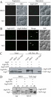
Figure 1.
Figure 1.
Atg9 self-associates independent of other Atg components or nutrient status. (A) Atg9 forms clusters independent of other Atg proteins under both nutrient-rich and nitrogen-starvation conditions. Wild-type (SEY6210; WT) or multiple-knockout (YCY123; MKO) strains were transformed with a plasmid expressing Atg9-GFP driven by the Atg9 native promoter. Cells cultured in nutrient-rich medium (Vegetative) or nitrogen-starved for 3 h (Starvation) were visualized by fluorescence microscopy. (B) Atg9 molecules colocalize. WT (CCH011) or MKO (CCH010) strains expressing integrated Atg9-3GFP and Atg9-3DsRed fusions were grown to midlog phase, subject to mild formaldehyde fixation, and imaged by fluorescence microscopy. The arrows mark examples of the sites where the two chimeras colocalize. (C) Atg9 is coprecipitated by Atg9-protein A (PA) independent of other Atg proteins. atg9Δ (JKY007) or MKO strains cotransformed with centromeric plasmids containing Atg9 and the Atg9-PA fusion driven by the Atg9 native promoter were used for affinity isolation. Cells were cultured in nutrient rich medium (SMD). A strain (UNY102) expressing chromosomally tagged TAP-Atg1 and plasmid-borne Atg9-GFP was used as a control in C and D. Total lysates (Total) and eluted polypeptides (Aff. Pur.) were separated by SDS-PAGE and blotted with anti-Atg9 antiserum in C and D. (D) Atg9 self-interaction in nutrient-deprived conditions is independent of other Atg components. The MKO strain expressing plasmid-borne, native promoter-driven Atg9 and Atg9-PA was used for affinity isolation. Cells were subjected to rapamycin treatment for 2 h (rap) or nitrogen starvation for 3 h (SD-N). T, total lysates; IP, immunoprecipitates. DIC, differential interference contrast. Scale bar, 2 μm.
Figure 2.
Figure 2.
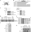
An Atg9 C-terminal mutant disrupts the ability to multimerize. (A) Atg9 self-interaction is mediated through its C terminus. Yeast two-hybrid cells (PJ69–4A) expressing full-length Atg9 with either full-length Atg9 (9 + 9), the Atg9 C terminus (9 + 9C) or the N terminus (9 + 9N), or expressing the Atg9 C terminus alone were grown for 6 d on plates lacking histidine. (B) Schematic representation of Atg9 truncation mutants. N, N terminus; TM, transmembrane domain; C, C terminus. (C) Atg9Δ870-997 and Atg9Δ787-997, but not Atg9Δ766-785, are coprecipitated with full-length Atg9. The MKO strain (YCY123) cotransformed with centromeric plasmids expressing native promoter-driven Atg9-PA and Atg9, Atg9Δ870-997, Atg9Δ787-997, or Atg9Δ766-785, were used for affinity isolation. Total lysates (Input) and eluted polypeptides (IP) were separated by SDS-PAGE and detected with anti-Atg9 antiserum. The MKO strain cotransformed with plasmids encoding Atg9 and PA alone was used as a control in C and D. (D) An Atg9 mutant with a five-amino acid deletion (Atg9Δ766-770) loses self-interaction. MKO cells expressing plasmid-borne Atg9-PA with Atg9, Atg9Δ781-785, or Atg9Δ766-770, driven by the Atg9 native promoter, were used for affinity isolation. (E) Alignment of Atg9 amino acids 766–785. Sequences from S. cerevisiae, Candida albicans, Pichia pastoris, Dictyostelium discoideum, Caenorhabditis elegans, Arabidopsis thaliana, and Mus musculus Atg9 were aligned using the ClustalW program. Amino acid identities and high and low similarities are highlighted in black, dark gray, and light gray, respectively. (F) Atg9Δ766-770 specifically loses self-interaction but not interactions with other Atg9-binding partners. The two-hybrid strain (PJ69–4A) was cotransformed with plasmids expressing the DNA-binding domain-fused wild-type Atg9 (WT) or Atg9Δ766-770 (mut) and a series of known Atg9 binding partners fused with the activation domain. Interactions were monitored by the ability of cells to grow on plates lacking histidine for 5 d. (G) Atg9Δ766-770 interacts with Atg23 similar to wild-type Atg9. An atg9Δ strain expressing an integrated Atg23-PA fusion (CCH020) was transformed with a 2-μm plasmid containing wild-type Atg9-triple hemagglutinin (3HA) or Atg9Δ766-770–3HA. Total lysates (T) and eluates (IP) were separated by SDS-PAGE and detected by anti-HA or anti-PA antibody. An atg9Δ strain expressing plasmid-borne Atg9-3HA and PA was used as a control.
Figure 3.
Figure 3.
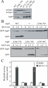
Atg9 self-interaction is required for autophagy activity in both nutrient-rich and starvation conditions. (A) Precursor Ape1 maturation is blocked in cells expressing Atg9Δ766-770. An atg9Δ strain (JKY007) was transformed with an empty vector, or a plasmid expressing wild-type Atg9 or a series of truncation mutants (Δ766-770, Δ771-775, Δ776-780, and Δ781-785) driven by the Atg9 native promoter. Protein extracts were analyzed by Western blotting using antiserum to Ape1 or Atg9. (B) GFP-Atg8 processing is impaired by deletion of residues 766–770 of Atg9. The atg9Δ strain (JKY007) was cotransformed with a GFP-Atg8 plasmid and an empty vector (−), or a plasmid expressing wild-type Atg9 or a series of truncation mutants (Δ766-785, Δ766-770, Δ781-785, and Δ787-997). Cells were grown in nutrient-rich medium to midlog phase then shifted to nitrogen-starvation conditions (SD-N). Aliquots were taken at the indicated time points and analyzed by Western blotting using anti-GFP antibody. (C) Pho8Δ60 activity is reduced in Atg9Δ766-770-expressing cells. The atg9Δ strain (CCH002) transformed with a plasmid expressing native promoter-driven wild-type Atg9 (WT), Atg9Δ766-770, Atg9Δ781-785, or an empty vector (vec), were grown in nutrient-rich medium (SMD) to midlog phase then shifted to starvation medium (SD-N) for 3 h. The Pho8Δ60 activity was measured according to Materials and Methods. Error bars, SD of three independent experiments.
Figure 4.
Figure 4.
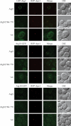
Atg9Δ766–770 is not defective in the PAS recruitment of other Atg proteins. An atg9Δ strain expressing integrated RFP-Ape1 (CCH025) was cotransformed with a plasmid expressing either CUP1 promoter-driven GFP-Atg2 or native ATG18 promoter-driven Atg18-GFP and a 2-micron plasmid expressing wild-type Atg9, Atg9Δ766-770, or an empty vector. An atg9Δ strain expressing chromosomally tagged Atg14-GFP and RFP-Ape1 (CCH026) was transformed with the above 2-μm plasmid expressing wild-type Atg9, Atg9Δ766-770, or an empty vector. Cells were cultured in nutrient-rich medium to midlog phase and imaged by fluorescence microscopy. DIC, differential interference contrast. Scale bar, 2 μm.
Figure 5.
Figure 5.
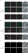
Atg9Δ766-770 is defective in PAS _targeting and phagophore formation. (A) Atg9Δ766-770 has a partial defect in anterograde transport from peripheral sites to the PAS during growth. The atg1Δ atg9Δ strain (CCH001) was cotransformed with a RFP-Ape1 plasmid and a plasmid expressing wild-type Atg9-GFP or Atg9Δ766-770-GFP driven by the CUP1 promoter. Cells were cultured in nutrient-rich medium to midlog phase and imaged by fluorescence microcopy. (B) Atg9Δ766-770 forms an abnormal fragmented phagophore. The cells in A were cultured to midlog phase and subject to nitrogen starvation for 3 h before imaging by fluorescence microscopy. Bottom right panels are enlarged images of the boxed regions. (C) The phagophore is colabeled with Atg9 and Atg8. The atg1Δ atg9Δ strain (CCH001) was cotransformed with plasmids expressing Atg9-GFP and RFP-Atg8. Cells were cultured to midlog phase and subject to nitrogen starvation for 2 h before imaging by fluorescence microscopy. DIC, differential interference contrast. Scale bar, 2 μm.
Figure 6.
Figure 6.
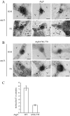
Atg9 localizes to the phagophore structure surrounding the Cvt complex. (A and B) The atg1Δ atg9Δ strain was transformed with a plasmid expressing CUP1 promoter-driven Atg9-GFP (A) or Atg9Δ766-770-GFP (B). Cells were grown to midlog phase, shifted to SD-N for 3 h, and prepared for electron microscopy using freeze substitution (FS) and stained with anti-GFP antibody followed by immunogold as indicated (IEM). Representative images are shown. Arrows mark Atg9 and membranous structures enwrapping the Cvt complex are indicated by arrowheads. V, vacuole. (C) The number of Atg9 or Atg9Δ766-770 molecules around Cvt complexes was quantified based on IEM images shown in A and B. n = 100; p < 0.0001. Error bars, SEM and statistical significance was analyzed by Student's two-tailed t test. Scale bar, 200 nm.
Figure 7.
Figure 7.
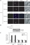
Atg9 functions in an oligomeric state at the PAS. (A) Overexpression of Atg11 rescues Atg9Δ766-770 transport to the PAS in both nutrient-rich and starvation conditions. The atg1Δ atg9Δ strain (CCH001) was cotransformed with centromeric plasmids containing CUP1 promoter-driven CFP-Atg11 and wild-type Atg9-GFP, or Atg9Δ766-770-GFP. Cells were cultured in nutrient-rich medium to midlog phase and imaged by fluorescence microscopy (Vegetative) or were shifted to starvation medium for 3 h before microscopy imaging (Starvation). DIC, differential interference contrast. Scale bar, 2 μm. (B) Atg11 overexpression does not rescue the Cvt pathway in cells expressing Atg9Δ766-770. The atg9Δ strain (JKY007) expressing plasmid-borne wild-type Atg9 or Atg9Δ766-770, with or without CUP1 promoter-driven Atg11, were grown to midlog phase, and protein extracts were analyzed by Western blotting using anti-Ape1 antiserum. (C) Atg11 overexpression does not rescue Atg9Δ766–770 function in bulk autophagy. The atg9Δ strain (CCH002) expressing plasmid-borne CUP1 promoter-driven wild-type Atg9 or Atg9Δ766-770, or an empty vector (vec), with or without CUP1 promoter-driven Atg11, were grown in SMD to midlog phase and shifted to SD-N for 3 h. The Pho8Δ60 activity was measured according to Materials and Methods. Error bars, SD of three independent experiments.
Figure 8.
Figure 8.
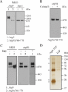
Biochemical characterization of the Atg9-containing complex. (A) Atg9Δ766-770 forms smaller complexes than wild-type Atg9 in the MKO strain. The MKO strain (YCY123) was transformed with a 2-μm plasmid expressing wild-type Atg9 or Atg9Δ766-770. Cell lysates were prepared under native conditions as described in Materials and Methods and analyzed on native gels in A–C. (B) Atg11 overexpression does not rescue the defect in complex formation resulting from Atg9 loss of self-interaction. The atg9Δ strain was cotransformed with plasmids expressing CUP1 promoter-driven HA-Atg11 and Atg9Δ766-770 as indicated. (C) Atg9 forms stable complexes in the wild-type strain during starvation. The MKO (YCY123) or atg9Δ (JKY007) strain was transformed with a 2-μm plasmid expressing wild-type Atg9 or Atg9Δ766-770. Cells were grown in SMD to midlog phase and treated with rapamycin for an additional 1.5 h if indicated. Native gels are detected with antiserum to Atg9 or Ape1 in A, and with Atg9 antiserum in B and C. (D) The Atg9 complex contains five different proteins. Two strains expressing the chromosomally tagged Atg9-TAP (CCH019) or an integrated TAP tag alone driven by the ATG9 promoter (CCH027) were used in tandem affinity purification as described in Materials and Methods. The eluates were analyzed on 10% SDS-PAGE gels and detected by silver staining. Proteins coprecipitated with Atg9 are marked by asterisks.

Similar articles

Cited by

References

    1. Abeliovich H., Zhang C., Dunn W. A., Jr, Shokat K. M., Klionsky D. J. Chemical genetic analysis of Apg1 reveals a non-kinase role in the induction of autophagy. Mol. Biol. Cell. 2003;14:477–490. - PMC - PubMed
    1. Baba M., Osumi M., Scott S. V., Klionsky D. J., Ohsumi Y. Two distinct pathways for _targeting proteins from the cytoplasm to the vacuole/lysosome. J. Cell Biol. 1997;139:1687–1695. - PMC - PubMed
    1. Cao Y., Cheong H., Song H., Klionsky D. J. In vivo reconstitution of autophagy in Saccharomyces cerevisiae. J. Cell Biol., 2008;182:703–713. - PMC - PubMed
    1. Cheong H., Nair U., Geng J., Klionsky D. J. The Atg1 kinase complex is involved in the regulation of protein recruitment to initiate sequestering vesicle formation for nonspecific autophagy in Saccharomyces cerevisiae. Mol. Biol. Cell. 2008;19:668–681. - PMC - PubMed
    1. Cheong H., Yorimitsu T., Reggiori F., Legakis J. E., Wang C.-W., Klionsky D. J. Atg17 regulates the magnitude of the autophagic response. Mol. Biol. Cell. 2005;16:3438–3453. - PMC - PubMed

Publication types

MeSH terms

LinkOut - more resources

  NODES
Association 1
twitter 2