. 2008 Sep 26;31(6):785-99.
doi: 10.1016/j.molcel.2008.09.003.

A piRNA pathway primed by individual transposons is linked to de novo DNA methylation in mice

Affiliations

A piRNA pathway primed by individual transposons is linked to de novo DNA methylation in mice

Alexei A Aravin et al. Mol Cell. .

Abstract

piRNAs and Piwi proteins have been implicated in transposon control and are linked to transposon methylation in mammals. Here we examined the construction of the piRNA system in the restricted developmental window in which methylation patterns are set during mammalian embryogenesis. We find robust expression of two Piwi family proteins, MIWI2 and MILI. Their associated piRNA profiles reveal differences from Drosophila wherein large piRNA clusters act as master regulators of silencing. Instead, in mammals, dispersed transposon copies initiate the pathway, producing primary piRNAs, which predominantly join MILI in the cytoplasm. MIWI2, whose nuclear localization and association with piRNAs depend upon MILI, is enriched for secondary piRNAs antisense to the elements that it controls. The Piwi pathway lies upstream of known mediators of DNA methylation, since piRNAs are still produced in dnmt3L mutants, which fail to methylate transposons. This implicates piRNAs as specificity determinants of DNA methylation in germ cells.


Figures
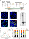
Figure 1
Figure 1. Expression of Piwi proteins though germ cell development
(A) A scheme of spermatogenesis is shown with the timing of expression of Mili, Miwi and Miwi2 indicated. After migration, primordial germ cells (PGCs) arrive at the gonad around 11.5 dpc and expand prior to undergoing cell cycle arrest at 15.5 dpc. The timing of cell cycle arrest coincides with establishment of de novo DNA methylation patterns on transposable elements and imprinted genes. Germ cells resume division after birth at around 3 dpp and initiate meiotic division at 10 dpp. The first cells at pachytene and haploid round spermatid stages appear at days ~14 and ~20, respectively. (B) Expression of GFP-MILI and GFP-MIWI2 transgenes in pre-natal (17.5 dpc) germ cells is shown. MILI exclusively expresses in germ cells and concentrates in perinuclear cytoplasmic granules in both developing testis and ovary. Miwi2 is absent in female germ cells and localizes in the nucleus as well as in cytoplasmic granules in male germ cells. Mili and Miwi2 are co-expressed in male germ cells at 17.5 dpc and are absent from somatic cells. (C) MILI, MIWI2 and MIWI complexes were immunoprecipiated from embryonic gonad at 16.5dpc (MILI and MIWI2) or adult (MIWI) testes. Associated piRNAs were 5’ labeled using sequential phosphatase/kinase reactions. (D) Size profiles are shown for MILI and MIWI2-bound piRNAs cloned from prenatal testis. (E) Small RNA libraries were prepared from size-selected (24–33 nt) total RNA and MILI and MIWI2-immunopurified complexes from the indicated developmental stages and tissue sources. Their small RNA content was classified as indicated.
Figure 2
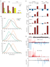
Figure 2. Repeat and gene-derived piRNAs
(A) Shown is the fraction of LINE1, LTR IAP and SINE B1 piRNA in total small RNA libraries at the indicated developmental time points. (B) The size distribution of LINE1, IAP and exon-derived piRNAs in total RNA, MILI and MIWI2 complexes is plotted. (C) The strand orientation of piRNAs derived from transposable elements and exons of protein-coding genes is shown as fold enrichment for sense (red) or antisense (blue) piRNAs. (D) The distribution of piRNAs on LINE1 retrotransposon consensus sequences is shown for the indicated piRNA libraries. piRNAs were mapped to LINE1-A consensus with up to three mismatches. The 5’ end of LINE1 consist of several ~200bp repetitive units.
Figure 3
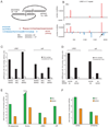
Figure 3. Ping-pong amplification in prenatal piRNAs
(A) A schematic ping-pong pair is shown. piRNAs generated in the ping-pong cycle are complementary to each other and have a 10 nt offset between their 5’ ends. Primary piRNAs have a bias for uridine at position 1 and do not have nucleotide bias at position 10. Secondary piRNAs, generated by primary piRNA guided cleavage have a bias for adenine at position 10 and do not have a bias at position 1. Below is an example of an actual ping-pong pair derived from the 5’ repeats of LINE1-A as shown in (B). (B) The distribution of MILI and MIWI2-associated piRNAs is shown across the 5’ repeats of LINE1-A. MILI has a preference for sense piRNAs, while MIWI2 is bound to both sense and antisense sequences. The most prominent ping-pong pair is indicated by arrow and its sequences are shown in (A). (C) The ping-pong interaction between piRNAs associated with each Piwi family member was measured as the number of piRNA pairs that have a 10 nt overlap between their 5’ ends normalized to the total number of sequences (arbitrary units). Comparison to a control set (overlap at position 2–11) shows that both Piwi proteins are involved in the ping-pong cycle. (D) The extent of the ping-pong interaction measured as in (C) was calculated for sense and antisense transposon piRNAs separately. (E) The ratio of primary (1U, no-10A) to secondary (no-1U, 10A) piRNA was calculated for MILI and MIWI2-bound piRNAs. (F) The correlation between strand orientation of transposon-derived piRNA and their processing category is displayed.
Figure 4
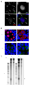
Figure 4. Interaction between MILI and MIWI2 complexes in germ cells
(A) Colocalization of MILI and MIWI2 granules is shown. Detection of MILI and MIWI2 in 17.5 dpc germ cells was performed in Myc-MIWI2 transgenic animals using anti-myc and anti-MILI antibodies. Note that MILI granules are smaller but more numerous as compared to MIWI2 granules. (B) MIWI2 localization depends on MILI. MIWI2 was detected in 17.5 dpc prenatal testes of heterozygous and homozygous MILI embryos (upper panels). MIWI2 is present in the germ cells of MILI mutants, but almost completely delocalizes from the nucleus to the cytoplasm where it is uniformly distributed. MILI localization in cytoplasmic granules and does not change in Miwi2-deficient animals (bottom panels).
Figure 5
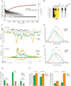
Figure 5. Genomic origins of prenatal piRNAs
(A) Prenatal MIWI2 piRNA clusters were identified by scanning the genome using a 1kB sliding window to find loci that produce at least 10 uniquely-mapped piRNAs per kB. More than 3000 piRNA clusters were thus identified and arranged by the number of uniquely-mapped piRNAs that they produce from left to right. Shown is the genomic size of each cluster (black diamonds) and the cumulative fraction of piRNAs contributed by clusters (red curve). (B) Expression patterns and features of the 8 most prominent piRNA clusters. Expression is calculated as a fraction of cluster-derived piRNAs in total RNA populations normalized for expression level at 16.5 dpc. Also shown is the genomic strand orientation of piRNAs produced by each cluster (strand asymmetry) and the fraction of cluster-derived piRNAs that matches the antisense strand of transposons. piRNA density and size profiles of piRNAs for clusters #1 and #3 are shown in (C), (D) and (E), (F), respectively. (C) piRNA density is shown for the most prominent piRNA cluster (chr. 7: 6526000-6612000). The cluster spans ~ 70 kB and is enriched in transposon sequences (LINE and LTR). The majority of transposons are located on the plus genomic strand and piRNAs are exclusively derived from the minus strand. Therefore, the majority of cluster-derived piRNAs are antisense to transposons. (D) The size profile of piRNAs derived from cluster #1 is shown. (E) piRNA density is plotted for cluster #3 (chr. 8: 48701000-48723000). This cluster is not significantly enriched in transposon sequences. (F) The size profile of piRNAs derived from cluster #3 is shown. (G) Processing features are displayed for piRNAs derived from clusters. The ratio of primary (1U, no-10A) to secondary (no-1U, 10A) piRNAs is shown for total MILI and MIWI2-bound populations and for piRNAs uniquely mapped to two piRNA clusters (#1, single-strand, (C) and #3, double-strand, (E)). For the double strand cluster #3, the ratio of primary to secondary piRNAs in both complexes is similar to that of total population. For the single-strand cluster #1, both MILI and MIWI2 are enriched in primary piRNAs. (H) Shown are fractions of repeat-derived piRNAs (left panel) and their sense/antisense ratio (right panel) for the two most prominent piRNA clusters. The genomic sequences of both clusters are enriched in transposable elements. The expected level of repetitive piRNAs and their sense/antisense ratios are shown (red line) if cluster sequences are randomly sampled.
Figure 6
Figure 6. Links between the DNA methylation and piRNA pathways
(A) MILI-piRNA complexes were immunoprecipitated from testes of 10 dpp wild type and Dnmt3L knock-out animals, and isolated RNAs were 5’ labeled. (B) piRNA populations were analyzed in Dnmt3L mutants. piRNAs isolated from MILI complexes shown in (A) were cloned, sequenced and annotated. Shown are the annotation (left panel) and genomic mapping (right panel) for RNAs from wild-type and Dnmt3L mutants. (C) Northern hybridization with LNA probe to detect an abundant IAP sense piRNA in 10 dpp testes of wild type and Dnmt3L KO mice. Hybridization with let-7 miRNA was used as a loading control. (D) The fraction of sense and antisense IAP piRNAs is shown for wild-type and Dnmt3L mutant libraries. (E) The distribution of piRNAs on the IAP retrotransposon consensus is shown for MILI complexes from wild-type and Dnmt3L mutant animals. (F) Primary and secondary IAP piRNA in Dnmt3L mutants. Shown is the ratio of primary (1U, no-10A) to secondary (no-1U, 10A) piRNAs in the indicated libraries.
Figure 7
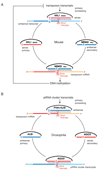
Figure 7. A comparison of ping-pong amplification in mouse and fly
The mouse (A) ping-pong cycle is reversed as compared to Drosophila (B). In Drosophila primary processing of long transcripts derived from piRNA clusters produces both sense and antisense piRNAs that enter a ping-pong cycle that involves AGO3 and AUB. AGO3 primarily binds sense secondary piRNAs and AUB binds primary antisense piRNAs. In mouse piRNA clusters are not the major source of primary piRNAs (Fig. 5A). mRNAs of active transposable elements likely represent the substrate for primary processing resulting in sense piRNAs that preferentially associate with MILI. In prenatal testis both MILI and MIWI2 participate in the amplification cycle. MIWI2 is specifically enriched in secondary antisense piRNAs as compared to MILI. Antisense piRNAs guide DNA methylation of transposable elements sequences in the nucleus probably through recognition of nascent transposon transcripts. After birth, when MIWI2 is no longer expressed, MILI continues to operate the cycle alone. If DNA methylation of transposon sequences is impaired due to downstream mutations in methyltransferase proteins, overexpression of transposon transcripts boosts primary processing and increases the fraction of primary sense piRNAs.

Similar articles

Cited by

References

    1. Aravin A, Gaidatzis D, Pfeffer S, Lagos-Quintana M, Landgraf P, Iovino N, Morris P, Brownstein MJ, Kuramochi-Miyagawa S, Nakano T, et al. A novel class of small RNAs bind to MILI protein in mouse testes. Nature. 2006;442:203–207. - PubMed
    1. Aravin AA, Hannon GJ, Brennecke J. The Piwi-piRNA pathway provides an adaptive defense in the transposon arms race. Science. 2007a;318:761–764. - PubMed
    1. Aravin AA, Sachidanandam R, Girard A, Fejes-Toth K, Hannon GJ. Developmentally regulated piRNA clusters implicate MILI in transposon control. Science. 2007b;316:744–747. - PubMed
    1. Bourc'his D, Bestor TH. Meiotic catastrophe and retrotransposon reactivation in male germ cells lacking Dnmt3L. Nature. 2004;431:96–99. - PubMed
    1. Brennecke J, Aravin AA, Stark A, Dus M, Kellis M, Sachidanandam R, Hannon GJ. Discrete small RNA-generating loci as master regulators of transposon activity in Drosophila. Cell. 2007;128:1089–1103. - PubMed

Publication types

Associated data

  NODES
Association 1
Note 1
Project 1
twitter 2