. 2009 Feb;19(2):221-33.
doi: 10.1101/gr.080861.108. Epub 2008 Dec 1.

H3K27me3 forms BLOCs over silent genes and intergenic regions and specifies a histone banding pattern on a mouse autosomal chromosome

Affiliations

H3K27me3 forms BLOCs over silent genes and intergenic regions and specifies a histone banding pattern on a mouse autosomal chromosome

Florian M Pauler et al. Genome Res. 2009 Feb.

Abstract

In mammals, genome-wide chromatin maps and immunofluorescence studies show that broad domains of repressive histone modifications are present on pericentromeric and telomeric repeats and on the inactive X chromosome. However, only a few autosomal loci such as silent Hox gene clusters have been shown to lie in broad domains of repressive histone modifications. Here we present a ChIP-chip analysis of the repressive H3K27me3 histone modification along chr 17 in mouse embryonic fibroblast cells using an algorithm named broad local enrichments (BLOCs), which allows the identification of broad regions of histone modifications. Our results, confirmed by BLOC analysis of a whole genome ChIP-seq data set, show that the majority of H3K27me3 modifications form BLOCs rather than focal peaks. H3K27me3 BLOCs modify silent genes of all types, plus flanking intergenic regions and their distribution indicates a negative correlation between H3K27me3 and transcription. However, we also found that some nontranscribed gene-poor regions lack H3K27me3. We therefore performed a low-resolution analysis of whole mouse chr 17, which revealed that H3K27me3 is enriched in mega-base-pair-sized domains that are also enriched for genes, short interspersed elements (SINEs) and active histone modifications. These genic H3K27me3 domains alternate with similar-sized gene-poor domains. These are deficient in active histone modifications, as well as H3K27me3, but are enriched for long interspersed elements (LINEs) and long-terminal repeat (LTR) transposons and H3K9me3 and H4K20me3. Thus, an autosome can be seen to contain alternating chromatin bands that predominantly separate genes from one retrotransposon class, which could offer unique domains for the specific regulation of genes or the silencing of autonomous retrotransposons.


Figures

Figure 1.
Figure 1.
H3K27me3 forms BLOCs covering silent genes and intergenic regions. A screen shot from the UCSC genome browser showing the distribution of histone modifications across a 650 kb region of mouse chr 17 (UCSC Mouse [mm6], March 2005) in mouse embryo fibroblasts (MEFs). Profiles for three active histone modifications (H3K4me3, H3K4me2, and H3K9Ac) and the repressive H3K27me3 are displayed. Active histone modifications form peaks detected by ChIPOTle (Buck et al. 2005) (short black bars), while H3K27me3 spreads over large regions (orange bars) called BLOCs (broad local enrichments) that are also detected as dense clusters of ChIPOTle peaks. Expression was determined by hybridizing polyA RNA to the tiling array (RNA chip track). Black and gray bars above the RNA chip track represent, respectively, expressed and silent genes (Supplemental Fig. 4). Positions of genes are shown by Ensembl predictions. Y-axis: log2 ChIP/input ratio or cDNA/input. X-axis: 50 bp oligonucleotides from repeat-masked sequence included on the NimbleGen custom mouse chr 17 tiling array.
Figure 2.
Figure 2.
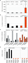
Overview of active and repressive marks on mouse chr 17 in MEFs. The analysis of modifications forming peaks (H3K4me2, H3K4me3, and H3K9Ac) was based on the sum of all chr 17 regions with a probe density of at least eight oligonucleotides per 1500 bp. The analysis of modifications forming BLOCs (H3K27me3) was based on the whole mouse chr 17 tiling array. Data from two independent MEF cell lines (MEFB1 and MEFF) were merged for all analyses (Supplemental Table 1). (A) Chromosome-wide analysis showing the average width of genomic regions enriched by four histone modifications on chr 17, as analyzed by ChIPOTle (all modifications) or the BLOCs algorithm (H3K27me3). Only regions found to be enriched in all technical and biological replicates (Supplemental Table 1) were used in this analysis. (B) The percentage of chr 17 covered by four histone modifications analyzed by ChIPOTle peaks (all modifications) or the BLOCs algorithm (H3K27me3). (C) The percentage of ChIPOTle peaks (all modifications) or BLOCs (H3K27me3) located at the gene body excluding the promoter (solid bar), promoters (diagonally striped bar), and intergenic regions (horizontally striped bar). (D) The significance of overlap between genomic regions enriched for different histone modifications was calculated by identifying Z-scores for all possible pairs of ChIPOTle peaks (all modifications) and BLOCs (H3K27me3) (details in Methods). A high Z-score identifies an overlap that occurs more often than expected compared to a randomized data set.
Figure 3.
Figure 3.
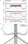
Silent and expressed genes cluster close to H3K27me3 BLOCs. (A) The combined log2 ChIP/input ratios from one MEFF chr 17 ChIP-chip replicate are shown for all expressed genes (top) all silent genes (middle), and silent genes in BLOCs (bottom) relative to the transcription start site (TSS), gene length, and flanking regions. All positions are relative and the length of the gene (black box: expressed/gray box: silent) is defined as 100% and the flanking regions are ±50% of the gene length. Orange line: continuous log2 ChIP/input values, black line: randomized data set. (B) The graph indicates the distance of all genes (dotted line) or of expressed (black line) or silent (gray line) genes relative to the closest H3K27me3 BLOC (gray shaded area) in the MEFF cell line. Distances were calculated separately for each gene and then combined into distance bins (black boxes underneath). The percentage of genes in each bin is indicated on the y-axis. Silent genes are inside and expressed genes are outside, but close to H3K27me3 BLOCs.
Figure 4.
Figure 4.
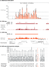
qPCR and ChIP-seq validation of H3K27me3 ChIP-chip BLOCs. (A) Scanning qPCR of one MEFF H3K27me3 ChIP-chip BLOC spanning 365.4 kb in a 650 kb region on mouse chr 17 (12.05–12.70 Mb, UCSC Mouse [mm8], February 2006) with 38 primers (orange bars, error bars indicate variation in three technical replicates) spaced ∼10 kb. Y-axis: %ChIP/input. Mock IP samples were lower than 10% of input with two exceptions (marked with X). Asterisk: low relative qPCR value. M: qPCR assay located in H3K27me3 peaks previously identified in this region by ChIP-seq (Mikkelsen et al. 2007). Lower dotted line indicates the cutoff for significant signals, upper dotted line indicates cutoff for enriched peaks. Primer sequences, qPCR assay details and Ct values are shown in Supplemental Table 2. (B) ChIP-seq of the H3K27me3 ChIP sample assayed by qPCR in A (details in Methods). The ChIP-seq sequence-tag abundance is displayed as 25 bp densities (high density). ChIP-seq BLOCs (horizontal dark red bar) in this region as well as significantly enriched regions (vertical dark red bars) are shown above the ChIP-seq track. Ensembl genes (expressed: black font, silent: gray font, see Supplemental Fig.4) are shown underneath. (C) H3K27me3 ChIP-chip profile (orange peaks) for the region analyzed in (A,B). The orange bar marks the BLOC identified in this region. Genes in this region (black font: expressed, gray font: silent, see Supplemental Fig. 4) are indicated with CG-poor promoters indicated underneath by a bar and CG-rich promoters indicated by a bar plus circle. (D) ChIP-seq BLOCs are identified on all mouse chromosomes in one MEFF data set. Box and whisker plots illustrate the size distribution for each chromosome. The number of BLOCs per Mb and % chromosome coverage by BLOCs is shown below. The ChIP-chip (orange) and ChIP-seq (black) BLOCs from one MEFF data set across chr 17 correlate well, as 82.3% of 1 kb windows show the same H3K27me3 BLOC state (BLOC or no BLOC). Note that MEFF cells are XO (data not shown) and thus have one active X chromosome.
Figure 5.
Figure 5.
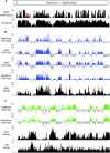
Histone modifications identify two types of chromosome bands that correlate with gene and repeat density. (A) Gene density (http://genome.ucsc.edu) and probe tiling density (repeat-masked, see Methods for details) on the whole chr 17 tiling array chip, were analyzed in 200 kb nonoverlapping windows. The red area marks the imprinted Igf2r cluster (shown in Fig. 4) that contains three small H3K9me3/H4K20me3 peaks and lacks H3K27me3 (Regha et al. 2007). (B) Cumulative ChIP-chip profiles (log2 ChIP/Input hybridization signal means from MEFF and MEFB1 cells, Supplemental Table 1) for H3K27me3, H3Ac, H4Ac, H4K20me1 (blue bars), and SINE density (black bars). Each vertical bar indicates an average signal from 200 kb nonoverlapping windows. (C) Cumulative ChIP-chip profiles for H3K9me3 and H4K20me3 (green bars) and LINEs and LTRs (black bars). Details as in (B).
Figure 6.
Figure 6.
Chromosome band features analyzed by hidden Markov modeling with Viterbi segmentations. (A, top) Giemsa annotated, shows the whole chr 17 Giemsa banding pattern obtained from http://genome.ucsc.edu that is derived from measurement of Giemsa bands relative to whole chromosome length. (Bottom) Predicted, shows the whole chr 17 banding pattern predicted from the enriched histone domains shown in (B) that is based on a nonsynchronized, mainly interphase cell population. Gray shading indicates the predicted relationship between the two binding patterns. Light bands: gene-rich domains enriched for H3K27me3, H3Ac, H4Ac, H4K20me1, and SINEs. Dark bands: gene-poor domains enriched for H3K9me3, H4K20me3, and LINEs/LTRs. Domains sizes larger than 2.5 Mbp are shown and an asterisk marks the imprinted Igf2r cluster analyzed in Figure 4. (B) Hidden Markov modeling with Viterbi segmentations (HMM-Seg) for H3K27me3, H3Ac, H4Ac and H4K20me1 ChIP-chip hybridizations shown as blue boxes, separately for MEFF and MEFB1 cells. Note that all four histone modifications identify the same domains in two independent MEF cell lines. HMM-Seg for H3K9me3 and H3K20me3 is shown as green boxes using the combined MEFF and MEFB1 data. HMM-Seg is shown separately for LINEs + LTRs and for SINEs (black boxes). Note that areas of LINE and LTR density are distinct from areas of SINE density. HMM-Seg for gene density is shown underneath as dark gray boxes. (C) Quantitative analysis comparing the similarity of the above sets of HMM-Seg analyses. Pie charts show the amount of overlap of two groups of segments (similar: black) and the amount of nonoverlap (different: gray). Blue: H3K27me3, H3Ac, H4Ac, and H4K20me1.
Figure 7.
Figure 7.
Summary of H3K27me3 distribution along an autosomal chromosome. (Top) Predicted Giemsa banding pattern in metaphase chr 17 predicted from the data obtained in Figure 6 that is based on a nonsynchronized, mainly interphase cell population. (Middle) Schematic showing enlarged detail of the histone modification profile in regions with a high gene and SINE density that are enriched for repressive H3K27me3 and multiple active histone modifications. At a 100 bp high resolution level, repressive H3K27me3 and active H3Ac/H4Ac histone modifications are mutually exclusive. Expressed genes (black boxes) are characterized by active histone modifications at promoters (blue line) or by H3K36me3 throughout the gene body (not shown). In contrast, silent genes (gray boxes) and nontranscribed intergenic regions are covered by large BLOCs of H3K27me3 (orange line). BLOCs have sharp boundaries that are immediately flanked by transcripts from expressed genes. (Bottom) Enlarged detail showing sites of initial H3K27me3 deposition with the highest levels of H3K27me3 (piled orange hexagons). H3K27me3 could then spread in an unknown manner, along the chromosome, but is excluded or erased by RNA Pol II (red circles) or blocked by boundary elements marked by active histone modifications (blue triangles).

Similar articles

Cited by

References

    1. Azuara V., Perry P., Sauer S., Spivakov M., Jorgensen H.F., John R.M., Gouti M., Casanova M., Warnes G., Merkenschlager M., et al. Chromatin signatures of pluripotent cell lines. Nat. Cell Biol. 2006;8:532–538. - PubMed
    1. Barski A., Cuddapah S., Cui K., Roh T.Y., Schones D.E., Wang Z., Wei G., Chepelev I., Zhao K. High-resolution profiling of histone methylations in the human genome. Cell. 2007;129:823–837. - PubMed
    1. Beisel C., Buness A., Roustan-Espinosa I.M., Koch B., Schmitt S., Haas S.A., Hild M., Katsuyama T., Paro R. Comparing active and repressed expression states of genes controlled by the Polycomb/Trithorax group proteins. Proc. Natl. Acad. Sci. 2007;104:16615–16620. - PMC - PubMed
    1. Bernstein B.E., Mikkelsen T.S., Xie X., Kamal M., Huebert D.J., Cuff J., Fry B., Meissner A., Wernig M., Plath K., et al. A bivalent chromatin structure marks key developmental genes in embryonic stem cells. Cell. 2006a;125:315–326. - PubMed
    1. Bernstein E., Duncan E.M., Masui O., Gil J., Heard E., Allis C.D. Mouse polycomb proteins bind differentially to methylated histone H3 and RNA and are enriched in facultative heterochromatin. Mol. Cell. Biol. 2006b;26:2560–2569. - PMC - PubMed

Publication types

MeSH terms

LinkOut - more resources

  NODES
Note 3
Project 1
twitter 2