. 2009 Apr 10;284(15):9781-7.
doi: 10.1074/jbc.M807849200. Epub 2009 Feb 10.

Synaptotagmin 2 couples mucin granule exocytosis to Ca2+ signaling from endoplasmic reticulum

Affiliations

Synaptotagmin 2 couples mucin granule exocytosis to Ca2+ signaling from endoplasmic reticulum

Michael J Tuvim et al. J Biol Chem. .

Abstract

Synaptotagmin 2 (Syt2) functions as a low affinity, fast exocytic Ca(2+) sensor in neurons, where it is activated by Ca(2+) influx through voltage-gated channels. _targeted insertion of lacZ into the mouse syt2 locus reveals expression in mucin-secreting goblet cells of the airways. In these cells, rapid Ca(2+) entry from the extracellular medium does not contribute significantly to stimulated secretion (Davis, C. W., and Dickey, B. F. (2008) Annu. Rev. Physiol. 70, 487-512). Nonetheless, Syt2(-/-) mice show a severe defect in acute agonist-stimulated airway mucin secretion, and Syt2(+/-) mice show a partial defect. In contrast to Munc13-2(-/-) mice (Zhu, Y., Ehre, C., Abdullah, L. H., Sheehan, J. K., Roy, M., Evans, C. M., Dickey, B. F., and Davis, C. W. (2008) J. Physiol. (Lond.) 586, 1977-1992), Syt2(-/-) mice show no spontaneous mucin accumulation, consistent with the inhibitory action of Syt2 at resting cytoplasmic Ca(2+) in neurons. In human airway goblet cells, inositol trisphosphate receptors are found in rough endoplasmic reticulum that closely invests apical mucin granules, consistent with the known dependence of exocytic Ca(2+) signaling on intracellular stores in these cells. Hence, Syt2 can serve as an exocytic sensor for diverse Ca(2+) signaling systems, and its levels are limiting for stimulated secretory function in airway goblet cells.


Figures

FIGURE 1.
FIGURE 1.
Syt2 is expressed in airway secretory (Clara) cells. A, Western-blotted lung homogenates from WT (+/+), heterozygous (+/–), and null (–/–) mutant mice were probed with rabbit polyclonal antibodies against Syt2. A single band was observed in WT and heterozygous mouse lung tissue at ∼65 kDa. B, bronchial airways of WT and heterozygous mutant mice with lacZ knocked into the syt2 locus were stained with X-gal (blue) and then counterstained with treosin (red). Blue staining is observed only in heterozygous mice in cells that do not contain ciliated tufts. Scale bar in all tissue sections = 10 μm. C, bronchial airways of WT and heterozygous mutant mice were stained with X-gal (blue) and then immunostained with rabbit polyclonal antibodies against mouse CCSP developed with diaminobenzidine (brown). Blue staining is observed in association with brown-stained Clara cells. D, bronchial airways of null mice were labeled with antibodies against β-galactosidase (red) and CCSP (green), and nuclei were labeled with 4′,6-diamidino-2-phenylindole (blue). Both red and green labeling are observed in Clara cells (arrowheads), alternating with ciliated cells that are not labeled with either antibody (arrows). Control WT mice show no red labeling (supplemental Fig. 1). E, bronchial airways of null mice were labeled with antibodies against β-galactosidase (red) or acetylated tubulin (green), and nuclei were labeled with 4′,6-diamidino-2-phenylindole (blue). Red labeling is observed in Clara cells that are not labeled with acetylated tubulin antibodies (arrowheads), whereas green labeling is observed in ciliated cells that are not labeled with β-galactosidase antibodies (arrows).
FIGURE 2.
FIGURE 2.
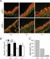
Syt2 mediates stimulated mucin secretion. A, bronchial airways of WT, heterozygous, and homozygous mutant P19 mice that had had been exposed 3 days earlier to aerosolized IL-13 to induce mucous metaplasia (top row), then exposed to aerosolized ATP to induce mucin secretion (bottom row), were stained for intracellular mucin with PAFS (orange). Scale bar, 10 μm. B, intracellular mucin content of bronchial airways from three experiments such as that in Fig. 2A, which included at least three pups of each genotype and five sections from each airway, was measured and analyzed. C, data from B are replotted as the percentage of intracellular mucin released for each genotype relative to WT.
FIGURE 3.
FIGURE 3.
Airway mucin secretion is impaired in heterozygous Syt2 mutant adult mice. A, mucous metaplasia was induced in the airways of WT and heterozygous adult mice of both sexes by intraperitoneal sensitization and aerosol challenge with ovalbumin. Three days later, secretion was stimulated by exposure to aerosolized ATP, and retained intracellular mucin was measured by PAFS staining as in Fig. 2. There was no difference in intracellular mucin content between heterozygous and WT mice in the absence of mucous metaplasia (∼1 nl/mm2, not shown) or in mice with mucous metaplasia prior to stimulation with the ATP aerosol (∼12 nl/mm2, black bars). However, heterozygous mice released only half as much mucin as WT mice after ATP stimulation (white bars). Shown are the results of a representative experiment that was performed three times with similar results. B, data from A are replotted as the percentage of intracellular mucin released for each genotype relative to WT.
FIGURE 4.
FIGURE 4.
ER apical to the nucleus of airway secretory cells is closely apposed to mucin secretory granules. A, frozen human lung section imaged by differential interference contrast (DIC) microscopy (left) and labeled with antibodies against the secretory granule glycoprotein mature MUC5AC (red) and the lumenal ER resident protein PDI (green). An overlay image and a scattergram of the outlined goblet cell indicating the relative intensity of red and green labels for each pixel show a lack of colocalization of the two antibodies. Basal (B), ciliated (C), and goblet (G) cells are marked. Scale bar, 16 μm. B, ER in airway goblet cells is predominantly rough ER. A frozen human lung section labeled with antibodies against the newly synthesized cargo protein immature (nonglycosylated) MUC5AC (red) that labels only rough ER and antibodies against PDI (green) that labels both smooth and rough ER (31) shows a high degree of colocalization in the overlay image and scattergram. Scale identical to A. C, electron micrograph of a goblet cell from normal human lung, with the nucleus (N) marked. Scale bar, 2.5 μm. D, higher magnification of the area indicated in C, with rough ER (RER) and a mucin secretory granule (SG) marked. E, neighboring area from C at the same magnification, with Golgi apparatus (Golgi) and a mucin secretory granule (SG) marked.
FIGURE 5.
FIGURE 5.
IP3-Rs are concentrated in apical ER adjacent to mucin secretory granules but not on granule membranes. A, frozen human lung section labeled with antibodies against IP3-R (red) and PDI (green), with the scattergram showing colocalization in the goblet cell outlined in the overlay image. Scale bar, 16 μm. DIC, differential interference contrast. B, mucin granule membrane and lumenal proteins are not resolved under the optical conditions of our experiments. Paraffin-embedded human lung section labeled with antibodies against the secretory granule surface protein Rab3D (red) and antibodies against the secretory granule lumenal protein mature MUC5AC (green), with the scattergram showing colocalization in the cell outlined in the overlay image. C, IP3-R are not detected on mucin granules. Paraffin-embedded human lung section labeled with antibodies against IP3-R (red) and mature MUC5AC (green) is shown, with the scattergram showing a lack of colocalization in the cell outlined in the overlay image (note the splaying of two clearly separated populations of points to their respective axes).
FIGURE 6.
FIGURE 6.
Juxtaposition of apical endoplasmic reticulum and mucin secretory granules. A, electron micrograph of the apical membrane region of a goblet cell, showing the juxtaposition of a mucin secretory granule (SG) and rough endoplasmic reticulum (rER). This human lung specimen was fixed ∼24 h after harvest for transplantation, and hence fixation is not optimal and internal membrane structures are not well preserved. Nonetheless, the arrays of ribosomes reveal the rough ER and show the intimate relationships between the plasma membrane, granule, rough ER, and mitochondria (M). B, pathway for activation of regulated secretion in airway goblet cells. Extracellular ligands in the airway surface liquid layer (bottom) bind to heptahelical receptors in the apical membrane that activate Gq and phospholipase (PLC) β1, generating the second messengers diacylglycerol (DAG) and inositol trisphosphate (IP3). Diacylglycerol activates the exocytic priming protein Munc13-2 (dotted blue arrow, left fork) and the exocytic regulator protein kinase Cε (data not shown). IP3 induces the release of Ca2+ from endoplasmic reticulum (ER) in the vicinity of mucin-containing secretory granules, activating Syt2 on the surface of secretory granules (dotted blue arrow, right fork) and coactivating Munc13 proteins (data not shown).

Similar articles

Cited by

References

    1. Knowles, M. R., and Boucher, R. C. (2002) J. Clin. Investig. 109 571–577 - PMC - PubMed
    1. Williams, O. W., Sharafkhaneh, A., Kim, V., Dickey, B. F., and Evans, C. M. (2006) Am. J. Respir. Cell Mol. Biol. 34 527–536 - PMC - PubMed
    1. Davis, C. W., and Dickey, B. F. (2008) Annu. Rev. Physiol. 70 487–512 - PubMed
    1. Zhu, Y., Ehre, C., Abdullah, L. H., Sheehan, J. K., Roy, M., Evans, C. M., Dickey, B. F., and Davis, C. W. (2008) J. Physiol. (Lond.) 586 1977–1992 - PMC - PubMed
    1. Ehre, C., Zhu, Y., Abdullah, L. H., Olsen, J., Nakayama, K. I., Nakayama, K., Messing, R. O., and Davis, C. W. (2007) Am. J. Physiol. 293 C1445–C1454 - PubMed

Publication types

LinkOut - more resources

  NODES
Association 1
INTERN 1
Note 1
twitter 2