. 2009 Sep;16(9):1279-88.
doi: 10.1038/cdd.2009.41. Epub 2009 Apr 17.

NEMO/IKKgamma regulates an early NF-kappaB-independent cell-death checkpoint during TNF signaling

Affiliations

NEMO/IKKgamma regulates an early NF-kappaB-independent cell-death checkpoint during TNF signaling

D Legarda-Addison et al. Cell Death Differ. 2009 Sep.

Abstract

TNF receptor 1 (TNFR1) ligation can result in cell survival or cell death. What determines which of the two opposing responses is triggered is not fully understood. The current model suggests that it is the activation of the NF-kappaB pathway and its induction of prosurvival genes, or the lack thereof, which determines the outcome. NF-kappaB essential modifier (NEMO)/IkappaB kinase-gamma (IKKgamma)-deficient cells are highly sensitive to apoptosis, and as NEMO is essential for NF-kappaB activation, it has been assumed that this is due to the lack of NF-kappaB. This study demonstrates that this assumption was incorrect and that NEMO has another antiapoptotic function that is independent of its role in the NF-kappaB pathway. NEMO prevents receptor interacting protein-1 (RIP1) from engaging CASPASE-8 before NF-kappaB-mediated induction of antiapoptotic genes. Without NEMO, RIP1 associates with CASPASE-8 resulting in rapid tumor necrosis factor (TNF)-induced apoptosis. These results suggest that there are two cell-death checkpoints following TNF stimulation: an early transcription-independent checkpoint whereby NEMO restrains RIP1 from activating the caspase cascade, followed by a later checkpoint dependent on NF-kappaB-mediated transcription of prosurvival genes.


Figures
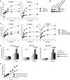
Figure 1
Figure 1. NEMO inhibits TNF-induced apoptosis in a NF-κB-independent manner
(A) Parental 3T8 Jurkat T cells transduced with retrovirus encoding either control protein or IκBαSR, and untransfected NEMO-null 8321 cells were treated with the indicated doses of TNF for 4 and 8 hours. Cell death was measured by staining with PE-conjugated Annexin V and FACS analysis. (B) Cell lysates from 3T8/Con, 3T8/IκBαSR, NEMO-null/IκBαSR, or NEMO-WT/IκBαSR Jurkat T cells were subject to Western blot analysis using anti-NEMO followed by anti-ZAP70 as a loading control. (C) 3T8 cells transfected with control protein or IκBαS, and NEMO-null or NEMO-WT Jurkat T cells transfected with IκBαSR were treated with the indicated doses of TNF for 4, 8 and 24 hours. Cell death was analyzed by staining with PE-conjugated Annexin V. The experiments shown are representative of three experiments that were conducted. (D) Cell death was analyzed as in (A) after stimulation with 10 ng/ml of TNF for 4, 8, and 24 hours. Each bar indicates the mean ± SD of quintuplicate samples treated and analyzed independently. T-test was used to calculate p-values, and significance was determined at p<0.001. (E) Cells were treated with 5 ng/ml of TNF for the indicated times. Cell death was measured by staining with PE-conjugated Annexin V.
Figure 2
Figure 2. NEMO-deficient cells undergo more caspase activation than NEMO-sufficient cells
(A, B) NEMO-null/ IκBαSR or NEMO-WT/ IκBαSR cells were treated with indicated doses of TNF for 2 and 4 hours (A and B, respectively). SDS-soluble cell extracts were subject to Western blot analysis using antibodies specific to CASPASE-8, CASPASE-3, PARP, and ZAP70 (loading control). The experiments shown are representative of three experiments that were conducted. (C) Cell lines were treated with 2 ng/ml of TNF for the indicated times. SDS-soluble cell extracts were subject to Western blot analysis using antibodies specific to CASPASE-8, CASPASE-3, PARP, and ZAP70. The experiments shown are representative of three experiments that were conducted. (C) Cell lines were treated with 2 ng/ml of TNF for the indicated times. SDS-soluble cell extracts were subject to Western blot analysis using antibodies specific to CASPASE-8, CASPASE-3, PARP, and ZAP70. The experiments shown are representative of three experiments that were conducted.
Figure 3
Figure 3. TNF-induced cell death in NEMO-deficient cells requires RIP1
(A) 3T8/IκBαSR and NEMO-null/IκBαSR cells were infected with retroviruses generated with pSUPERretro-GFP vector encoding shRNA directed against non-silencing _target (si-NS) or RIP1 (si-RIP1). Transduced cells were selected by cell sorting for high GFP expression. Knockdown of RIP1 expression was determined by Western blot analysis for RIP1. Anti-ZAP70 blotting was performed as a loading control. (B) The four cell lines used in (A) were treated with the indicated doses of TNF for 4, 8, and 24 hours. Cell death was measured by staining with PE-conjugated Annexin V and FACS analysis. The experiments shown are representative of three experiments that were conducted. (C) Cell death was analyzed as in (B) after stimulation with 10 ng/ml of TNF for 4, 8, and 24 hours. Each bar represents the mean ± SD of quintuplicate samples treated and analyzed independently. P-values <0.001 are indicated.
Figure 4
Figure 4. NEMO requires its ubiquitin-binding domain for its NF-κB-independent anti-apoptotic function
(A) Cell lysates from NEMO-null/IκBαSR, NEMO-WT/IκBαSR, NEMO-Y308S/IκBαSR, NEMO-F312A/IκBαSR, NEMO-L329P/IκBαSR cells were subject to Western blot analysis using anti-NEMO followed by anti-ZAP70 as a loading control. (B) Cell lines from (A) were treated with 5 or 10 ng/ml of TNF for 4, 8, and 24 hours. Cell death was measured by staining with PE-conjugated Annexin V and FACS analysis. Each bar represents the mean ± SD of triplicate samples treated and analyzed independently. T-test was used to calculate p-values and asterisks indicate p<0.01 when compared to values from the NEMO-WT/IκBαSR cell line. (C) Cell lines from (A) were treated with 100 ng/ml of TNF for 15 minutes. FLAG-tagged wild-type NEMO or NEMO mutants were immunoprecipitated from total cell lysates using anti-FLAG M2 affinity beads. The immunoprecipitates were sequentially blotted with anti-RIP1 (first and second panels) and anti-FLAG (third panel) antibodies. Blotting with the two antibodies on 1% of the total cell extracts are shown in the bottom two panels. The experiment shown is representative of three experiments that were conducted.
Figure 5
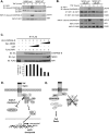
Figure 5. NEMO inhibits RIP1 from associating with CASPASE-8
(A) NEMO-null and NEMO-WT Jurkat T cells were treated with 25 ng/ml TNF for 1 and 2 hours. RIP1 was immunoprecipitated from the lysates using anti-RIP1 antibody. The protein complexes were eluted and subject to sequential Western blot analysis using anti-cleaved CASPASE-8 and anti-RIP1 antibodies. (B) NEMO-null/IκBαSR and NEMO-WT/IκBαSR cells were treated as in panel (A). Blotting was performed sequentially with anti-cleaved CASPASE-8, anti-FADD and anti-RIP1 antibodies. (C) 293EBNA cells were transfected with FLAG-RIP1, and HA-CASPASE-8 in the presence of increasing amounts of myc-NEMO or the negative control GST. Anti-FLAG immunoprecipitations were eluted with the FLAG peptide and sequentially blotted with anti-HA, anti-myc and anti-FLAG. The intensities of the CASPASE-8 bands were determined by densitometry and normalized to the corresponding RIP1 band. The ratios of the intensity in the different samples are presented relative to the ratio in the sample with no GST or NEMO. (D) Model of TNFR1 signaling showing two cell death checkpoints. Upon ligation of TNFR1, ubiquitination of RIP1 occurs rapidly leading to its association with NEMO and other components of the IKK signalsome. This sequesters RIP1 away from CASPASE-8 and serves as the early transcription-independent cell death checkpoint. The formation of the IKK signalsome subsequently leads to the induction of NF-κB-dependent anti-apoptotic genes and this serves as a second cell death checkpoint, which provides for a longlasting genetically programmed protection from death. (E) If the early cell death checkpoint fails, such as when ubiquitination of RIP1 is prevented or if a regulatory component such as NEMO is absent, RIP1 can engage CASPASE-8 and death rapidly ensues. This early NF-κB independent cell death checkpoint serves to determine whether the survival or death pathway is triggered following TNFR1 ligation.

Similar articles

Cited by

References

    1. Aggarwal BB. Signalling pathways of the TNF superfamily: a double-edged sword. Nat Rev Immunol. 2003;3:745–56. - PubMed
    1. Micheau O, Tschopp J. Induction of TNF receptor I-mediated apoptosis via two sequential signaling complexes. Cell. 2003;114:181–90. - PubMed
    1. Yamaoka S, Courtois G, Bessia C, Whiteside ST, Weil R, Agou F, et al. Complementation cloning of NEMO, a component of the IkappaB kinase complex essential for NF-kappaB activation. Cell. 1998;93:1231–40. - PubMed
    1. Rudolph D, Yeh WC, Wakeham A, Rudolph B, Nallainathan D, Potter J, et al. Severe liver degeneration and lack of NF-kappaB activation in NEMO/IKKgamma-deficient mice. Genes Dev. 2000;14:854–62. - PMC - PubMed
    1. He KL, Ting AT. A20 inhibits tumor necrosis factor (TNF) alpha-induced apoptosis by disrupting recruitment of TRADD and RIP to the TNF receptor 1 complex in Jurkat T cells. Mol Cell Biol. 2002;22:6034–45. - PMC - PubMed

Publication types

  NODES
Association 1
twitter 2